Copyright © 2000, American Society for Microbiology. All Rights Reserved.
Strain Identification of
Trichophyton rubrum
by Specific
Amplification of Subrepeat Elements in the
Ribosomal DNA Nontranscribed Spacer
COLIN J. JACKSON,1RICHARD C. BARTON,2* STEVEN L. KELLY,1ANDE. GLYN V. EVANS2
Institute of Biological Sciences, University of Wales Aberystwyth, Aberystwyth, Wales SY23 3DA,1and
Division of Microbiology and Mycology Reference Centre, University of Leeds, and General Infirmary, Leeds LS2 9JT,2United Kingdom
Received 19 June 2000/Returned for modification 19 July 2000/Accepted 16 September 2000
Trichophyton rubrumis the commonest cause of dermatophytosis of skin and nail tissue. Molecular
char-acterization of the T. rubrum ribosomal DNA nontranscribed-spacer region revealed two novel tandemly
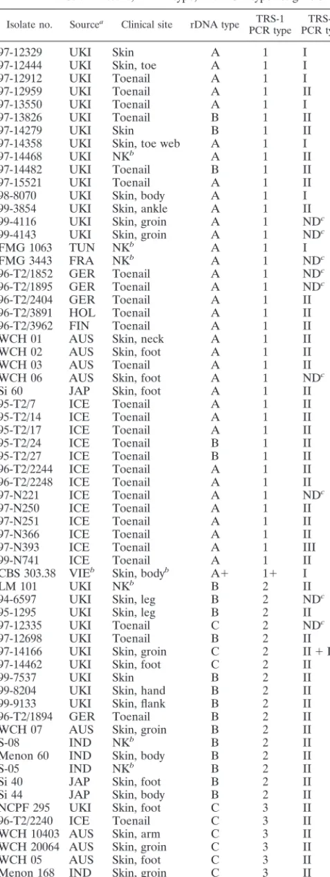
repetitive subelements (TRSs): TRS-1, containing a 27-bp palindromic sequence, and TRS-2. Specific ampli-fication of TRS-1 produced strain-characteristic banding patterns (PCR types), with 21 TRS-1 PCR types recognized from 101 clinical isolates. Four simple patterns representing 1 to 4 copies of TRS-1 accounted for 75 (75%) of all 101 strains, whereas more complex patterns were observed for 21 (20%) of the 101 isolates. The copy number of TRS-2 was 0 to 3 repeats per cistron, with a majority of isolates having two copies of this element. Eleven isolates were polymorphic for TRS-2, and in combination, 23 separate PCR types were recognized by amplification of both TRS-1 and TRS-2. The PCR patterns from both elements were stable and
reproducible. Elements with homology to TRS-1 were present in three phylogenetically related species,
Tricho-phyton violaceum,Trichophyton gourvilii, andTrichophyton soudanense, but these elements were not identified in
other dermatophyte taxa. There was no clear correlation of PCR type with specimen (skin or nail tissue), but
certain PCR types appeared to show a bias in geographic distribution. This new method of typingT. rubrum
will enable important questions about pathogenesis and epidemiology of this fungus to be addressed.
The dermatophytes are keratinophilic fungi belonging to three genera,Trichophyton,Microsporum, andEpidermophyton. The anthropophilic speciesTrichophyton rubrumis an impor-tant etiologic agent of tinea (ringworm) infections, including onychomycosis (nail infection), tinea cruris (groin infection), and tinea pedis (“athlete’s foot”). These conditions are rela-tively benign and easy to treat, except in immunocompromised patients (6), but therapy is costly and can fail in a significant proportion of cases (14, 19).
A lack of adequate methods for strain identification in T. rubrum has impeded efforts to identify strain-related differ-ences in infective potential or transmissibility. A typing system would be useful to determine, for example, whether certain strains are more likely to cause onychomycosis than skin in-fection. In cases of recurrent posttreatment onychomycosis (19), strain fingerprinting could determine whether the original isolate is responsible for reinfection (i.e., whether treatment failed) or a new strain has been acquired. There are several potential epidemiological applications for typing, including identifying strains endemic to a particular area and determin-ing common sources of infection.
Attempts to differentiate isolates ofT. rubrumusing molec-ular methods (7, 10, 12, 21) have been largely unsuccessful, and the extent of interstrain genomic variation within this appar-ently clonal species appears limited (7). However, we have recently identified substantial polymorphism in the ribosomal DNA (rDNA) repeat region ofT. rubrum(9). In this report we describe two novel tandemly repetitive subelements (TRSs), TRS-1 and TRS-2, located in theT. rubrumrDNA
nontran-scribed spacer (NTS). Specific amplification of the NTS region identified strain-specific variations in the copy number of these different subrepeat elements. The characteristic fingerprints generated by this PCR assay provided a rapid, stable, and reproducible molecular typing system to study the epidemiol-ogy and pathogenicity ofT. rubruminfection.
MATERIALS AND METHODS
Dermatophyte isolates.The strains ofT. rubrumand their origins are listed in Table 1. Isolates were recovered from a range of clinical lesions. There were no evident epidemiological links between any of the strains in the study, with the exception that one pair of isolates (WCH 01 and WCH 02) was derived from two different sites on a single patient. All isolates were identified to species level using standard mycological procedures (15), and species identity was confirmed by restriction analysis of the amplified internal transcribed spacer (ITS) regions I and II (9).
Additional dermatophyte species were kindly provided by Gillian Midgley, Institute of Dermatology, St. Thomas’ Hospital, London, United Kingdom.
Extraction ofT. rubrumgenomic DNA from fungal plate cultures.The nucleic acid extraction procedure described previously (9) was modified to allow DNA extraction directly from primary plate cultures. Approximately 2 mg of hyphal tissue was placed in a 0.6-ml microcentrifuge tube containing 500 mg of 0.45- to 0.5-mm-diameter sterile glass beads (B. Braun Biotech International GmbH, Melsungen, Germany). Three hundred microliters of lysis buffer (400 mM Tris-HCl [pH 8.0], 60 mM EDTA, 150 mM NaCl, 1% sodium dodecyl sulfate, 50g of proteinase K per ml) was added, and two freeze-thaw cycles at⫺80°C were carried out. The sample was vortexed for 3 min and incubated for 1 h at 60°C. The base of the microcentrifuge tube was pierced with a sterile needle and placed in a 1.5-ml microcentrifuge tube, and the liquid contents were transferred to the larger tube by centrifugation (13,000⫻g, 30 s). Fifty microliters of 5 M sodium perchlorate was added, and incubation continued for a further 15 min at 60°C. Nucleic acids were phenol-chloroform extracted, ethanol precipitated, and washed as described previously (9), and the purified DNA was resuspended in 30
l of sterile water.
PCR amplification of the rDNA NTS region ofT. rubrumNCPF 295.Two primers were designed from conserved regions of fungal 25S and 18S genes to amplify the NTS region of T. rubrum. The 25S consensus primer 25SCON2 (5⬘-TAGACCGTCGTGAGACAG-3⬘) was designed from an alignment of DNA sequences from the 3⬘ends of five fungal 25S genes obtained from the GenBank (1) database. This primer corresponds to bases 2,969 through 2,986 of the
* Corresponding author. Mailing address: Mycology Reference Cen-tre, Division of Microbiology, University of Leeds, Leeds LS2 9JT, England. Phone: 44 113 233 5598. Fax: 44 113 233 5640. E-mail: micrb @leeds.ac.uk.
4527
on May 15, 2020 by guest
http://jcm.asm.org/
Candida albicans25S sequence (GenBank accession no. X70659). The second primer, NS1-R (5⬘-GAGACAAGCATATGACTAC-3⬘) was constructed using the reverse complement sequence of a universal fungal 18S rDNA primer, NS-1 (21).
Genomic DNA ofT. rubrumNCPF 295 was prepared as described previously (9) and treated with RNase ONE ribonuclease (Promega Corp, Madison, Wis.) according to the manufacturer’s instructions to remove RNA. The NTS region of
T. rubrumNCPF 295 was amplified using the Expand Long Template PCR system (Boehringer Mannheim UK Ltd., Lewes, East Sussex, United Kingdom). Master Mix 1 contained 500M deoxynucleoside triphosphates (dNTPs; dATP, dCTP, dGTP, and dTTP) and 300 nM upstream (25SCON2) and downstream (NS1-R) primers, made up in a total volume of 90l with sterile water. Three 30-l aliquots of Mix 1 were distributed into thin-walled PCR tubes. Three microliters of dilutedT. rubrumNCPF 295 genomic DNA was added to give approximately 10 and 20 ng of template in two of the tubes, and 3l of water was placed in the third tube.
Master Mix 2 contained 1⫻Expand Long PCR buffer 3 (Boehringer Mann-heim; patent formulation) with magnesium chloride at 2.25 mM and 3l of PCR enzyme mix (Boehringer Mannheim; patent formulation) in a total volume of 100l. Immediately prior to thermal cycling, 33l of Mix 2 was added to the three 33-l aliquots of Mix 1. The amplification reaction was carried out on a Primus 96 thermal cycler (MWG-BIOTECH GmbH, Ebersberg, Germany) with an initial denaturation for 2 min at 93°C, followed by 30 cycles of primer annealing at 51°C for 0.5 min, extension at 68°C for 12 min, and denaturation at 93°C for 0.5 min. A terminal extension step of 68°C for 7 min completed the PCR.
Subcloning and sequencing of the NTS region ofT. rubrumNCPF 295.The PCR produced a single 3-kb product. This fragment was gel fractionated and
Si 42 JAP Skin, foot E 5 II
97-12332 UKI Toenail F 6 II
96-T2/3964 FIN Toenail G 7 II
S-9-01 IND NKb O 8 II
S-9-02 IND NKb O 8 II
97-15477 UKI Toenail M 9 II
96-8533 UKI Toenail H 10 II
96-T2/2833 GER Toenail P 10 NDc
99-7824 UKI Skin M 10 NDc
95-T2/20 ICE Toenail L 11 NDc
97-12496 UKI Toenail N 11 NDc
95-764 UKI Skin I 12 II
98-5693 UKI Toenail J 12 0
97-12790 UKI Toenail M 13 II
Si 19 JAP Skin, foot M 13 II
S-06 IND NKb K 14 II
LM 102 UKI NKb I 15 II
97-14581 UKI Skin, foot K 15 II
Si 62 JAP Skin, body Q 16 II
98-1973 UKI Toenail Q 17 II
Si 8 JAP Skin, foot R 18 II
98-9514 UKI Toenail S 19 II
S-900008 IND NK T 20 III
aStrain origins: AUS, Australia; FIN, Finland; FRA, France; GER, Germany; HOL, Holland; ICE, Iceland; IND, India; JAP, Japan; TUN, Tunisia; UKI, United Kingdom; VIE, Vietnam; NCPF 295, National Collection of Pathogenic Fungi, PHLS Mycology Reference Laboratory, Bristol, United Kingdom; CBS 303.38, Centraalbureau voor Schimmelcultures, Baarn, The Netherlands.
bNK, not known. cND, not determined.
98-8070 UKI Skin, body A 1 I
99-3854 UKI Skin, ankle A 1 II
99-4116 UKI Skin, groin A 1 NDc
99-4143 UKI Skin, groin A 1 NDc
FMG 1063 TUN NKb A 1 I
FMG 3443 FRA NKb A 1 NDc
96-T2/1852 GER Toenail A 1 NDc
96-T2/1895 GER Toenail A 1 NDc
96-T2/2404 GER Toenail A 1 II
96-T2/3891 HOL Toenail A 1 II
96-T2/3962 FIN Toenail A 1 II
WCH 01 AUS Skin, neck A 1 II
WCH 02 AUS Skin, foot A 1 II
WCH 03 AUS Toenail A 1 II
WCH 06 AUS Skin, foot A 1 NDc
Si 60 JAP Skin, foot A 1 II
95-T2/7 ICE Toenail A 1 II
95-T2/14 ICE Toenail A 1 II
95-T2/17 ICE Toenail A 1 II
95-T2/24 ICE Toenail B 1 II
95-T2/27 ICE Toenail B 1 II
96-T2/2244 ICE Toenail A 1 II
96-T2/2248 ICE Toenail A 1 II
97-N221 ICE Toenail A 1 NDc
97-N250 ICE Toenail A 1 II
97-N251 ICE Toenail A 1 II
97-N366 ICE Toenail A 1 II
97-N393 ICE Toenail A 1 III
99-N741 ICE Toenail A 1 II
CBS 303.38 VIEb Skin, bodyb A⫹ 1⫹ I
LM 101 UKI NKb B 2 II
94-6597 UKI Skin, leg B 2 NDc
95-1295 UKI Skin, leg B 2 II
97-12335 UKI Toenail C 2 NDc
97-12698 UKI Toenail B 2 II
97-14166 UKI Skin, groin C 2 II⫹III
97-14462 UKI Skin, foot C 2 II
99-7537 UKI Skin B 2 II
99-8204 UKI Skin, hand B 2 II
99-9133 UKI Skin, flank B 2 II
96-T2/1894 GER Toenail B 2 II
WCH 07 AUS Skin, groin B 2 II
S-08 IND NKb B 2 II
Menon 60 IND Skin, body B 2 II
S-05 IND NKb B 2 II
Si 40 JAP Skin, foot B 2 II
Si 44 JAP Skin, body B 2 II
NCPF 295 UKI Skin, foot C 3 II
96-T2/2240 ICE Toenail C 3 II
WCH 10403 AUS Skin, arm C 3 II
WCH 20064 AUS Skin, groin C 3 II
WCH 05 AUS Skin, foot C 3 II
Menon 168 IND Skin, groin C 3 II
95-T2/11 ICE Toenail D 3 II
95-T2/253 FIN Toenail D 3 II
Si 10 JAP Skin, body C 3 II
Continued
on May 15, 2020 by guest
http://jcm.asm.org/
[image:2.612.306.549.77.430.2] [image:2.612.57.296.81.714.2]purified using a StrataPrep DNA gel extraction kit (Stratagene Cloning Systems, La Jolla, Calif.). It was then subcloned into the pGEM-T vector system (Pro-mega) and transformed into theEscherichia colihost strain XL1-Blue (Strat-agene). The recombinant plasmid pGEM-tTrNTS was purified by sodium dode-cyl sulfate lysis and matrix binding using a Qiagen plasmid miniprep kit (Qiagen GmbH, Hilden, Germany). The cloned insert was sequenced in both orientations using a primer walking strategy. Sequencing was carried out using dye terminator chemistry on an Applied Biosystems 377 automated sequencer. Repetitive re-gions of DNA were identified by using a nucleic acid dot matrix self-comparison program found at http://arbl.cvmbs.colostate.edu/molKit/dnadot/.
PCR assay for strain characterization.Two primer pairs were designed from sequences flanking the two repetitive elements, TRS-1 and TRS-2. Primers TrNTSF-2 (5⬘-ACCGTATTAAGCTAGCGCTGC-3⬘) and TrNTSR-4 (5⬘-TGCCA CTTCGATTAGGAGGC-3⬘) were used to amplify TRS-1, and primers TrNTSR-1 (5⬘-CTCAGTCGAACCGTGAGGC-3⬘) and TrNTSC-1 (5⬘-CGAGACCACGT GATACATGCG-3⬘) amplified TRS-2.
For amplification of TRS-1, a reaction mixture was made containing reaction buffer (50 mM KCl, 10 mM Tris-HCl [pH 9.0], 0.1% Triton X-100), 1.5 mM magnesium chloride, a 0.25 mM concentration of each dNTP (dATP, dCTP, dGTP, and dTTP), 34g of bovine serum albumin (BSA), 30 pmol each of primers TrNTSF-2 and TrNTSR-4, 5 U of Taqpolymerase (Promega), and approximately 10 ng of diluted template DNA. The final reaction volume was made up to 100l with pure water. The amplification reaction was carried out on a Primus 96 thermal cycler (MWG-BIOTECH GmbH) with an initial denatur-ation for 2 min at 94°C, followed by 30 cycles of primer annealing at 58°C for 0.5 min, extension at 72°C for 3 min, and denaturation at 94°C for 0.5 min. A terminal extension step of 72°C for 10 min completed the PCR. Amplification of TRS-2 was carried out using the same reaction mixture as TRS-1 but without BSA. Cycling conditions for this reaction included denaturation for 1 min at 94°C, followed by 30 cycles of primer annealing at 55°C for 0.5 min, extension at 72°C for 2 min, denaturation at 94°C for 0.5 min, and a terminal extension step of 72°C for 10 min. Amplification products were separated by electrophoresis in 2% agarose gels, visualized by staining with ethidium bromide, and photo-graphed.
PCR types 1 (strain 96-T2/2248) and 3 (strain NCPF 295) were run as size markers to assist in the scoring of simple PCR types 1 through 4. The designation of complex PCR types was made by visual comparison of the separate pattern types run contemporaneously on agarose gels. Multiple comparisons were made to differentiate similar patterns with minor band variations. Samples were am-plified de novo for each comparative gel, and all bands, including minor ones, were reproducible.
Confirmation of assay specificity.A single copy each of the TRS-1 repeat and flanking regions was amplified from strain 96-T2/2248 using primers TrNTSF-2 and TrNTSR-4. The product was labeled by incorporation of digoxigenin-labeled dNTPs using a random primed DNA labeling kit (Boehringer Mannheim UK Ltd.). Genomic DNA was prepared from 22 dermatophyte species and two isolates each ofC. albicansandAspergillus fumigatus, as described previously (9).
EcoRI restriction digests of the genomic DNAs were electrophoresed, trans-ferred to a nylon membrane, and hybridized with the single TRS-1 repeat probe. Conditions were as previously described (9), except that the two 15-min stringent washes were carried out using increased salt (2⫻SSC [1⫻SSC is 0.15 M NaCl plus 0.015 M sodium citrate] instead of 0.1⫻SSC) and a lower wash temperature (20°C instead of 68°C) to reduce the stringent conditions. Specificity was also confirmed by preparing a 1/25 dilution of genomic DNA from each species and using it as template in the PCR strain-typing assay above.
rDNA typing.An rDNA type was determined for allT. rubrumisolates by Southern hybridization using the methods described previously (9).
The species and GenBank accession numbers of the five fungal 25S sequences used to design the 25SCON2 consensus primer were C. albicans (X70659),
Saccharomycopsis fibuligera(U09238),Cryptococcus neoformans(L14067), Pneu-mocystis carinii(M86760), andArxula adeninivorans(Z50840).
Nucleotide sequence accession numbers.The nucleic acid sequence data for the NTS regions and/or subrepeat elements ofT. rubrumstrains NCPF 295, 96-T2/2248, 97-12332, and CBS 303.38 have been deposited in the GenBank/ EMBL/DDBJ databases with the accession numbers AF222887, AF222888, AF222889, and AF222890.
RESULTS
Sequence characterization of the NTS region ofT. rubrum
NCPF 295 and identification of subrepeat elements.The
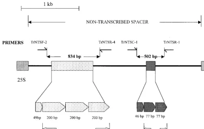
pu-tative NTS region was 2,486 bp in length and contained two sets of TRSs (Fig. 1). The first repeat locus was located 480 bp downstream from the putative 3⬘terminus of the large-subunit (25S) RNA gene. Three tandemly repetitive copies of a 200-bp element (T. rubrumTRS-1) were identified, with an adjacent 49-bp partial repeat. A truncated, highly degenerate copy of a single repeat unit was located on the immediate downstream (18S) side of this repeat region. A second set of shorter sub-FIG. 1. Schematic representation of the NTS region ofT. rubrumNCPF 295, showing the location of two sets of repetitive subelements, TRS-1 and TRS-2. The locations of the primer pairs used to amplify these repetitive regions for strain typing are indicated. The expanded section indicates the length in base pairs (bp) and the positions of the five complete and two partial repetitive elements.
on May 15, 2020 by guest
http://jcm.asm.org/
[image:3.612.95.514.75.339.2]repeat elements (T. rubrum TRS-2) was located 590 bp up-stream from the predicted 5⬘end of the 18S gene. This repeat unit was present as two tandem copies, 77 bp in length, fol-lowed by a third 46-bp partial repeat (Fig. 1).
Nucleotide homology searches (using Basic Local Align-ment Search Tool Nucleotide [BLASTN] software) found no significant similarities between theT. rubrumNTS region and other sequences in the GenBank database. A number of pos-sible open reading frames were identified from the NTS se-quence, encoding predicted proteins up to 148 amino acids in length. None of these showed significant homology to any sequences deposited in SWISS-PROT or other protein data-bases, nor was a putative 5S gene identified. A region of sec-ondary structure, with multiple homopolymer tracts up to 19 residues in length, was located immediately downstream of the predicted 3⬘terminus of the 25S gene.
Sequence analysis of TRS-1 repeat units from T. rubrum
isolates of different rDNA types.Length variations in the NTS
region ofT. rubrumwere identified during a previous study (9). Sequence analysis was carried out to confirm that these poly-morphisms were primarily due to variations in the copy num-ber of TRS-1. The first strain analyzed,T. rubrumNCPF 295, represented an rDNA type C strain (9) and contained three complete copies of TRS-1. The sequence of an rDNA type A strain, 96-T2/2248, contained a single complete copy of TRS-1. Incomplete sequence data from an rDNA type F strain (strain 97-12332) suggested the presence of up to seven copies of this element. Strain CBS 303.38 was a variant rDNA type A strain, and analysis of the single TRS-1 element present showed it had undergone major rearrangements, including loss of 19 bp from
the central region (Fig. 2). In addition, one copy of the smaller TRS-2 subrepeat elements had been lost in this isolate.
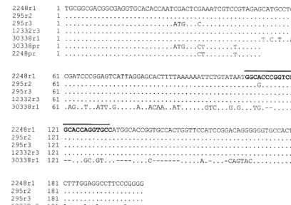
Sequence alignments showed that most copies of TRS-1, both within and between strains, were identical (Fig. 2) or nearly so, with two repeats showing minor G3A or C3A transitions or transversions. However, the third TRS-1 element fromT. rubrumNCPF 295 plus two of the partial 49-bp elements had a specific alteration in bases 28 through 30 (CGA3ATG) followed by a G3C transversion at position 34. These changes are also present in the incomplete sequence obtained from a fourth copy of TRS-1 in strain 97-12332 (data not shown). Finally, an almost perfect palindromic sequence of 27 bp, high-lighted in Fig. 2, is present in each full copy of TRS-1, as well as in the partial and degenerate TRS-1 sequences described above.
Strain typing ofT. rubrum by specific amplification of the
TRS-1 and TRS-2 repeat regions. Primers TrNTSF-2 and
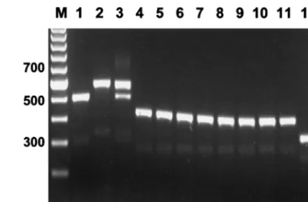
TrNTSR-4 were used to amplify the TRS-1 repeat regions from sixT. rubrumstrains of rDNA types A through F. The major fragment amplified from each rDNA type showed an incremental length variation equivalent to one TRS-1 repeat unit, except the variation in length between types E and F, which may have represented seven rather than six copies of the subrepeat. A ladder of minor bands ranging in size from 634 to 1,434 bp and stepping up in increments of 200 bp was also present. These simple patterns were designated PCR types 1 through 6 (Fig. 3, lanes 1 through 6).
[image:4.612.93.502.72.360.2]The assay was then used to examine 101 random clinical isolates ofT. rubrum(Table 1). The majority of these isolates produced band patterns characteristic of PCR types 1 through FIG. 2. Sequence variation in the TRS-1 elements ofT. rubrumstrains 96-T2/2248, NCPF 295, 97-12332, and CBS 303.38. Dots indicate residues which are identical to the 2248rl sequence, and dashes represent bases which are absent from 30338rl, the truncated TRS-1 element of strain CBS 303.38. The sequences of five TRS-1 elements, rl from NCPF 295 and rl, r2, r5, and r6 from 97-12332, are identical to the 2248 rl sequence (data not shown). The 49-bp partial TRS-1 element represented by 2248pr is identical in strains 96-T2/2248, NCPF 259, and 97-12332. The partial element 30338pr from strain CBS 303.38 has the CGA3ATG change also present in 295r3. The 27-bp palindromic sequence is shown in bold and overlined. Dots indicate residues which are identical to the 2248rl sequence, and dashes represent bases which are absent from 30338rl, the truncated TRS-1 element of strain CBS 303.38
on May 15, 2020 by guest
http://jcm.asm.org/
6, with the PCR type usually but not always related to the corresponding rDNA type (Table 1). Forty strains (40%) were PCR type 1, 17 (17%) were PCR type 2, and 13 (13%) were PCR type 3. Altogether, 80 strains (80%) were PCR types 1 through 6 (Fig. 3, lanes 1 through 6). The remaining 21 strains (20%) had complex PCR patterns, consisting of multiple bands in several different registers. The assignment of individual PCR types to these strains was complicated by difficulties in accurately scoring the presence of minor bands and in estimat-ing band intensities. These complex patterns were derived from isolates that also had multiple polymorphic bands on rDNA typing.
The NTS region containing TRS-2 was amplified using prim-ers TrNTSR-1 and TrNTSC-1. A single 502-bp product (Fig. 1), representing two complete copies of TRS-2, was obtained from the majority of strains tested (75 of 87 [86%]). In seven isolates, the fragment appeared smaller by a length equivalent to one 77-bp TRS-2 repeat unit (Fig. 4). All of these strains had a single copy of TRS-1 (PCR type 1). Strain CBS 303.38, which had a degenerate form of TRS-1, also had a single TRS-2 element. In strains 97-N393 and S-900008, the fragment length (577 bp) suggested the presence of three copies of
TRS-2, while the product amplified from isolate 98-5693 (an rDNA type J/PCR type 12 strain) had lost a fragment equiva-lent in size (144 bp) to both copies of TRS-2 (Fig. 4). Strain 97-14166 (PCR type 2) contained two forms of the TRS-2 locus, representing three and two copies of TRS-2, respec-tively.
Individual PCR types did not show statistically significant associations with specific geographic regions, but no strains of the commonest PCR type (type 1) were present in 7 samples originating from the Indian subcontinent (Table 1), and only one type 1 strain was present in 11 strains from Japan. There are no clear relationships between PCR type and tissue type (e.g., skin or nail) or site of a clinical lesion.
Reproducibility and specificity of PCR strain typing. The
PCR types remained constant throughout different batches of primers and PCR reagents and using different thermal-cycling machines. The addition of BSA to the reaction mixture in-creased the efficiency of amplification but did not affect the size or number of bands detected. DNA extracted fromT. rubrum
NCPF 295 subcultured on Sabouraud agar on four separate occasions over a 2-year period gave identical PCR types. The use of a high-quality DNA template was important for consis-tent results, and the rapid DNA extraction procedure gave identical results to those for a template purified by the original method (9). The rapid DNA extraction procedure gave best results for fresh mycelial growth, although successful strain typing was achieved using template extracted from 4-month-old plate cultures.
The specificity of PCR was determined by carrying out a standard PCR typing reaction on 22 different dermatophyte species, plus 2 clinical isolates each of C. albicans and A. fumigatus. Specific amplification products were obtained from
T. rubrum,Trichophyton gourvilii,Trichophyton violaceum, and
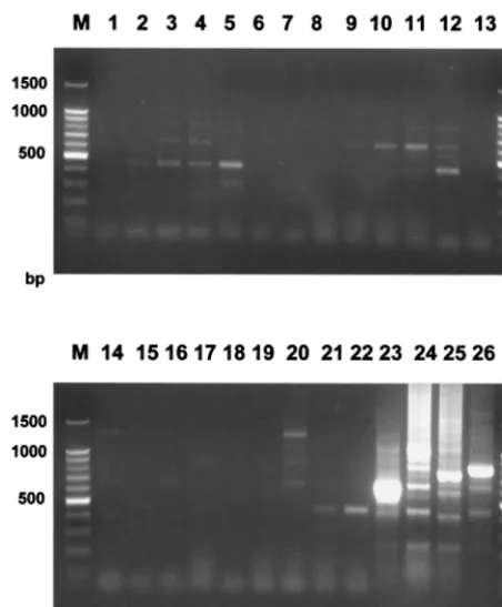
Trichophyton soudanense. All other species either produced weak bands which were interpreted as nonspecific or there were no visible products (Fig. 5). The specificity was further confirmed by probing Southern blots of these species using a single, labeled TRS-I element. The probe hybridized to the same four dermatophyte taxa that produced specific amplifi-cation products, although some weak hybridization to other species was detected at low stringency (data not shown).
[image:5.612.320.542.70.216.2]FIG. 3. Amplification of the TRS-1 subrepeat element produced 21 PCR types from 101 clinical isolates ofT. rubrum. (PCR type 1⫹from strain CBS 303.38 is not shown). PCR types 1 through 6 represent strains with 1 to 6 copies of TRS-1 in the NTS. The copy number and chromosomal distribution of TRS-1 in strains with complex pattern types 7 through 20 are undetermined. M, mo-lecular weight marker.
FIG. 4. Amplification of the TRS-2 subrepeat element, using primers TrNTSR-1 and TrNTSC-1. A 502-bp product (lane 1) representing two complete copies of TRS-2 was amplified from 86% of all isolates. Variation in the TRS-2 fragment occurred mainly in strains with a single copy of TRS-1 (PCR type 1). Lanes: 1, NCPF 295; 2, 97-12329; 3, 97-12444; 4, 97-12912; 5, 97-13550; 6, 97-14358; 7, 98-8070; 8, CBS 303.38; 9, FMG 1063; 10, 97-N393; 11, 97-14166; 12, 98-5693. M, molecular weight marker.
on May 15, 2020 by guest
http://jcm.asm.org/
DISCUSSION
Strain identification in the dermatophyte fungusT. rubrum
has been attempted using a range of molecular-typing proce-dures. These investigations have mainly involved the use of random amplified polymorphic-DNA methods and typically identified just a single strain (7, 10, 12) or a small number of strain lineages (21). The lack of differentiation and problems of reproducibility associated with random amplified polymorphic-DNA procedures have prompted us to evaluate alternative genotyping approaches. We previously described a method for differentiating strains of this species by analysis of restriction fragment length polymorphisms in the nontranscribed region of the rDNA spacer (9). In this study, we confirmed these polymorphisms and demonstrated the presence of two novel subrepeat elements, TRS-1 and TRS-2, in the NTS region. We further showed that variations in the copy number of these elements are responsible for the NTS length polymorphisms detected previously. Specific amplification of the NTS regions containing these elements provides a simple, rapid, and dis-criminatory method for strain typing ofT. rubrum.
A similar PCR assay has been described for typing strains of
tion of this species from foci of endemicity in Southeast Asia and West Africa, which may have occurred as recently as the mid-nineteenth century (5, 13). This global spread ofT. rubrum
may have resulted from the rapid proliferation of a limited number of comparatively undifferentiated clones, reflecting the restricted number of prevalent PCR types reported here. Strains of these common PCR types may demonstrate specific characteristics of pathogenicity or infectivity that have en-hanced their spread.
The extensive diversity present in the rDNA NTS contrasts with a recent account of the strictly clonal nature ofT. rubrum
(7). The variations in copy number of the NTS subrepeats presumably result from the process of unequal sister chromatid exchange at mitosis (17). Polymorphisms in tracts of repetitive DNA generated by unequal crossing-over are localized and are not linked to variation due to recombination and mutation elsewhere in the genome. The localized nature of micro- and minisatellite polymorphisms means that they may not be de-tected by random PCR-based methods that index whole-ge-nome variation or by isoenzyme analysis (multilocus enzyme electrophoresis). Unusually, therefore, we have demonstrated the presence of at least one variable minisatellite locus in the
T. rubrumgenome, which otherwise appears to be clonal (7). The short evolutionary history and the apparent clonality of this species suggest that such markers may represent the pri-mary or indeed only variable loci. Our results have encouraged us to seek other cryptic, polymorphic repetitive sequences for strain differentiation in T. rubrum. The identification of such additional unlinked variable markers would also permit allelic analysis using current population genetic methods (18).
Amongst isolates in the United Kingdom, the largest num-ber of strains were TRS-1 type 1, the second largest type 2, etc. One explanation for this pattern assumes that multiplication in TRS-1 copy number by unequal crossover is a rare event. Thus, if all strains with multiple copies of TRS-1 are derived initially from type 1 isolates, then there will be progressively smaller numbers of strains with increased copy numbers of this repeat. There may also be coincidental selection for strains with a single copy or low copy numbers of TRS-1 among clinical isolates. Further work on other genetic markers and strains from patients with “carrier status” may enable these questions to be answered. We were surprised to discover that variation in copy number of the shorter TRS-2 element occurs predomi-nantly in strains which have a single copy of TRS-1. Although two copies of TRS-2 are found in most isolates, the coinciden-tal presence of two or more copies of TRS-1 seems to result in unequal crossover occurring preferentially between the larger elements.
[image:6.612.60.286.70.344.2]While the structure of these repeat elements was stable in a strain cultured in vitro over a period of 1 year, it remains possible that in vivo recombination and changes in copy num-ber occur more frequently, particularly in strains with large
FIG. 5. Specificity of the PCR. Amplification was performed using primers TrNTSF-2 and TrNTSR-4 of 22 different dermatophyte species, plus two labo-ratory isolates ofA. fumigatusandC. albicans. Lanes: 1,A. fumigatusstrain MRL 2-1; 2,A. fumigatusstrain MRL 2-5; 3,C. albicansstrain MRL 10-1; 4,C. albicans
strain MRL 12-2; 5,Epidermophyton floccosumstrain DS.EF; 6,Microsporum audouiniistrain SJ.EM 4875; 7,Microsporum canisstrain 97/12400; 8, Microspo-rum gypseumstrain SJ.EM 3928; 9,Microsporum persicolorstrain NCPF 356; 10,
Trichophyton concentricumstrain NCPF 600; 11,Trichophyton erinaceistrain NCPF 375; 12,Trichophyton equinumstrain SJ.A1; 13,T. mentagrophytesvar.
interdigitale strain LM 43; 14, T. mentagrophytes var. mentagrophytes strain DS.TMvM; 15, Arthroderma benhamiaestrain PV3000; 16,Arthroderma simii
strain CBS448.65; 17,Arthroderma vanbreuseghemiistrain A27961; 18, Tricho-phyton quinckeanumstrain LM 54; 19,Trichophyton schoenleiniistrain SJ.EV 327; 20,Trichophyton terrestrestrain LM 39; 21,Trichophyton tonsuransstrain SJ.EM 6717; 22,Trichophyton verrucosumstrain 98/4545; 23,T. gourviliiNCPF 5019; 24,T. rubrumstrain NCPF 295; 25,T. soudanensestrain 98/7676; 26,T. violaceumstrain SJ.EM 7115; M, molecular weight marker.
on May 15, 2020 by guest
http://jcm.asm.org/
copy numbers of TRS-1. Finally, it should be noted that the process of crossover fixation could produce identical events in strains that do not share a common ancestry. Consequently, epidemiological interpretations placed on strains which share the same PCR type have to be made with care and with due regard to additional factors such as the temporal and geo-graphical relationships between isolates.
Among the simple types (PCR types 1 through 6), there are often ladders of minor bands which appear to be PCR artifacts caused by the loss of entire copies of the TRS-1 repeat during amplification. We identified a 27-bp palindromic sequence in TRS-1 (Fig. 2) which has the potential to initiate hairpin for-mation during PCR, resulting in “looping out” of the inter-palindromic region. Hence, in a strain with three copies of TRS-1, hairpin formation between different repeats would pro-duce minor fragments representing one or two copies of TRS-1 in addition to the main amplification product representing three copies of the element. The use of hot starting and inclu-sion of dimethyl sulfoxide and detergents such as Triton X-100 in the PCR mixture were ineffective in eliminating or reducing these amplification artifacts.
The molecular basis and identification of the common PCR types (PCR types 1 through 6) are straightforward, but they are more difficult to pinpoint in strains with more complex banding patterns (PCR types 7 through 20). These multiple bands may represent the presence of rRNA cistrons with different TRS-1 and -2 copy numbers, rearrangements within NTS regions to duplicate primer sites, or even heterokaryons.
We also noted several discrepancies (Table 1) between the PCR type and rDNA type for some isolates. In certain strains, the rDNA type determined by Southern hybridization was one size register greater than the corresponding PCR type; for example, a PCR type 1 appeared as an rDNA type B. These anomalies may be due to misinterpretation of the rDNA type, as the 200-bp increments representing each rDNA type occur inEcoRI fragments of 4 to 5 kb in overall length. In addition, the 77-bp size difference caused by loss of one copy of TRS-2 introduced minor incremental differences in the fragment size of some rDNA type A strains.
Several isolates with multiple band patterns had identical PCR types but different rDNA types (Table 1). These anom-alies probably arise from the influence of various technical differences between PCR and probe hybridization methodol-ogies. For instance, in strains with complex rDNA and PCR patterns, the distribution and copy number of TRS-1 and/or TRS-2 between different cistrons of the rDNA repeat may not be the same. These heterogeneous repeats may be evident by probe hybridization, but they may not be detected by the PCR assay, particularly if the amplified fragments are relatively long and the heterogeneous repeat copy numbers low. These expla-nations do not account for all of the discrepancies in the PCR and rDNA results of the complex types. Technical differences between the two methods and the extent to which either typing system is able to render variation visible are potential sources of inconsistency. Resolution of these issues requires the se-quencing of the NTS regions of complex types in order to determine the most reliable method of assay. While this work is ongoing, use of both PCR and rRNA analysis by Southern blotting to type complex types is recommended.
The TRS-1 sequence variations found in strain CBS 303.38 (Fig. 2) are also difficult to understand. This strain was first isolated from a case of tinea corporis in Vietnam in 1938, and it is therefore one of the oldest isolates ofT. rubrumstudied (4). This isolate may represent a progenitor of “modern”T. rubrumstrains, which subsequently evolved to infect nail and skin tissue in shod feet. Alternatively, the strain may be one of
an original group of isolates that did not spread to other parts of the world due to characteristics of low transmissibility or pathogenicity (5, 13). Further characterization of strain CBS 303.38 and of isolates with multiple banding patterns may provide answers to some of these questions.
The primers used in theT. rubrumassay allow specific am-plification from three closely related species, T. gourvilii, T. soudanense, andT. violaceum(8, 11, 16), but not from other dermatophyte species. We have recently found that isolates of the dermatophytesTrichophyton mentagrophytesvar. mentagro-phytesandT. mentagrophytesvar.interdigitalealso show exten-sive polymorphism in the NTS region (T. Mochizuki, C. J. Jackson, R. C. Barton, M. Moore, and E. G. V. Evans, unpub-lished data). The specificity of theT. rubrumPCR assay and the lack of hybridization of the TRS-1 probe with members of the T. mentagrophytes group indicate that the variable NTS region of this dermatophyte complex bears little genetic ho-mology to the TRS-1 region ofT. rubrum.
There was no clear correlation between strain PCR type and the clinical source of the isolate, i.e., skin or nail specimens. However, the distribution of PCR types was not the same across all geographic regions. For example, there was only a single type 1 strain among 11 Japanese isolates examined, whereas in the United Kingdom and most other countries, type 1 was by far the most common PCR type seen. Further isolates from this and other regions are required to confirm these divergences, and they may enable us to map the spread ofT. rubrumthat has occurred over the last 150 years.
The analysis of subrepeat elements in the rDNA of T. rubrumaffords a valuable technique for strain identification in this genetically invariant fungus. Variable repetitive sequences in this and otherTrichophytonspecies will continue to provide important tools for dermatophyte strain typing and epidemi-ology. This new approach will enable us to reassess our current knowledge of the epidemiology of dermatophyte infections and also provide new insights into pathogenicity and the pat-terns of disease caused by these organisms.
ACKNOWLEDGMENTS
Strains were generously provided by D. Ellis, Mycology Unit, Wom-ans and Childrens Hospital, North Adelaide, Australia; A. Chakrabarti, Department of Medical Microbiology, Postgraduate Institute for Medical Education and Research, Chandigarh, India; T. Mochizuki, Department of Dermatology, Kanazawa Medical University, Ishikawa, Japan; and R. Grillot, Department of Medical and Molecular Mycology, Faculte´ de Me´decine de Grenoble, France.
We thank the Janssen Research Foundation, Beerse, Belgium, for their financial support for this project.
REFERENCES
1.Benson, D. A., M. S. Boguski, D. J. Lipman, J. Ostell, B. F. F. Ouellette, B. A. Rapp, and D. L. Wheeler.1999. GenBank. Nucleic Acids Res.27:12–17. 2.Carlotti, A., T. Srikantha, K. Schro¨ppel, C. Kvaal, J. Villard, and D. R. Soll.
1997. A novel repeat sequence (CKRS-1) containing a tandemly repeated sub-element (kre) accounts for differences betweenCandida kruseistrains fingerprinted with the probe CkF1,2. Curr. Genet.31:255–263.
3.Carlotti, A., F. Chaib, A. Couble, N. Bourgeois, V. Blanchard, and J. Villard.
1997. Rapid identification and fingerprinting ofCandida kruseiby PCR-based amplification of the species-specific repetitive polymorphic sequence CKRS-1. J. Clin. Microbiol.35:1337–1343.
4.Catanei, A.1938. On the classification ofTrichophyton rubrum: observation and experimental investigation of a new strain of this fungus. Arch. Inst. Pasteur16:227–231.
5.Charif, M. A., and B. Elewski.1997. A historical perspective on onychomy-cosis. Dermatol. Ther.3:43–45.
6.Cribier, B., M. L. Mena, D. Rey, M. Partisani, V. Fabien, J. M. Lang, and E. Grosshans.1998. Nail changes in patients infected with human immunode-ficiency virus: a prospective controlled study. Arch. Dermatol.134:1216– 1220.
7.Gra¨ser, Y., J. Ku¨hnisch, and W. Presber.1999. Molecular markers reveal
on May 15, 2020 by guest
http://jcm.asm.org/
14. Suhonen, R. E., P. R. Dawber, and D. H. Ellis.1999. Fungal infections of the skin, hair and nails. Martin Dunitz, London, United Kingdom.
15. Summerbell, R. C., and J. Kane.1997. Physiological and other special tests
21. Zhong, Z., R. Li, D. Li, and D. Wang.1997. Typing of common dermato-phytes by random amplification of polymorphic DNA. Jpn. J. Med. Mycol.
38:239–246.