0095-1137/05/$08.00⫹0 doi:10.1128/JCM.43.4.1604–1611.2005
Copyright © 2005, American Society for Microbiology. All Rights Reserved.
Discrimination within Phenotypically Closely Related Definitive Types
of
Salmonella enterica
Serovar Typhimurium by the Multiple
Amplification of Phage Locus Typing Technique
Ian L. Ross and Michael W. Heuzenroeder*
Infectious Diseases Laboratories, Institute of Medical and Veterinary Science, Adelaide, South Australia, Australia
Received 21 September 2004/Returned for modification 18 November 2004/Accepted 6 December 2004
Multilocus sequence typing (MLST) is a relatively new high-resolution typing system employed for
epide-miological studies of bacteria, includingSalmonella. Discrimination based on MLST of housekeeping genes
may be problematical, due to the high identity of gene sequences of closely relatedSalmonellaspecies. The
presence of genomic sequences derived from stable temperate phages inSalmonellaoffers an alternative for
MLST ofSalmonella. We have used MLST of prophage loci inSalmonella enterica serovar Typhimurium to
discriminate closely related isolates of serovar Typhimurium. We have compared these results to MLST of five housekeeping genes, as well as pulsed-field gel electrophoresis (PFGE). The presence or absence of prophage loci in the 73 serovar Typhimurium isolates tested, as well as allelic variation as detected by sequencing, provided greater discrimination between isolates than either MLST of housekeeping genes or PFGE. Ampli-fication of prophage loci alone separated serovar Typhimurium isolates into 27 groups comprising multiple isolates or individual strains. Sequencing of isolates found within the clusters separated isolates even further. By contrast, PFGE could only divide the 73 isolates into five distinct groups. MLST using housekeeping genes did not provide any significant separation of isolates in comparison to amplification or MLST of prophage loci. The results demonstrate that the amplification and sequencing of prophage loci provides a high-resolution, objective method for the discrimination of closely related isolates of serovar Typhimurium. It is proposed that multiple amplification of phage locus typing may provide sufficient discrimination for epidemiological pur-poses without recourse to MLST.
Nontyphoidal Salmonella species are a common causative agent of bacterial food-borne disease worldwide (4, 12). Clas-sical typing methods for identification of isolates involves se-rotyping based on the Kauffmann-White serological scheme that targets the cell surface O and H antigens. This system currently identifies more than 2,000 serotypes worldwide (19). Phage typing systems have been developed for further discrim-ination of serovars commonly associated with disease such as
Salmonella entericaserovar Typhimurium (1) andSalmonella entericaserovar Enteritidis (35). More recently, molecular typing methods have been assessed and adopted for further discrimina-tion of Salmonella isolates (for a review of methodologies for typingSalmonella, see reference 36). Of the molecular-based sys-tems, pulsed-field gel electrophoresis (PFGE) is currently consid-ered by many to be the “gold standard” for molecular typing of
Salmonella(21). More recently, amplified-fragment length poly-morphism (AFLP) has been developed and adopted by research-ers because of its potential to discriminate between closely related isolates, flexibility with variable selective primers, and choices of restriction enzyme systems (9, 28, 33).
There are currently a number of potential problems with both classical and most molecular-based methods for typing
Salmonella. Phage typing requires a well-maintained phage library, precise methodology, and experience in interpretation of results (8, 30). Interpretation of plaque patterns is often
subjective and prone to variation between laboratories, espe-cially between phage types with similar reaction patterns. For example, serovar Typhimurium definitive type 12 (DT12) and DT108 are phenotypically similar phage types with regard to lysis patterns of serovar Typhimurium and are separated on the Anderson typing panel by the sensitivity of DT108 with bacteriophages A21, A27, and A35. In many Australian iso-lates of DT108, there is no observed reaction with phage A21. Furthermore, there are cases where there are very weak reac-tions with phages A27 and A35.
Perceived problems with phage typing are further com-pounded, as a significant number of isolates can be untypable by this method. Often, one phage type may predominate in a geographical area, making discrimination between outbreak and nonoutbreak isolates difficult (21, 30). Molecular methods such as PFGE, although less subjective than phage typing, at times do not provide clear discrimination between isolates. Furthermore, some isolates are not typable by PFGE, for ex-ample,Salmonella entericaserovar Kentucky strains and Sal-monella entericasubsp.salamaeserovar Sofia (8, 22).
Multilocus sequence typing (MLST) is a relatively new high-resolution typing system developed for both evolutionary and epidemiological studies of microorganisms (6, 8, 10). MLST involves the nucleotide sequencing of approximately 400-bp re-gions of at least seven genes, usually housekeeping genes. Al-though multilocus enzyme electrophoresis (MLEE) is similar to MLST in that both systems target enzymes or their genes, MLST has advantages over MLEE. MLST will detect single-nucleotide polymorphisms, especially third-base redundancy within a codon, whereas MLEE relies on nonconservative changes in the amino
* Corresponding author. Mailing address: Infectious Diseases Lab-oratories, Institute of Medical and Veterinary Science, Box 14, Rundle Mall, P.O. Adelaide, S.A. 5000, Australia. Phone: 618 8222 3275. Fax: 618 8222 3543. E-mail: [email protected].
1604
on May 16, 2020 by guest
http://jcm.asm.org/
acid sequence of a target enzyme that result in a change of its electrophoretic mobility on a starch gel (28). For example, Kotetishvili et al. (8) found that the ratio of nonsynonymous (amino acid substitution) to synonymous base changes (no change in amino acids) in threeSalmonellahousekeeping genes was⬎1. This meant that there was a low incidence of amino acid substi-tutions that would have affected enzyme mobility in MLEE stud-ies.
Sequencing ofSalmonellagenes has generally been restricted to comparing sequences of housekeeping genes between the ma-jor groups ofSalmonellaor serovars ofS.entericasubsp.enterica, including serovars Typhimurium and Enteritidis. Sequence vari-ation can be demonstrated for geographically and epidemiologi-cally unrelated isolates (5, 8, 16, 34). Consequently, MLST is a useful tool for studying evolution and global epidemiology of the salmonellae. However, for investigations of outbreaks, the fine discrimination of closely related isolates based on MLST of housekeeping genes may be not be possible, due to the high identity of the housekeeping gene sequences.
The presence of genomic sequences derived from temperate phages inSalmonellamay offer an alternative target for MLST ofSalmonella(25, 26). Although a typical prophage genome only constitutes approximately 1% of the bacterial chromo-some, these loci are subject to mutational events at a higher rate than housekeeping genes. Furthermore, recombination between phages has resulted in an array of mosaic genetic structures of many bacteriophages, both within individual genes and in the overall genome organization (3, 17, 24, 32). Indeed, it has been stated that prophage genes account for most of the genetic diversity among closely relatedSalmonella
strains (7). The presence of immunity exclusion genes such as thecIrepressor gene of theimmCregion of some phages may confer a selective advantage for the host cell, rendering the cell immune to superinfection by lytic phage (20). This suggests that prophage genes, both in their presence or absence in a host cell and in their nucleotide sequence, vary considerably within a serovar. Consequently, heterogenicity of prophage genes is likely to be greater than that of host housekeeping genes, making them superior targets for MLST.
Previous studies by our laboratory have identified two bac-teriophages (ST64T and ST64B) induced from a strain of serovar Typhimurium DT64. These two temperate phages were sepa-rated from each other by a cesium chloride gradient. The two bacteriophages were fully sequenced, and the sequences were deposited in GenBank (www.ncbi.nlm.nih.gov) under accession numbers AY052766 (ST64T) and AY055382 (ST64B) (14, 15). Southern hybridization studies have suggested that these bacte-riophages are present in a number of serovar Typhimurium phage types as well as otherS.entericasubsp.entericaserovars, including Enteritidis, Virchow, Heidelberg, and Hadar (13, 31). Based on the widespread nature of these phages, we used this sequence data to design a number of primer sets to analyze sequence variations of prophage loci. As well as using locus sequences from bacteriophages ST64B and ST64T, we also examined a range of similar loci from the well-characterized bacteriophage P22 (18, 32). A number ofSalmonellahousekeeping genes were also as-sessed to compare their level of sequence identity to the level of sequence identity of the prophage loci. We used this data to discriminate DT12 and DT108 strains of serovar Typhimurium phenotypically that were closely related by lysis pattern, in
addi-tion to DT126-related food poisoning by MLST and by amplifi-cation of phage loci alone.
MATERIALS AND METHODS
Strains and culture conditions.A total of 73 serovar Typhimurium isolates were used in this study. Thirty-six isolates were a selection of mostly epidemio-logically unrelated DT108 (18 isolates, including 2 isolates [03-108-022 and 03-108-023] from an outbreak in New South Wales in 2003), DT12 (9 isolates), and DT12a (3 isolates). Six isolates comprised serovar Typhimurium isolates of various phage types (one isolate each of DT64, DT9, DT135, and DT185 and two DT170 isolates).
Thirty-seven DT126 isolates were also included in the study. This group com-prised 13 isolates from a 2001 restaurant outbreak in New South Wales, Aus-tralia. Another 10 DT126 isolates were obtained during an outbreak in 2003. Four of these isolates were designated DT126 var, due to a variation in reaction to the Anderson typing panel. A further 14 DT126 isolates were epidemiologi-cally unrelated.
All serovar Typhimurium isolates used in this study were provided by the AustralianSalmonellaReference Centre, Institute of Medical and Veterinary Science, Adelaide, South Australia. Serotyping had previously been undertaken using the Kaufmann-White scheme, and bacteriophage typing was performed using the Anderson scheme of 31 bacteriophages (1), both by the Australian
SalmonellaReference Centre.
Amplification of phage loci.PCR ofSalmonellawas performed directly from cell lysates. Isolates were grown overnight in Luria-Bertani broth at 37°C with gentle shaking. A 30-l PCR mixture was prepared as follows: 3.0l of 10⫻
MgCl2-free buffer, 1.0M each forward and reverse primer, 3.0l of 200M
each deoxynucleoside triphosphate, 1.5 mM MgCl2, 1.0 U ofTaqpolymerase,
and 2.0l of overnight cell culture. The volume was made up to 30l with H2O.
Buffer, MgCl2, andTaq polymerase were supplied by Roche. Primers were
designed based on published sequences (Table 1.). All primers were supplied by Geneworks, Adelaide, South Australia. The deoxynucleoside triphosphates were supplied by Amersham Biosciences, Piscataway, N.J..
Touchdown PCR was performed in a Corbett Research (Sydney, Australia) PC-960G gradient thermal cycler as follows: 94°C (10 min), then 40 cycles comprising 94°C double-stranded DNA melting for 30 s and 72°C elongation for 60 s. Primer annealing temperatures were as follows: 59°C (1 cycle), 58°C (1 cycle), 57°C (2 cycles), 56°C (3 cycles), 55°C (5 cycles), 54°C (8 cycles), 53°C (10 cycles), and 52°C (10 cycles). All annealing steps were for 30-s duration each. A final elongation step at 72°C for 5 min was also performed.
The amplification product was detected and prepared for sequencing by run-ning 5.0l of PCR product on a 2.0% agarose gel (Progen, Darra, Queensland) in 1.0⫻TBE buffer (23) at 5.0 V/cm with pUC19 digested with HpaII (Biotech, Belmont, Western Australia) as a marker. Bands were visualized with UV light after being stained with ethidium bromide. The remaining 25-l PCR product of positive samples was prepared for sequencing by passage through a QIAquick PCR purification column (QIAGEN, Hilden, Germany) and collection in 30l of elution buffer per the manufacturer’s instructions. Amplicons were stored at
⫺20°C prior to sequencing.
MLST.Sequencing was performed in both directions with Big Dye Termina-tor, version 3-1 (Applied Biosystems, Foster City, Calif.). A 20-l reaction mixture comprised 4.0l of BD3-1 master mix, 1.5l of 3.0 mM either forward or reverse primer, 12.5l of H2O, and 2.0l of template. Sequencing was
performed with a Corbett Research PC-960G gradient thermal cycler and com-prised 25 cycles with the following parameters: 96°C (30 s), 50°C (15 s), and 60°C (4.0 min). The sequence product was precipitated, washed with 75% isopropanol, and then dried. Sequencing was performed with an Applied Biosystems 3700 DNA analyzer.
PFGE.The protocol for PFGE followed that of Maslow et al. (11). Briefly, cells grown overnight in brain heart infusion broth (Oxoid, Basingstoke, United Kingdom) were embedded in agarose and lysed by incubation of the plug in 4 ml of lysis buffer supplemented with 4 mg of lysozyme (Roche, Mannheim, Ger-many) and 80g of RNase/ml. Plugs were then digested with proteinase K and washed, and the DNA was digested overnight with XbaI restriction endonuclease (New England BioLabs, Beverley, Mass.). The plugs were placed into the wells of a 1% agarose gel prepared with PFGE-grade agarose (Bio-Rad Laboratories, Hercules, Calif.) in 0.5⫻TBE buffer.Staphylococcus aureusstrain NCTC 8325 digested with SmaI was used as a molecular size marker (29). The PFGE was run on a Bio-Rad CHEF-DR III system for 19 h at 6.0 V cm⫺1
at 4°C, with an initial switch time of 2.0 s and a final switch time of 50 s. After being run, the gel was stained in ethidium bromide, destained in water, and photographed under UV light.
on May 16, 2020 by guest
http://jcm.asm.org/
TABLE 1. Primers used in this study
Source of sequencea Locus Primer name Primer sequence (5⬘–3⬘)b sequenceSize of (bp)
Location in GenBankc Bacteriophage ST64B immC cl(5⬘) (BIM1) Forward: BIM1F1 ATGGTGGCCTTGTCGACGC 433 28028–28502
Reverse: BIM1R1 GCTAACGTGAAGGATTTGTTCCG
immC cl(3⬘) (BIM2) Forward: BIM2F1 CCATTACCGGCGCTTGCAC 410 28441–28893 Reverse: BIM2R1 TAACGTATAACCATGCGATTTCCG
immC cro(BIM3) Forward: BIM3F1 GCGATATACGCAAAAGAAGGAGG 475 28819–29336 Reverse: BIM3R1 TGGCTACTGAATGTGCCAGG
immC putc2(BIM4) Forward: BIM4F1 GCTGGTACTGCAACGTGCC 526 29284–29851 Reverse: BIM4R1 CGAATGACATGGACATAAAGTCC
ORF SB6 Forward: SB6F1 ACGACAAGCGCGTTGAGGC 512 4394–494
Reverse: SB6R1 GCTTCCACGTTGAAGAAGGC
ORF SB26 Forward: SB26F1 GACACCATCAATGTATGGATCGC 477 19466–19989 Reverse: SB26R1 AGGTTATCTATAATTCCGACCTGG
ORF SB28 Forward: SB28F1 TGCAGTCAAGAGGACGTCC 589 21366–21994
Reverse: SB28R1 TGCCGATATGCTGATCTGGC
ORF SB37 Forward: SB37F1 TGGTAGTGAATTGGTTAGCTGCG 439 26841–27322 Reverse: SB37R1 CGGAAAGCTGTTACAGCAGG
ORF SB46 Forward: SB46F1 CATTGATGGTATCGAAGTTCGCC 448 33640–34130 Reverse: SB46R1 CCTGGAGTTTCTGGCACGC
Bacteriophage ST64T gene9(5⬘)d
Forward: G9F1 GCRATTCCTTGCATCTGGAGC 562 38606–39208 Reverse: G9R1 GCAATGCGGGAACCTTTGCC
gene9(3⬘)d
Forward: G9F2 TACCGTAGAAGATTGCGCTGG 464 39995–40501 Reverse: G9R2 GGRACAAATGGTATCTCTGCCC
gene17d
Forward: F17A GGCTGTYGTTTCTTCTTTCAGGC 264 10609–10921 Reverse: R17A AGGAAATATGAAATTACGTGTCTGGC
gtrAd
Forward: GTRAF1 AGACCTTTCCGAATCCGCTG 292 2449–2784 Reverse: GTRAR1 TAATTGCCGAGAAAGTGATAAGGG
gtrBd
Forward: GTRBF1 CTTTCTCGGCAATTAGCCTG 383 2041–2463 Reverse: GTRBR1 TTAGCCAGCACCATATCCGC
gtrCd
Forward: GTRCF1 CTACTACTCGCTATTCTTTGCGC 492 152–689 Reverse: GTRCR1 CATTAACACCTCTGACCACATCC
mnt Forward: TMNTF1 GAGTAAAGCCCGGTTCGCC 280 38228–38552
Reverse: TMNTR1 TATAACCAGTAGATCATATGATGCCG
c2 Forward: TC2F1 GGAATTGTTAGAGGCCTTGCC 362 12886–13287
Reverse: TC2R1 GATTTCCCCTGATTAGCTGGG
cro Forward: TCROF1 CCATCCTGAGGAGATATACCG 222 13653–13917
Reverse: TCROR1 GGTTCAGATTGGTAAAGAGCGG
c1c
Forward: PTC1F1 CTTTACCAATCTGAACCGCCG 385 13901–14332 Reverse: PTC1R1 CTGAGTTGTTTTGGCATAATTACTCC
Bacteriophage P22 mnt Forward: PMNTF1 TTATAAGTAGTCAATATGGCCCAGG 275 37946–38269
Reverse: PMNTR1 AATACACTAACTTGGAGTGATGGC
int Forward: INTPF1 CATTTCCTGCAATACCGAAATCGG 461 3257–3717
Reverse: INTPR1 GCTGGCTTGAGCCTCACG
cro Forward: PCROF1 AGTGTTCTTTAATTTCGGAGCGAG 195 13468–13708
Reverse: PCROR1 CGGTTCAGATTGGTAAAGAGCG
c3 Forward: PC3F1 CTGCACAAGGATGGTTCCG 251 9902–10193
Reverse: PC3R1 ATTGATTGGTATAGCGAGTGCC
sieA Forward: SIEAF1 GCGCTATAAGCCAAGGACGG 462 36788–37292
Reverse: SIEAR1 TGAGTTATGCTGTGCTAGTTGCC
sieB Forward: SIEBF1 CGATGAACAACTCATGGTGGC 568 11507–12117
Reverse: SIEBR1 AGCGAGGTAAGGTATTTGTCG
Salmonellahousekeeping genes fhuA Forward: FHUAF1 AGAAGAAACCATTACCGTAACCG 402 223851–224296 Reverse: FHUAR1 TGCTAACCATCGAAATGATACCG
sucA Forward: AROCF1 GCACCGAAGAGAAACGCTG 600 802274–802915
Reverse: AROCR1 GGTTGTTGATAACGATACGTAC
tonB Forward: TONBF1 AGAATCTGTACATTTTCCACTCGC 433 1831399–1831867 Reverse: TONBR1 CAGCGCAGCCTATCACGG
manBe
Forward: MANBF1 GGCAGCTACAGACAAATCAGC 508 2187413–2187961 Reverse: MANBR1 GCCATAAATGGCATCCTCCG
glnA Forward: FHUAF1 GTTATCGACCCGTTCTTCGC 579 4216166–4216783
Reverse: FHUAR1 GTTGGTGCCGTTCTTCGCC
aPrimer sequences from bacteriophages andSalmonellahousekeeping genes derived from the following GenBank accession numbers ST64B (AY055382), ST64T (AY052766), P22 (NC_002371), andSalmonellahousekeeping genes (NC_003197) (www.ncbi.nlm.nih.gov).
bY, C or T; R, A or G.
cLocation in published GenBank sequence includes primers.
dThe primers derived from bacteriophage ST64T are also used to detect similar regions in bacteriophage P22. eThe phosphomannomutase genemanBis designatedcpsGin the referred GenBank accession number NC_003197.
on May 16, 2020 by guest
http://jcm.asm.org/
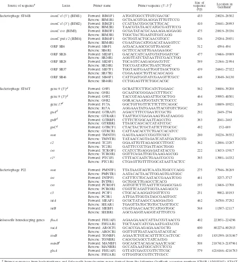
Data analysis.Isolates were initially separated based on PCR results. Each primer set was given a number, and data were entered into a spreadsheet consisting of all positive results for each strain. The spreadsheet was imported into GelCompar IV (Applied Maths, Kortrijk, Belgium) with appropriate for-matting. A dendrogram of isolate PCR profiles was generated by a Dice coeffi-cient and clustering by unweighted-pair group method using average linkages (UPGMA) (Fig. 1).
Nucleotide sequences were analyzed in both directions with GeneBase, ver-sion 1.0 software (Applied Maths).
RESULTS
Multiple amplification of phage locus typing (MAPLT).
Twenty-two of the 25 prophage-derived primer sets generated amplified product from at least one of the 73 serovar Typhi-murium isolates (Table 2). The most frequently amplified re-gions were the loci from bacteriophage ST64B. A range of P22 and ST64T prophage loci were routinely found in non-DT126 isolates; these included the 5⬘and 3⬘regions of the9P22/ST64T,
sieBP22/ST64T, gtrCP22/ST64T, intP22/ST64T, and mntST64T genes.
Amplified product from other primer sets gtrAP22/ST64T,
gtrBP22/ST64T, andmntP22was obtained with only a small
num-ber of isolates. The three prophage primer sets that failed to amplify with any isolate weresieAP22/ST64T,croST64T, andc3P22.
Only one isolate (01-126-114) failed to generate amplified product with any of the 25 prophage primer sets.
Analysis of the PCR profiles of the 72 isolates that tested positive for prophage loci is summarized in Fig. 1. Six separate clusters, each containing isolates with identical amplification profiles, were identified (Fig. 1). Four separate clusters of non-DT126 isolates with identical PCR profiles were observed.
Different phage types were represented in each of these four clusters. Eighteen non-DT126 isolates (including the DT135 isolate) were all separated from each other by differences in their PCR profiles. Cluster 6 contained 29 DT126 isolates plus the DT9 isolate and included 12 of the 13 2001 outbreak isolates and the 6 DT126 isolates from the 2003 outbreak. A further cluster (cluster 5) consisted of the four DT126 var isolates from the 2003 outbreak. These four isolates were sep-arated from the other DT126 isolates from the same outbreak, as well as the other DT126 isolates as they did not contain the SB28ST64Bloci (putativeintgene). The DT126 isolates, as well
as the DT9 and DT135 isolates, had PCR profiles significantly different from the other serovar Typhimurium phage types tested. All but three DT126 isolates (01-126-101, 02-126-122, and 02-126-123) failed to produce a PCR product with primers from P22 and ST64T.
MLST analysis. All PCR products for all relevant primer
sets were sequenced with both forward and reverse primers, and the number of different alleles for each primer set was determined (Table 2). Most bacteriophage-derived primer sets produced at least two different alleles when sequenced, except those primer sets where only a small number of strains gener-ating a PCR product were observed. Some primer sets pro-duced only one allele even when a significant number of iso-lates produced PCR product, for example, SB28ST64Band the
mntST64T gene. In a few cases where significant numbers of
[image:4.585.131.456.70.322.2]isolates produced product, only one or two isolates contained an allele distinct from the majority of isolates. For example, a
FIG. 1. Dendrogram of PCR profiles for separation of serovar Typhimurium isolates based on MAPLT. The dendrogram was generated by Dice coefficient with clustering by UPGMA, based on the presence or absence of amplified product. A total of six clusters of isolates with identical PCR profiles were generated, as well as a total of 21 isolates with unique profiles. Boldface numbers in the dendrogram refer to cluster numbers. Cluster 1 comprises the eight non-DT126 isolates, and cluster 5 comprises the four DT126 var isolates. Abbreviations: N.S.W., New South Wales; N.T., Northern Territory; Qld., Queensland; S.A., South Australia; Vic., Victoria.
on May 16, 2020 by guest
http://jcm.asm.org/
second SB26ST64Ballele was present in only one isolate,
01-9-001.
The housekeeping genesfhuA,glnA, andsucAamplified in all 73 isolates. In contrast, 46 and 15 isolates yielded product for manB and tonB, respectively. More than one allele was present only inglnAandmanB. Isolate 01-126-114 possessed a
manB allele different from that of the other 45 that were positive with manBprimers. Six DT126 and four DT126 var isolates from the 2003 outbreak had a characteristicglnAallele. MLST of phage loci was undertaken to further discriminate between isolates that clustered as depicted in Fig. 1. Separa-tion of the eight isolates in cluster 1 produced three subgroups (Table 3). Separation was based on a single allele in each case, with the two-outbreak strains possessing a SB37ST64B allele
different from that of the other six isolates. Isolate 02-12-002 could be separated from the other seven isolates in this cluster based upon theimmCST64Bc15⬘sequence.
In cluster 2, isolate 02-108-001 could be separated from the other three isolates based upon the immCST64B region. In
cluster 3, no further separation could be achieved. Cluster 4 comprised two isolates, which could be separated based upon different alleles within the ST64BimmCregion and SB37ST64B.
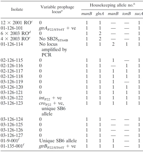
MLST of the DT126 isolates indicated a lower degree of variation than observed above (Table 4). No allelic variation
[image:5.585.302.540.89.265.2]was observed for the 13 isolates from the 2001 restaurant outbreak or the 10 2003 outbreak isolates for prophage loci. However, separation of the 2003 isolates could be achieved based uponglnA. Of the remaining DT126 isolates only one
TABLE 3. The separation of MAPLT-clustered non-DT126 isolates by sequencing
Clustera Isolate Differing allele(s)b PFGEc
1 02-108-004 3
03-108-014 3
02-12-001 3
02-12-005 3
02-12-006 2
02-12-002 c1ST64B(5⬘) 3
03-108-022 SB37ST64B 3
03-108-023 SB37ST64B 3
2 02-108-001 immCST64B 3
03-108-015 5
03-108-016 5
02-12-004 3
3 02-170-001 3
02-170-002 3
02-108-005 3
4 01-108-008 1
02-12a-002 immCST64B, SB37ST64B 3
aBased on clusters as depicted in Fig. 1.
bUnique allele(s) which separate an isolate(s) within each cluster. cPFGE clusters as depicted in Fig. 2.
TABLE 4. Separation of DT126 and DT126 var isolates by MAPLT and sequencing
Isolate Variable prophage locusa
Housekeeping allele no.b
manB glnA manB tonB sucA
12⫻2001 ROc 0 1 1 — — 1
01-126-101 gtrAP22/ST64T⫹ve 1 1 — — 1
6⫻2003 ROd 0 1 2 — — 1
4⫻2003 ROe No SB28
ST64B 1 2 — — 1
01-126-114 No locus amplified by PCR
1 1 2 1 1
02-126-115 0 1 1 1 — 1
02-126-116 0 1 1 — 1 1
02-126-117 0 1 1 1 1 1
02-126-118 0 1 1 1 1 1
03-126-119 0 1 1 1 — 1
03-126-120 0 1 1 1 1 1
03-126-121 0 1 1 1 1 1
03-126-122 intP22⫹ve 1 1 1 1 1
03-126-123 croP22⫹ve, unique SB6 allele
1 1 1 1 1
03-126-124 0 1 1 — — 1
03-126-125 0 1 1 — — 1
03-126-126 0 1 1 — — 1
03-126-127 0 1 1 — — 1
01-9-001f Unique SB6 allele 1 1 1 — 1
01-135-001f gtrB
P22/ST64T⫹ve 1 1 1 — 1 aVariation from the prophage PCR profile of the 29 DT126 isolates (see cluster 6 in Fig. 1) where only the nine ST64B loci were detected. 0, no variation detected. bAllelic number as determined by sequencing, allelic numbers are assigned for comparative purposes only. —, no amplification product detected.
cTwelve isolates out of thirteen from the 2001 restaurant outbreak (RO). Isolate 01-126-101 is the thirteenth isolate.
dSix DT126 isolates from the 2003 restaurant outbreak (RO). eFour DT126 var isolates from the 2003 restaurant outbreak (RO).
[image:5.585.42.284.89.400.2]fSingle DT9 and DT135 isolates were included as phenotypic outliers with regard to DT.
TABLE 2. The incidence of loci, number of alleles, and their distribution in 73S.entericaserovar Typhimurium isolates
Prophage or gene group Locus No. positivea
No. of allelesb
No. with most-common
allelec
ST64B c1: 5⬘ 72 3 56
c1: 3⬘ 70 2 62
SB39cro 71 3 62
SB40c2 70 5 63
SB6 68 3 59
SB26 55 2 54
SB28 63 1 63
SB37 68 3 53
SB46 62 5 49
ST64T and P22 gene9: 5⬘ 33 3 30
gene9: 3⬘ 36 3 25
gene17 9 2 6
gtrA 4 1 4
gtrB 7 2 6
gtrC 31 4 25
c1 1 1 1
ST64T mnt 29 1 29
c2 1 1 1
cro 0 0 NA
P22 mnt 4 1 1
cro 1 1 1
int 34 4 30
c3 0 0 NA
sieA 0 0 NA
sieB 33 2 30
Housekeeping genes fhuA 73 1 73
glnA 73 2 63
manB 46 2 45
tonB 15 1 15
sucA 73 1 73
a
Number of isolates with amplified product for each primer set. b
Number of different alleles as detected by sequencing of amplified product. c
Number of isolates containing the most commonly observed allele. NA, not applicable.
on May 16, 2020 by guest
http://jcm.asm.org/
[image:5.585.300.540.390.645.2]isolate, 01-126-123, exhibited different prophage alleles from the other DT126 isolates.
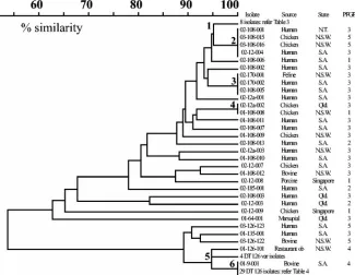
PFGE.The 73 serovar Typhimurium isolates generated five
different PFGE profiles (Fig. 2). PFGE profiles 2 and 3 dis-played⬎90% similarity, suggesting that isolates within these profiles are closely related. All DT126 isolates were exclusively in profiles 4 or 5. The remaining isolates could be found in all profiles, although profile 3 contained the majority of isolates (25 of 36 non-DT126 isolates). No relationship was observed between phage type and pulsed-field profile.
DISCUSSION
Phage typing ofSalmonellacan be subjective and may fail to provide sufficient discrimination between isolates. Discrimina-tion by molecular methods such as PFGE is often unsatisfac-tory, due to the clonal nature ofSalmonella. Although MLST ofSalmonellahousekeeping genes provides a satisfactory level of discrimination for diverse isolates of Salmonella (8), this method may not be suitable for closely related isolates within a serovar, due to sequence identity of their housekeeping genes. Prophages are genetically variable and are widespread within the genusSalmonella(7). The primary aim of this paper was to investigate the potential of prophage loci as suitable targets for the discrimination of serovar Typhimurium phage types closely related by lysis pattern.
Amplification of prophage loci provided the first level of typing of the serovar Typhimurium isolates based on the pres-ence or abspres-ence of PCR product. We term this method mul-tiple amplification of phage locus typing (MAPLT). PCR of prophage loci in serovar Typhimurium generated 27 distinct profiles (Fig. 1), compared to only 5 profiles generated by PFGE (Fig. 2). The MAPLT profiles comprised either single isolates or clusters of isolates with identical PCR profiles within each cluster. Some MAPLT profiles were closely related with the only product from a single primer set separating them;
for example, clusters 1 and were separated by the absence of the SB26ST64Bgene amplification in isolates of cluster 2.
DT126 isolates (as well as the single DT9 and DT135 iso-lates) were clearly different from the other phage types ana-lyzed. There were six MAPLT profiles for DT126, in compar-ison to PFGE, which only generated two profiles, even though the isolates were from diverse sources. These results demon-strate the limitations of PFGE and the potential of MAPLT as an epidemiological tool when typing closely related strains.
It was also observed that most DT126 isolates contained only ST64B sequences. The apparent lack of other phage-related sequences in this phage type reduced the ability of MAPLT to discriminate between isolates. This observation may partially explain the susceptibility of this phage type to almost all (except A6 and A8) of the bacteriophages in the Anderson typing panel, since no P22- and ST64T-like immu-nity genes were amplified. In contrast, DT12 and DT108 con-tain a mosaic of sequences that are related to phages other than ST64B, including genes related to immunity. It is signif-icant that DT12 and DT108 are susceptible to only three and six members of the Anderson panel, respectively. It is this lack of diversity of susceptibility of certain phage types of serovar Typhimurium to the typing panel that can prove problematical in classical phage typing. Typing is then dependent on the more subjective assessment of lysis intensity. The likely pres-ence of a mosaic of phage loci in these DTs makes MAPLT a suitable method for fine discrimination between isolates, as a high number of suitable targets will be present for PCR.
MLST was undertaken with all PCR products, to separate clustered isolates as illustrated in Fig. 1 and to determine which prophage loci exhibit the greatest sequence variability. This information is important, since nucleotide sequence may sometimes be required for further discrimination of isolates. It was observed that the more unrelated isolates by MAPLT showed greater variability of prophage sequences (data not shown). Interestingly, DT170 and DT108 in cluster 3 could not
FIG. 2. Separation of serovar Typhimurium isolates based on PFGE. Dendrogram generated by Dice coefficient with clustering by UPGMA. Five different profiles were generated. Profiles 2 and 3 had⬎90% similarity, suggesting isolates with these profiles are genetically closely related. Serovar Typhimurium DT126 isolates were either profile 4 or 5; the remaining serovar Typhimurium isolates were found in all five profiles. Marker sizes are in kilobases⫻103.
on May 16, 2020 by guest
http://jcm.asm.org/
be separated further (Table 3); these DTs can be difficult to separate using the Anderson typing panel with only variable reactions, with phages A14 and A21 providing separation of the two phage types. It could be postulated that these three isolates do indeed represent the same phage type.
Limited separation of the DT126 isolates was achieved by sequencing of prophage loci (Table 4). In addition, PFGE and MAPLT suggest that DT126 was more clonal than the other DTs tested. In 2001, DT126 became the most frequently iso-lated phage type in Australia and was associated with a number of food-associated outbreaks (2). After 2001, the frequency of DT126 isolation declined to pre-2001 numbers. It is possible that the DT126 isolates isolated during and after 2001 were from a common clone from within Australia or an imported clone. This clone found its way into the food chain, resulting in the increased number of outbreak incidences.
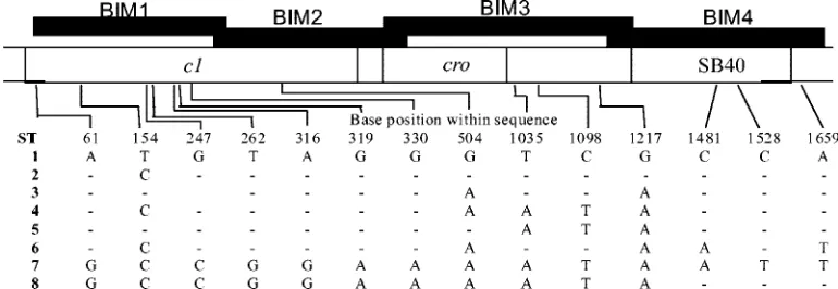
The sequences derived from bacteriophage ST64B have been found to be highly variable in the non-DT126 isolates tested in both amplification and nucleotide sequence analyses (Table 2) (Fig. 3). Genomic sequences of ST64B are wide-spread in serovars ofS.entericasubsp.enterica, including se-rovar Typhimurium (13, 31). The apparent widespread inci-dence of ST64B genomic material in serovar Typhimurium observed with MAPLT and sequence data suggests that loci from ST64B will make excellent targets for isolate discrimina-tion within serovar Typhimurium. It is also likely that these sequences could be of use in otherSalmonellaserovars (C. P. Tucker and M. W. Heuzenroeder, unpublished data). In con-trast, the incidence of ST64T and P22 loci was generally re-stricted to the non-DT126 isolates (Tables 2 and 4). Although discrimination of some isolates was enhanced by targeting ST64T and P22 loci, it should be noted that these loci (a) occur at lower frequency or (b) have limited allelic variation. For example, mntST64T and sieBP22, both of which occur in high
frequency in non-DT126 isolates, were found to have little sequence variation (Table 2). While these loci provided less discrimination between isolates tested in this study than those derived from ST64B, they may be potentially useful for isolate discrimination in other DTs or serovars that are yet to be tested.
MLST of the fiveSalmonellahousekeeping genes provided little discrimination between isolates, either by amplification or
sequencing (Table 2). For three of five loci, all isolates that generated PCR product had identical sequences. A different
glnAallele was found only in the 10 DT126 isolates from the 2003 outbreak, while only a single isolate contained a different
manB allele. In addition to three of the genes described by Kotetishvili et al. (8) used in this study, other genes have been described in the literature for MLST, including the 6-phos-phophogluconate dehydrogenase (gnd) (16), malate dehydro-genase (mdh) (5), and isocitrate dehydrogenase (icd) (34) genes. In these studies, strains were representative of all the describedSalmonellasubspecies and some serovars. As a con-sequence of the broad range of strains employed, allelic vari-ation was not unexpected and demonstrated that sequencing of
Salmonella housekeeping genes is a useful taxonomic tool. However, MLST of housekeeping genes may not be sufficient for separation within a serovar or phage type in real outbreak scenarios; the data presented here reinforce this concept.
Like PFGE, MAPLT and subsequent sequencing of loci did not separate the DT12 and DT108 isolates into groups based on phage type. MAPLT provided a greater level of separation of isolates than PFGE, regardless of phage type. It can be difficult to discriminate between DT108 and DT170, since they react with six and eight members of the typing panel, respec-tively, and share five reactions, with three being variable or weak. These results suggest that DT170 isolates may be distin-guished from the majority of DT108 isolates by MAPLT when phage typing delivers ambiguous results. Further testing with more DT170 isolates will be required to confirm this initial observation.
[image:7.585.98.483.70.203.2]Ten DT126 isolates were obtained from an outbreak in a restaurant in 2003. MAPLT and sequencing analyses indicated that these isolates were similar to all the other DT126 isolates tested, with the exception of theglnAsequence. When phage typed, four of the isolates did not react with phages A12 and A13, which normally produce semiconfluent lysis on DT126. These four isolates were designated DT126 var, based upon this difference. MAPLT separated DT126 var from the other 2003 outbreak isolates. It could be postulated that DT126 var is the result of phage type conversion or some other genetic event by a mobile element rendering the DT126 var isolates resistant to A12 and A13 typing phages. This demonstrates that when multiple phage-derived primers are used, MAPLT
FIG. 3. Sequence types (ST) within theimmCregion of phage ST64B. The base position is the position within the sequenced region; hence, position 1 is position 28047 in GenBank accession number AY055382, which includes ST1. Eight different sequence types including ST1 were detected in the 73 serovar Typhimurium isolates. Where there was no nucleotide difference to ST1, this is indicated by a⫺. The filled boxes indicate regions covered by primers BIM1 to BIM4.
on May 16, 2020 by guest
http://jcm.asm.org/
has the power to discern underlying genetic relationships be-tween strains, even when a phage type conversion or acquisi-tion of a mobile element occurs during an outbreak.
In conclusion, this pilot study has shown the potential of utilizing prophage loci for MLST of phenotypically closely related phage types by lysis patterns of serovar Typhimurium. It is clear from the data that phage type does not necessarily reflect a close genetic relationship between isolates. The re-sults show that the genetic variation of prophage genomes makes them excellent targets for PCR amplification and se-quencing, compared to housekeeping genes. The variation of frequency of occurrence of many of the loci selected indicates that discrimination of isolates can often be achieved by MAPLT alone, without the need to determine the nucleotide sequence. As this is the first report of the utilization of pro-phage loci for typing, further work is required to obtain a minimal number of primer sets that will give the maximum level of isolate discrimination. This will mean the examination of genes from other Salmonella phages as a source of new primer sets, for example, A12 and A13, which are genetically distinct from the P22- and ES18-related phages in the Ander-son typing panel (27).
ACKNOWLEDGMENTS
We thank Dianne Davos, Helen Hocking, and the staff of the Aus-tralian SalmonellaReference Centre, Adelaide, for providing typed strains for this study. We also thank Wendy Hart for critical reading of the manuscript.
This project was undertaken with the generous assistance of the Rural Industries Research and Development Corporation (Chicken Meat Program).
REFERENCES
1.Anderson, E. S., L. R. Ward, M. J. de Saxe, and J. D. de Sa.1977. Bacteri-ophage-typing designations ofSalmonella typhimurium.J. Hyg.78:297–300. 2.AustralianSalmonellaReference Centre.2001. 2001 annual report. Institute
of Medical and Veterinary Science, Adelaide, South Australia.
3.Baker, J., R. Limberger, S. J. Schneider, and A. Campbell.1991. Recombi-nation and modular exchanges in the genesis of new lamboid phages. New Biol.3:297–308.
4.Bell, C., and A. Kyriakides.2002.Salmonella—a practical approach to the organism and its control in foods. Blackwell Science, Ltd., London, United Kingdom.
5.Boyd, E. F., K. Nelson, F.-S. Wang, T. S. Whittam, and R. K. Selander.1994. Molecular genetic basis of allelic polymorphism in malate dehydrogenase (mdh) in natural populations ofEscherichia coliandSalmonella enterica.
Proc. Natl. Acad. Sci. USA91:1280–1284.
6.de Boer, P., B. Duim, A. Rigter, J. van der Plas, W. Jacobs-Reitsma, and J. Wagenaar.2000. Computer-assisted analysis and epidemiological value of genotyping methods forCampylobacter jejuniandCampylobacter coli.J. Clin. Microbiol.38:1940–1946.
7.Figueroa-Bossi, N., and L. Bossi.2004. Resuscitation of a defective prophage inSalmonellacocultures. J. Bacteriol.186:4038–4041.
8.Kotetishvili, M., O. C. Stine, A. Kreger, J. G. Morris, Jr., and A. Su-lakvelidze.2002. Multilocus sequence typing for characterization of clinical and environmentalSalmonellastrains. J. Clin. Microbiol.40:1626–1635. 9.Lindstedt, B.-A., E. Heir, T. Varund, and G. Kapperud.2000. A variation of
the amplified-fragment length polymorphism (AFLP) technique using three restriction endonucleases, and assessment of the enzyme combinationBgl
II-MfeI for AFLP analysis ofSalmonella entericasubsp.entericaisolates. FEMS Microbiol. Lett.189:19–24.
10.Maiden, M. C. J., J. A. Bygraves, E. Feil, G. Morelli, J. E. Russell, R. Urwin, Q. Zhang, J. Zhou, K. Zurth, D. A. Caugant, I. M. Feavers, M. Achtman, and B. G. Spratt.1998. Multilocus sequence typing: a portable approach to the identification of clones within populations of pathogenic microorganisms. Proc. Natl. Acad. Sci. USA95:3140–3145.
11.Maslow, J. N., A. M. Slutsky, and R. D. Arbeit.1993. Application of pulsed-field electrophoresis to molecular epidemiology, p. 563–572.InD. H. Pers-ing, T. F. Smith, F. C. Tenover, and T. J. White (ed.), Diagnostic molecular
microbiology: principles and applications. American Society for Microbiol-ogy, Washington, D.C.
12.Mead, P. S., L. Slutsker, V. Dietz, L. F. McCaig, J. S. Bresee, C. Shapiro, P. M. Griffen, and R. V. Tauxe.1999. Food-related illness and death in the United States. Emerg. Infect. Dis.5:607–625.
13.Mmolawa, P. T., R. Willmore, C. J. Thomas, and M. W. Heuzenroeder.2002. Temperate phages inSalmonella entericaserovar Typhimurium: implications for epidemiology. Int. J. Med. Microbiol.291:633–644.
14.Mmolawa, P. T., H. Schmieger, C. P. Tucker, and M. W. Heuzenroeder.
2003. Genomic structure of theSalmonella entericaserovar Typhimurium DT 64 bacteriophage ST64T: evidence for modular genetic architecture. J. Bacteriol.185:3473–3475.
15.Mmolawa, P. T., H. Schmieger, and M. W. Heuzenroeder.2003. Bacterio-phage ST64B, a genetic mosaic of genes from diverse sources isolated from
Salmonella entericaserovar Typhimurium DT 64. J. Bacteriol.185:6841– 6845.
16.Nelson, K., and R. K. Selander.1994. Intergeneric transfer and recombina-tion of the 6-phosphogluconate dehydrogenase gene (gnd) in enteric bacte-ria. Proc. Natl. Acad. Sci. USA91:10227–10231.
17.Oberto, J., S. B. Sloan, and R. A. Weisberg.1994. A segment of the phage HK022 chromosome is a mosaic of other lamboid chromosomes. Nucleic Acids Res.22:354–356.
18.Pedulla, M. L., M. E. Ford, T. Karthikeyan, J. M. Houtz, R. W. Hendrix, G. F. Hatfull, A. R. Poteete, E. B. Gilcrease, D. A. Winn-Stapley, and S. R. Casjens.2003. Corrected sequence of the bacteriophage P22 genome. J. Bacteriol.185:1475–1477.
19.Popoff, M. Y.2001. Antigenic formulas of theSalmonellaserovars, 8th rev. World Health Organization Collaborating Centre for Reference and Re-search onSalmonella, Institut Pasteur, Paris, France.
20.Ptashne, M., A. Jeffrey, A. D. Johnson, R. Maurer, B. J. Meyer, C. O. Pabo, T. M. Roberts, and R. T. Sauer.1980. How the lambda repressor and Cro work. Cell19:1–11.
21.Ridley, A. M., E. J. Threlfall, and B.Rowe.1998. Genotypic characterization ofSalmonella enteritidisphage types by plasmid analysis, ribotyping, and pulsed-field gel electrophoresis. J. Clin. Microbiol.36:2314–2321. 22.Ross, I. L., R. Willmore, and M. H. Heuzenroeder.2003. A fluorescent
amplified fragment length polymorphism study ofSalmonella enterica sero-var Sofia, the majorSalmonellaserovar isolated from chickens in Australia. Int. J. Med. Microbiol.293:371–375.
23.Sambrook, J., and D. W. Russell.2001. Molecular cloning: a laboratory manual, 3rd ed. Cold Spring Harbor Laboratory Press, Cold Spring Harbor, N.Y.
24.Schicklmaier, P., and H. Schmieger.1997. Sequence comparison of the genes for immunity, DNA relication, and cell lysis of the P22-related Sal-monellaphages ES18 and L. Gene195:93–100.
25.Schicklmaier, P., E. Moser, T. Weiland, W. Rabsch, and H. Schmieger.1998. A comparative study on the frequency of prophages among natural isolates ofSalmonellaandEscherichia coliwith emphasis on the generalized trans-ducers. Antonie Leeuwenhoek73:49–54.
26.Schicklmaier, P., and H. Schmieger.1995. Frequency of generalized trans-ducing phages in natural isolates of theSalmonella typhimuriumcomplex. Appl. Environ. Microbiol.61:1637–1640.
27.Schmieger, H.1999. Molecular survey of theSalmonellaphage typing system of Anderson. J. Bacteriol.181:1630–1635.
28.Scott, F., J. Threlfall, and C. Arnold.2002. Genetic structure ofSalmonella
revealed by fragment analysis. Int. J. Syst. Evol. Microbiol.52:1701–1713. 29.Tenover, F. C., R. D. Arbeit, R. V. Goering, P. A. Mickelsen, B. E. Murray,
D. H. Persing, and B. Swaminathan.1995. Interpreting chromosomal DNA restriction patterns produced by pulsed-field gel electrophoresis: Criteria for bacterial strain typing. J. Clin. Microbiol.33:2233–2239.
30.Threlfall, E. J., and J. A. Frost.1990. The identification, typing and finger-printing ofSalmonella: laboratory aspects and epidemiological applications. J. Appl. Bacteriol.68:5–16.
31.Tucker, C. P., and M. W. Heuzenroeder.2004. ST64B is a defective bacte-riophage inSalmonella entericaserovar Typhimurium DT64 that encodes a functional immunity region capable of mediating phage-type conversion. Int. J. Med. Microbiol.294:59–63.
32.Vander Byl, C., and A. M. Kropinski.2000. Sequence of the genome of
Salmonellabacteriophage P22. J. Bacteriol.182:6472–6481.
33.Vos, P., R. Hogers, M. Bleeker, M. Reijans, T. van de Lee, M. Hornes, A. Frijters, J. Pot, J. Peleman, M. Kuiper, M. Zabeau.1995. AFLP: a new technique for DNA fingerprinting. Nucleic Acids Res.23:4407–4424. 34.Wang, F-S., T. W. Whittam, and R. K. Selander.1997. Evolutionary genetics
if the isocitrate dehydrogenase gene (icd) inEscherichia coliandSalmonella enterica.J. Bacteriol.179:6551–6559.
35.Ward, L. R., J. D. H. de Sa, and B. Rowe.1987. A phage-typing scheme for
Salmonella enteritidis.Epidemiol. Infect.99:291–294.
36.Winokur, P. L.2003. Molecular epidemiolgical techniques forSalmonella
strain discrimination. Front. Biosci.8:14–24.