Journal of Chemical and Pharmaceutical Research
__________________________________________________
ISSN No: 0975-7384 CODEN(USA): JCPRC5
J. Chem. Pharm. Res., 2010, 2(5):656-681
656
Study of the optimized molecular structures and vibrational
characteristics of neutral L-Ascorbic acid and its anion and cation
using density functional theory
Priyanka Singha, N. P. Singha, R. A. Yadavb
a
Lasers and Spectroscopy Laboratory Department of Physics, U P (PG) Autonomous College, Varanasi, India
b
Department of Physics, Banaras Hindu University, Varanasi, India
______________________________________________________________________________
ABSTRACT
FTIR spectra of the neutral L-Ascorbic acid (vitamin C) (L-AA) have been recorded in the range 50-4000 cm-1 on a Varian spectrometer model 3100 using KBr and Nujol optics with 2 cm-1 resolution. The computations were carried out by employing the RHF and DFT methods to investigate the optimized molecular geometries, atomic charges, thermodynamic properties and harmonic vibrational frequencies along with intensities in IR and Raman spectra and depolarization ratios of the Raman bands for the neutral L-AA and its singly charged anionic (L-AA-) and cationic (L-AA+) species. All the 54 normal modes of the L-AA molecule have been assigned and discussed in details in the present study. The bond lengths in the lactone ring for the 2 (C-O) bonds in L-AA- are found to increase whereas for L-AA+ these are found to be decrease as compared to the neutral molecule. The bond angles α(C-O-H) decreases in L-AA- but increases for L-AA+ as compared to the neutral molecule. The dihedral angle H-C-C-H increases by 12.4° while reverse change is noticed for the other H-C-C-H dihedral anglewhich decreases by 14.3° in going from L-AA to L-AA- . The magnitudes of the calculated frequencies for the δ(C-H) modes ν38 and ν34 decrease by 37 and 27 cm
-1
for L-AA- whereas increase 15 and 33 cm-1 for L-AA+ with respect to the neutral molecule. The radicalization of the neutral molecule shifts the magnitude of the frequency of the ν(C-OH) mode ν32 by ~30 cm-1 for L-AA- and by 20 cm-1 for L-AA+ and the IR intensity
for the ν32 mode decreases in going from L-AA to L-AA -
to L-AA+.
Keywords: ab initio and DFT studies; optimized molecular geometries; APT charges; vibrational
657
INTRODUCTION
L-Ascorbic acid (also called vitamin C), hereafter abbreviated as L-AA; is one of the most essential vitamins for both pharmaceutical and food processing industries. In view of its nutritional significance, varied uses in food and high daily doses necessary for optimum health, L-AA is a very significant vitamin for better public health [1]. It has been reported that large doses of vitamin C increases greatly the rate of production of lymphocytes under antigenic stimulation and it is well established that such a high rate of lymphocyte blastomogenesis is associated with a favourable prognosis of cancer [2-9]. L-AA is known to kill HIV-positive cells and to be useful in HIV-positive patients as a consequence of the potentiating the immune system [10].
L-AA is a six-carbon keto-lactone, a strong reducing agent and serves as an antioxidant. The hydrogen donation from L-AA is considered to be primarily responsible for the antioxidant properties attributed to this molecule. It contains four OH groups (two enol OH groups on lactone ring carbons and two OH groups on the side chain C atoms). It can be very easily oxidized and changed to dehydroascorbic acid. Its four hydroxyl (OH) groups play important role in its antioxidant property.
The crystal structure of this compound was studied by different workers [11-13]. Al-Laham et al. [14] performed conformational analysis of AA by forcing geometry of the ring to be constant and optimizing only the conformers of the side chain while Mora and Melendez [15] optimized 36 conformers of AA at the RHF/6-31G, RHF/6-31G(d,p), RHF/6-311+G(d,p) and MP2/6-31G(d,p) levels. The fully optimized gas phase structure [15] was found to be closer to the so called crystallographic B structure of L-AA molecule. The structural and vibrational studies of the L-AA molecule were carried out by number of workers [16-21]. We have recently [22] made a comprehensive structural and vibrational studies of this molecule using DFT and ab initio methods and the reported experimental structural and vibrational spectral data.
In the present paper the optimized geometries, APT charges, thermodynamic properties and harmonic vibrational frequencies along with their IR intensities and Raman activities and depolarization ratios of the Raman lines of singly charged positive and negative radicals of the L-AA molecule have been computed and the results for the neutral L-AA molecule and its anion and cation are compared.
EXPERIMENTAL SECTION
The compound L-Ascorbic acid (L-AA) was purchased from Sigma-Aldrich Chemical Company, (USA) with a purity ≥ 99%. This is a white solid at room temperature. It was used as such without further purification for recording the spectra. IR spectra have been recorded in KBr pellets and Nujol mull using Varian FTIR-3100 spectrometer in the spectral range 50-4000 cm-1 with the following experimental parameters:
658
Fig.-1. Experimental FTIR spectrum of L-AA in KBr pellet/Nujol mull
Fig.-2. Experimental Far IR spectrum of L-AA in KBr pellet/Nujol mull
Theoretical Computations
ab initio and DFT computations of the molecular structures, atomic charges and vibrational
659
molecule was taken as the input structure and the calculations were performed by taking the charge as +1 and multiplicity as 2. The assignments of the normal mode of vibration for the neutral molecule and its radical anion and cation species were made by visual inspection of the individual mode using the Gauss View software [24]. The observed IR frequencies corresponding to the fundamental modes have been correlated to the calculated fundamental frequencies.
RESULTS AND DISCUSSION
4.1. Molecular structures
The optimized geometrical structures for the L-AA molecule and its radical anion and cation calculated at the B3LYP/6-31++G** level along with the experimental parameters are collected in Table-1. The atomic numbering for these molecules are shown in Figs 3-5. As expected the neutral molecule and its radical anion and cation possess non-planar structures with C1 point group symmetry.
As can be seen from the Table-1, the optimized bond lengths of the two single C-C bonds, C1-C5 and C3-C4 in the lactone ring are calculated to be 1.499 Å and 1.457 Å respectively for the neutral molecule. For the neutral molecule shortening of the C3-C4 bond as compared to the C1 -C5 bond is noticed which could be due to attachment of the O atom at the site C3. For the molecule, the calculated values of all the four C-C bonds (including the lactone ring and the side chain) are found to agree with the corresponding experimental values [18] within 0.002 Å - 0.005 Å. The two C-O bond lengths C1-O2 and O2-C3 in the lactone ring are found to be ~1.45 Å and 1.37 Å. In this case also the shorter bond length r (O2-C3) as compared to the bond length r(C1-O2) is a consequence of the attachment of the O atoms at the site C3. The bond length r (C5 -O9) has slightly reduced value for the neutral molecule containing OH group(s). However, there is no such difference for the bond length r(C4-O7). The r(C1-H11) and r(C12-H13) bond lengths appear to be unaffected due to substitution as long as there is at least two identical C-H bonds attached to the site C1/C12. All the four calculated O-H bond lengths are found to agree with the corresponding experimental values [18] within 0.017 Å – 0.039 Å. The bond angles α(O2-C1-C5) and α(O2-C3-C4) are found to be 103.9° and 108.3° respectively, whereas the α(C3-C4-O7) and
α(C4-C5-O9) are found to be 123.1° and 131.3° respectively.
No experimental data for the geometrical structures for radical ions are available. In the L-AA -species due to addition of an electron, the oxygen atom strongly pulls the electron cloud of other atoms towards itself in the lactone ring. The ab initio electron density analysis shows that the removal of electron is delocalized mostly in the ring portion of the radical cation (L-AA+). This is reflected in the relative changes of the bond distances as well as bond angles in both the radical ions as compared to the neutral molecule. The calculated bond length r(C4-C5) of L-AA -(0.052 Å) and L-AA+ (0.065 Å) is longer than those of the L-AA molecule. The calculated bond length in the lactone ring r(C1-C5) and r(O2-C3) increase in going from L-AA to L-AA- by ~0.31 Å and decrease in going from L-AA to L-AA+ by ~0.046 Å. Both the carbonyl bond lengths r(C4-O7) and r(C5-O9) of L-AA- are increased whereas for L-AA+ these are found to decrease as compared to the neutral molecule. As a result, the bond angles α(O2-C1-C12), α(H11-C1-C12) and
660
whereas the bond angle α(O2-C3-C4) decreases in both radical ions as compared to the neutral molecule.
It can be seen from the Table-1, which all the dihedral angles of the lactone moiety (consisting of atoms C1 to H10) are found to be either ± 0° or ±180° within ±0.03°. The value of the dihedral angles C1-C12-C16-H17/C1-C12-C16-H18 increase considerably (14.1°/12.2°) in going from the L-AA to L-L-AA- molecules whereas these decrease by 8.2°/8.7° in going from the L-AA to L-AA+ molecules due to the attachment of an OH group at the site C13. The dihedral angle O14-C12-C16 -H17 increases slightly (by 1.9°) while the angle O14-C12-C16-H18 decreases considerably by 13.8° in going from the L-AA to L-AA- molecules. However, the magnitude of the dihedral angle O14 -C12-C16-H17/O14-C12-C16-H18 is found to decrease by ~12.0° and increase by 12.5° in going from the L-AA to L-AA+ molecules. The value of the dihedral angles C16-C12-O14-H15 and H13-C12 -O14-H15 increase in going from L-AA to L-AA- and decreases in going from L-AA- to L-AA molecules. The magnitude of the dihedral angle H13-C12-C16-H18increases by 12.4° while reverse effect is calculated for the dihedral angles C1-C12-C16-O19 andH13-C12-C16-H17, which decrease by 15.4° and 14.3° respectively, in going from the L-AA to L-AA- while the value of the dihedral angles C1-C12-C16-O19 and H13-C12-C16-H17 increases in going from the L-AA- to L-AA+. Different bond lengths, bond angles and dihedral angles along with their values are shown in figs. 6(a), 6(b) and 6(c), respectively, for the three molecules L-AA, L-AA- and L-AA+. It can be seen from fig. 6(a) that there is variation in the bond lengths for (seven bonds) only due to radicalization. However, in bond angles (Fig. 6b) and dihedral angles (Fig. 6c) variations are noticeable for many more cases.
661
Fig.-5: L-AA+
Figures: 3-5. Atomic labeling scheme for L-AA and its radical ions
Table-1: Computed and experimental geometrical parametersb of L-AA and its radical ions
Parameters L-AA L-AA- L-AA+ Cal. Obsc. Cal. Cal. r(C1-O2) 1.450 1.444 1.450 1.451
r(C1-C5) 1.499 1.491 1.506 1.496
r(C1-H11) 1.096 1.011 1.112 1.093
r(C1-C12) 1.543 1.521 1.538 1.560
r(O2-C3) 1.377 1.355 1.432 1.351
r(C3-C4) 1.457 1.452 1.405 1.493
r(C3-O6) 1.204 1.216 1.236 1.193
r(C4-C5) 1.340 - 1.392 1.405
r(C4-O7) 1.355 1.361 1.386 1.293
r(C5-O9) 1.343 1.326 1.396 1.291
r(O7-H8) 0.968 0.929 0.968 0.981
r(O9-H10) 0.966 0.949 0.975 0.975
r(C12-C16) 1.538 1.521 1.535 1.546
r(C12-H13) 1.096 1.137 1.097 1.096
r(C12-O14) 1.413 1.427 1.422 1.408
r(O14-H15) 0.968 0.937 0.966 0.973
r(C16-H17) 1.097 1.107 1.103 1.094
r(C16-H18) 1.091 1.065 1.094 1.092
r(C16-O19) 1.424 1.431 1.424 1.417
r(O19-H20) 0.967 0.945 0.970 0.964
αααα(O2-C1-C5) 103.9 104.2 105.0 104.4
αααα(O2-C1-H11) 107.4 - 108.5 108.5
662
αααα(C5-C1-H11) 111.1 110.5 109.0 112.5
αααα(C5-C1-C12) 114.9 114.8 118.7 109.6
αααα(H11-C1-C12) 109.0 110.6 107.6 111.6
αααα(C1-O2-C3) 109.5 109.5 108.2 111.8
αααα(O2-C3-C4) 108.3 109.5 107.2 107.4
αααα(O2-C3-O6) 123.6 121.4 120.8 127.5
αααα(C4-C3-O6) 128.1 129.1 131.7 125.1
αααα(C3-C4-C5) 108.8 107.8 111.7 107.9
αααα(C3-C4-O7) 123.1 124.6 121.1 125.3
αααα(C5-C4-O7) 128.1 127.5 126.7 126.8
αααα(C1-C5-C4) 109.5 109.5 105.4 107.7
αααα(C1-C5-O9) 119.3 116.9 117.4 121.8
αααα(C4-C5-O9) 131.3 133.7 125.6 130.4
αααα(C4-O7-H8) 108.0 106.1 105.0 111.3
αααα(C5-O9-H10) 109.8 117.7 108.7 114.2
αααα(C1-C12-C16) 111.9 112.7 111.9 113.4
αααα(C1-C12-H13) 106.2 108.1 106.9 106.0
αααα(C1-C12-O14) 111.6 107.6 112.1 107.5
αααα(C16-C12-H13) 109.0 - 107.4 109.4
αααα(C16-C12-O14) 110.4 106.9 111.4 111.0
αααα(H13-C12-O14) 107.5 - 106.7 109.3
αααα(C12-O14-H15) 106.8 109.0 106.7 107.3
αααα(C12-C16-H17) 110.6 108.1 108.1 111.5
αααα(C12-C16-H18) 109.0 107.4 108.8 107.5
αααα(C12-C16-O19) 111.4 108.0 114.8 110.3
αααα(H17-C16-H18) 108.3 108.7 107.9 108.2
αααα(H17-C16-O19) 111.7 107.3 110.5 112.7
αααα(H18-C16-O19) 105.7 110.7 106.5 106.3
αααα(C16-O19-H20) 108.3 110.5 107.8 110.9
δδδδ(H11-C1-O2-C3) 119.4 - 103.0 129.6
δδδδ(C12-C1-O2-C3) 122.0 - 140.8 108.1
δδδδ(H11-C1-C5-C4) 116.3 - 100.5 125.9
δδδδ(H11-C1-C5-O9) 64.0 - 44.9 57.3
δδδδ(C12-C1-C5-C4) 119.4 - 136.0 109.3
δδδδ(C12-C1-C5-O9) 60.3 - 78.6 67.5
δδδδ(O2-C1-C12-C16) 56.1 - 67.3 57.2
δδδδ(O2-C1-C12-H13) 175.0 - 175.4 177.2
δδδδ(O2-C1-C12-O14) 68.1 - 58.8 65.9
663
b: Bond lengths(r) in Angstrom as (Å), bond angles(α) and dihedral angles(δ) in degrees as (°). c: Ref. [28]
Fig.-6(a) Fig.-6(b) Fig.-6(c)
Figs. 6(a-c): The bond lengths, bond angles and dihedral angles differences from theoretical approaches of the neutral L-AA molecule and its radical ions.
Atomic Charges
APT charges at the various atomic sites of the neutral L-AA molecule and its radical ions calculated are collected in Table-2. The calculated atomic charges at different atomic sites are plotted in Fig.-7 for the neutral molecule and its radical ions. L-AA is a dibasic acid with an
δδδδ(C5-C1-C12-H13) 68.1 - 56.3 68.6
δδδδ(C5-C1-C12-O14) 48.8 - 60.2 48.3
δδδδ(H11-C1-C12-C16) 61.6 - 49.5 63.3
δδδδ(H11-C1-C12-H13) 57.3 - 67.9 56.8
δδδδ(H11-C1-C12-O14) 174.2 - 175.5 173.6
δδδδ(C1-C5-O9-H10) 175.5 - 112.3 174.7
δδδδ(C4-C5-O9-H10) 4.0 - 25.4 1.2
δδδδ(C1-C12-O14-H15) 94.4 - 37.7 109.0
δδδδ(C16-C12-O14-H15) 30.8 - 88.7 15.6
δδδδ(H13-C12-O14-H15) 149.5 - 154.4 136.4
δδδδ(C1-C12-C16-H17) 46.3 - 60. 4 38.1
δδδδ(C1-C12-C16-H18) 165.2 - 177.4 156.5
δδδδ(C1-C12-C16-O19) 78.7 - 63.3 88.0
δδδδ(H13-C12-C16-H17) 70.9 - 56.6 80.0
δδδδ(H13-C12-C16-H18) 48.0 - 60.4 38.4
δδδδ(H13-C12-C16-O19) 164.3 - 179.6 154.0
δδδδ(O14-C12-C16-H17) 171.2 - 173.1 159.2
δδδδ(O14-C12-C16-H18 ) 69.9 - 56.1 82.4
δδδδ(O14-C12-C16-O19) 46.3 - 63.1 33.2
δδδδ(C12-C16-O19-H20) 65.5 - 35.3 93.2
δδδδ(H17-C12-O19-H20) 58.7 - 87.2 32.2
664
enediol group built into a five membered heterocyclic lactone ring. The molecule is stabilized by delocalization of the π-electron over the conjugated carbonyl and enediol system. All the oxygen atoms are seen to possess negative charges due to their electron-withdrawing nature. However, for the lactone ring, the O2 and O7 atom(s) the value of the charges decrease by 0.1100 and -0.0674 in L-AA- and by -0.1406 and -0.0399 in L-AA+ as compared to the neutral molecule. The APT charges at the sites O14 and O19 increase in going from the L-AA+ to L-AA- and L-AA+ to L-AA due to radicalization. In the lactone ring, all the four C atoms possess positive charges but in L-AA-, C4 and C5 are negative because they are strongly affected by bond character. The maximum positive charge on the atom C3 due to presence of the two electronegative O atoms attached to the C3 site. The charge at the sites C12 and C13 decrease in going from L-AAto L-AA -by 0.0523/0.0261 but increases -by 0.0836/0.0187 in going from L-AA- to L-AA+ due to radicalization. For the neutral and anionic species, the increasing magnitude of charge on the carbon atoms of the side chain in the order C13>C12 due to the attachment of the hydroxyl group while as a result of cationic radicalization of L-AA, the charges are found to be in the reverse order i.e, C13<C12.
[image:9.595.68.566.490.721.2]The charges computed for the H8 and H10 atoms are found to be decrease in going from the neutral species to the anionic species whereas these increase in going from the anionic species to cationic species due to the bond character (see, Figs. 3-5). The charge is more negative in the anionic species and less negative in the cationic species at the sites H17/H18 as compared to the neutral species. Thus, in the case of electron addition the most of the negative charge is concentrated at the O19 atom. The charge at the site H15 decreases by -0.0210 in going from the neutral species to the anionic species while increases by 0.0364 in going from the anionic to the cationic species. However, reverse effect is observed at the site H20, whose charge increases by 0.0208 in going from the L-AA to L-AA- and decreases by -0.0149 in going from the AA to L-AA+ species.
Table - 2: Calculated APT charges at various atomic sites for L-AA and its radical ions
Atoms
L-AA L-AA- L-AA+ APT charges APT charges ∆∆∆∆q = q- - q APT charges ∆∆∆∆q = q+ - q q q- q+
C1 0.2858 0.4511 0.1653 0.0193 -0.2665
O2 -0.7618 -0.6581 0.1100 -0.6212 0.1406
C3 1.2806 1.7394 0.4588 1.0167 -0.2639
C4 0.0861 -0.2124 -0.2985 0.4323 0.3462
C5 0.5882 -0.4218 -1.0100 0.8161 0.2279
O6 -0.8662 -1.4211 -0.5549 -0.6141 0.2521
O7 -0.6427 -0.5753 0.0674 -0.6028 0.0399
H8 0.3355 0.2270 -0.1085 0.4202 0.0847
O9 -0.7100 0.2402 0.9502 -0.6734 0.0366
H10 0.3072 -0.1674 -0.4746 0.4036 0.0964
H11 -0.0311 -0.1978 -0.1667 0.0748 0.1059
C12 0.4116 0.3593 -0.0523 0.4429 0.0313
665
Fig. 7: APT atomic charges at various atoms of the L-AA molecules and its radical ions.
Vibrational Assignments Neutral Molecule
The computed IR and Raman spectra for the neutral L-AA molecule and its radical ions are shown in Figs. 8-13. The calculated fundamental frequencies along with their IR intensities, Raman activities, depolarization ratios for the Raman bands and the proposed vibrational assignments are collected in Table-3. The experimentally observed FTIR frequencies for the neutral molecule are also included in the Table-3. The normal mode assignments were made with the help of Gauss View software. In order to make consistent assignments for all the fundamental modes of the L-AA molecule help has been taken from the normal mode assignments made for the DHF, DHMF, DHHMF and DHHEF molecules [22], calculated IR intensities and Raman activities as well as the Raman depolarization ratios along with the experimental IR and Raman spectral studies [18-21] for the L-AA molecule.
There are several inconsistencies in the assignments of the fundamentals of the neutral L-AA molecule [18-21]. Dimitrova [18] distributed the 54 normal modes of L-AA as: the torsional modes-16; the deformation modes-19 and the stretching modes-19 whereas Panicker et al.[19] have assigned only thirty-eight normal modes based on their FT-IR, FT-Raman and SERS spectral studies. Obviously the distribution reported earlier [19] is incorrect and that given by Panicker et al. is incomplete. As the L-AA molecule is a twenty-atomic molecule, its 54 normal modes of vibration are : 20-stretching modes (ν) corresponding to the twenty bonds, 8-deformation modes (δ) as the 2 δ(C1-C12), 2 δ(C12-C16), 2 δ(C1-H11)and 2 δ(C12-H13) modes, 8-O14 -0.5992 -0.5729 0.0263 -0.5567 0.0425
H15 0.2969 0.2759 -0.0210 0.3333 0.0364
C16 0.4211 0.3950 -0.0261 0.4137 0.0094
H17 -0.0523 -0.0713 -0.0190 -0.0085 0.0438
H18 -0.0175 -0.0534 -0.0359 0.0137 0.0312
O19 -0.6142 -0.6256 -0.0114 -0.5819 0.0323
666
angle bending modes (α) as the 4 α(C-O-H),2 α(C-C-O) and 2 angle bending of the lactone ring, 3-in–plane (with respect to the lactone ring) bending modes (β) as the 2 β(C-OH) and 1 β(C=O), 5-out-of plane (with respect to lactone ring) modes (γ/φ) as the 2 γ(C-OH), 1 γ(C=O) and 2 out-of plane lactone ring deformation φ, 7-torsional modes (τ) as the 4 τ(C-OH), 1 τ(CH2 ), 1 τ(C1-C12) and 1 τ(C12-C14), 1-CH2 scissoring mode (σ), 1-CH2 rocking mode (ρ) and 1-CH2 wagging mode (ω). As this molecule possesses C1 symmetry all of its 54 normal modes of vibration are IR as well as Raman active.
O-H modes (12 modes)
There are 4 ν(OH), 4 α(C-O-H) and 4 τ(C-OH) modes due to the four OH groups two being attached to the ring and two to the side chain. The modes ν54, ν53, ν52, ν51, ν43, ν42, ν41, ν35, ν15,
ν14, ν13 and ν11 correspond to these modes.
For the L-AA molecule the O-H stretching region exhibits four strong IR bands in the region 3216 cm-1- 3626 cm-1 [18-19]. In the present case the recorded IR spectra contain four strong bands in the range 3200-3600 cm-1, namely, 3220 (vs), 3317(vs), 3412(vs) and 3528(vs) cm-1. The first three frequencies agree with the earlier reported frequencies [28-29], however, the fourth frequency agrees only with that reported by Panicker et al.[18]. The ν(O9-H10) mode is found to have higher frequency than the mode ν(O7-H8) which can be explained as follows: the H10 atom is hydrogen bonded with the O7 atom whereas the H8 atom is hydrogen bonded with the O6 atom. Since the O6 atom bears more negative charge as compared to the O7 atom, the O6…H8 distance is smaller than the O7…H10, as a consequence of which the distance r(O9-H10) is larger than the distance r(O7-H8) giving rise to higher force constant corresponding to ν(O9-H10) as compared to that corresponding to ν(O7-H8). Similarly, the frequency ν(O19-H20) is higher than the frequency ν(O14-H15) which could be due to more negative charge on O19 as compared to that on the O14 atom. Thus, on the basis of APT charges only the order of frequencies of the four OH stretching modes should be ν(O7-H8) < ν(O9-H10) < ν(O14-H15) < ν(O19-H20). However, the four
ν(O-H) modes of the L-AA molecule are found in the order ν(O9-H10) > ν(O19-H20)>ν(O7 -H8)>ν(O14-H15) (see Table-3, ν51-ν54) which could be due to complexity of hydrogen bonding in the lactone ring and the side chain. The presently observed frequencies 3220 (vs), 3317(vs), 3412(vs) and 3528(vs) cm-1 are correlated to the modes ν(O9-H10), ν(O19-H20),ν(O7-H8) and
ν(O14-H15) respectively.
The ν11, ν13, ν14 and ν15 modes correspond to the four τ(OH) modes. Assignment of these τ(OH) modes is a difficult task as these are strongly coupled amongst themselves and with many other modes. The lowest magnitude (357 cm-1) mode ν11 is found for the τ(O7-H8) mode which is coupled with the τ(O9-H10) mode with the corresponding IR frequency 356 cm−1 observed with very strong intensity. Earlier [17] this mode was correlated to the observed frequency at 350 cm -1
. The τ(O9-H10) mode is calculated to be 420 cm-1 and appears to be coupled with the τ(O7-H8) mode. The observed IR frequency 366 cm-1 was earlier correlated to this mode by Hvoslef [17], which seems to be quite a low frequency for this mode. No observed frequency could be correlated to this mode in the present case. The τ(O14-H15) mode, strongly coupled with the
τ(O19-H20) and τ(O9-H10) modes has a magnitude of 424 cm-1. The calculated frequency for the
667
observed frequencies corresponding to the OH torsional modes ν14 and ν15 have magnitudes 447 cm-1 and 494 cm-1 and appear to have medium and very weak IR intensities respectively.
Assignment of the angle bending modes α(C-O-H) is also complicated by coupling of these modes amongst themselves and with other modes. The mode ν35 arises due to α(C4-O-H) and is calculated to be 1302 cm-1 with the corresponding observed Raman and IR frequencies at 1258 cm-1 and 1246 cm-1 [19], respectively and it appears to be strongly coupled with the ν(C5-OH),
δ(C1-H11) and δ(C12-H13) modes. The mode ν43 originates due to α(C5-O-H) and is strongly coupled with α(C4-O-H) mode and is calculated to have the frequency 1448 cm-1 which could be correlated to the observed IR frequency 1443(m) in agreement with the earlier [17] work. The angle bending mode ν41 arising due to α(C12-O-H) shows coupling with the δ(C12-H13) mode and it is calculated to have a magnitude 1415 cm-1 with the corresponding observed frequency 1388 cm-1 in the IR spectrum. The α(C16-O-H) mode (ν42) with the calculated frequency 1431 cm-1 and coupled with the α(C12-O-H) mode was earlier [19] correlated to the observed IR frequency 1385 cm-1 by which seems to be relatively a lower magnitude. No experimental frequency could be observed corresponding to this mode in the present case.
CH2 modes (6 modes)
The CH2 group has six normal modes of vibration as: an anti-symmetric stretching
mode-νas(CH2), a symmetric stretching mode-νs(CH2), a scissoring mode-σ(CH2), a wagging
mode-ω(CH2), a torsional mode-τ(CH2) and a rocking mode-ρ(CH2). The modes ν50, ν47, ν44, ν39, ν33 and ν26 correspond respectively to these modes. The CH2 anti-symmetric and symmetric stretching modes (ν50 andν47) do not couple with any other modes, except the C1-H11 stretching mode which couples with the νs(CH2) mode. Panicker et al. [19] observed three frequencies each in the IR (3030, 2917 and 2907 cm-1) and Raman (3004, 2919 and 2879 cm-1) spectra in the range 2850-3150 cm-1 corresponding to the C-H/CH2 stretching modes; however, they did not correlate these frequencies to specific modes arising due to the C-H/CH2 stretching modes. Dimitrova [18] correlated all the four C-H/CH2 stretching frequencies to a single frequency observed at 2915 cm-1 and labeled the four C-H stretching modes as pure single bond C-H stretching modes [18]. The present calculations place the four C-H/CH2 stretching frequencies at 3100, 3046, 3034 and 3011 cm-1 the first (3100 cm-1) and the last (3011 cm-1) frequencies of the above four correspond to the modes νas(CH2) and νs(CH2) while the second (3046 cm-1) and third (3034 cm-1) frequencies arise due to the in-phase (ip) and out-of-phase (op) coupling of the C12 -H13 and C1-H11 stretching modes. As the calculated frequencies corresponding to the νas(CH2) and νs(CH2) modes differ by ~90 cm-1, the frequencies 3030 cm-1 and 2879 cm-1 could be correlated to these modes. In the present case the observed frequency 3036 cm−1 is found to appear strongly in the IR spectra and could be correlated to the νas(CH2) mode.
668
which could be correlated to the observed [19] frequency 1364 cm-1 in the IR spectrum. The CH2 torsion mode (ν33) is calculated to be 1221 cm-1 with medium and weak intensities in the IR and Raman spectra, respectively, corresponding to the observed frequencies 1199 (s) and 1193 (w) cm-1 in the IR and Raman spectra [19]. The calculated frequency 964 cm-1 corresponding to the CH2 rocking mode (ν26) is found to have medium and very weak IR and Raman intensities, respectively, which could be correlated to the observed [19] frequencies 990 (s) cm-1 in the IR and 984 (w) cm-1 in the Raman spectra. From the present IR spectral study the IR bands are seen at 1492, 1361, 1198 and 987 cm−1 are correlated to the CH2 scissoring, wagging, torsional and rocking modes with the corresponding correlated frequencies 1501, 1374, 1221 and 964 cm−1 respectively.
C-H modes (6 modes)
Stretching of the two C-H bonds C1-H11 and C12-H13 gives rise to two coupled C-H stretching modes as the in-phase(ip) coupled (ν48) and out–of-phase(op) coupled (ν49) C-H stretching modes which are calculated to be 3046 cm-1 and 3034 cm-1. It is to be noted here that the ip and op coupled ν(C-H) modes should correspond to the νs(CH2) and νas(CH2) modes in magnitude, however, reverse of this is found in the present case which could be due to location of the C1-H11 and C12-H13 bonds at two different sites C1 and C12.As the two modes ν48 and ν49 have calculated frequencies lying between the frequencies due to the νas(CH2) and νs(CH2) modes, the observed frequencies due to the ν(C1-H11) and ν(C12-H13) modes should also lie between the observed frequencies due to the νas(CH2) (3030 cm-1) and νs(CH2) (2879 cm-1). Therefore, the frequencies 2915 cm-1 and 2907 cm-1 [17,19] are correlated to the two C-H stretching modes ν48 and ν49 respectively. The presently observed frequency 2917 cm-1 corresponds to the ν(C1-H11) mode and has medium intensity in the IR spectra.
The present calculations place the two δ modes (ν40 and ν36) due to the C1-H11 bond at the frequencies 1388 and 1314 cm-1, the former of which corresponds to the observed frequency ~1372 cm-1 [18, 19] while the latter one is coupled with the α(C4-OH) mode and corresponds to the observed IR frequency ~1276 cm-1 [17,19]. The remaining two δ modes (ν38 and ν34) arising due to the C12-H13 bond have calculated frequencies 1364 and 1240 cm-1 and are strongly coupled with the two δ(C1-H11) modes. These modes (ν38 and ν34) could be correlated to the observed frequencies 1344 cm-1 [17] and ~1221 cm-1 [17, 19]. In the present case the observed frequencies 1273 and 1221 cm-1 have been assigned to the modes ν36 and ν34 and these are found to appear with medium intensities in the IR spectra.
Lactone ring mode (9 modes)
669
cm-1. The ring stretching mode ν30 involves mainly stretching of the O2-C3 bond and is calculated to be 1112 cm-1 corresponding to the observed frequency 1113 cm-1[19]. This mode shows strong coupling of the ring stretching with the ν(C4-OH) and α(C5-OH) modes. The ring stretching mode ν28 with calculated frequency 1048 cm-1, involves mainly stretching of the C1 -O2 and C3-O2 bonds with slight contributions from the α(C4-OH) and α(C5-OH) modes. The observed frequency ~1045 cm-1 [19] could be assigned to the above mode. The ring stretching mode ν27 involves stretching of the C1-C5, C3-C4 and C1-O2 bonds and has the calculated frequency 1027 cm-1. This modeis found to arise due to coupling of the ring stretching motions with the α(C4-O-H) and α(C5-O-H) modes. The observed IR and Raman frequencies at ~1025 cm-1 [17, 19] could be correlated to the mode ν27. In the present case the experimentally observed frequency 1026 cm-1 in the IR spectrum is correlated to the mode ν27.The last ring stretching mode ν23 having the calculated frequency 825 cm-1 is strongly coupled with the α(ring) mode corresponding to the observed IR and Raman frequencies at ~820 cm-1 [17, 19]. The IR frequency 822 cm-1 observed with medium intensity is correlated to the mode ν23.
The planar-ring deformation mode ν18 is calculated to be 693 cm-1 with the corresponding observed frequency at ~690 cm-1 [19]. The other planar-ring deformation mode ν17 appears to arise due to ring deformation strongly coupled with the τ(O14-H15)and τ(O19-H20)modes and is found to have the frequency564 cm-1. The two non-planar ring deformation modes ν20 and ν16 are calculated to be 614 and 580 cm-1 with the corresponding observed frequencies 581 cm-1 [19] and ~565 cm-1 [17,19]. The planar-ring deformation mode ν17 and the non-planar ring deformation modes ν20 are correlated to the experimentally observed IR frequencies 567 and 683 cm-1 respectively.
C=O modes (3 modes)
The C=O group gives rise to three normal modes of vibration as a C=O stretching (ν), a C=O in-plane bending (β) and a C=O out-of-plane bending (γ) modes. The modes ν46, ν21 and ν19 correspond respectively, to these modes. The present calculation shows that the C=O stretching mode (ν46) with the calculated frequency at 1836 cm-1 having strong IR intensity and weak Raman intensity is strongly coupled with the ν(C=C) mode. The observed frequencies at ~1760 cm-1 with strong IR and weak Raman intensities were earlier [18, 19] correctly correlated to the
ν(C=O) mode. The C=O in-plane bending vibration (β) is calculated to be 634 cm-1 (ν19) with weak intensity in both the spectra with the corresponding observed frequency 621 cm-1[19] and it is found to couple with the α(C5-C1-C12) mode. The C=O out-of-plane bending mode (γ) is found to have calculated frequency 751 cm-1 (ν21) with the observed frequency 722 cm-1 [17, 19]. The presently observed IR frequencies 1755(vs) cm-1, 630(m) cm-1 and 721(m) cm-1 are correlated to the modes ν46, ν21 and ν19 respectively.
C-OH modes (12 modes)
670
to the following six modes: ν{C12-O(H)}, ν{C16-O(H)}, α{C1-C12-O(H)}, α{C12-C16-O(H)},
τ{C1-C12-O(H)} and τ{C12-C16-O(H)}.
The ν(C4-OH) mode (ν37) coupled with the α(C5-O-H) and ω(CH2) modes is calculated to be 1331 cm-1 with the corresponding observed IR and Raman frequencies ~1320 cm-1 [19]. The
ν(C5-OH) mode (ν32) is calculated to be 1172 cm-1 corresponding to the observed frequency ~1140 cm-1[17, 19] and appears to be strongly coupled with the α(C4-O-H) mode. The ν(C12 -OH) mode (ν31) does not appear to couple with any other mode(s) and could be correlated to the observed IR frequency 1121 cm-1 [19]. The mode ν29 arising due to the C16-OH stretching vibration is calculated to be 1084 cm-1 with the corresponding observed frequency at ~1080 cm-1 [19]. The IR frequencies observed in the present case corresponding to the modes ν37, ν32, ν31 and ν29 are 1321, 1136, 1119 and 1072 cm-1 respectively.
Out of the two planar modes β(C4-OH) (ν6) and β(C5-OH) (ν9) calculated to be at 230 and 307 cm-1 the former could be correlated to the observed frequency 180 cm-1 [17] but no observed frequency could be correlated to the latter fundamental. The γ(C4-OH) mode (ν10) is calculated to be 330 cm-1 and it is found to strongly couple with the γ(C5-OH) and α(C1-C12-H13) modes. No observed frequency could be correlated to this mode. The calculated frequency 148 cm-1 corresponding to the γ(C5-OH) mode (ν4) is found to have weak IR and Raman intensities and the observed Raman frequency 122 (w) cm-1 [19] could be correlated to this mode.
The calculated frequency 280 cm-1 having weak IR and Raman intensities corresponding to the
α(C1-C12-O14) mode (ν8) is correlated to the observed Raman frequency 273(w) cm-1 [17] which also agrees with the presently observed IR frequency 273 cm-1. The α(C12-C16-O19) mode (ν7) is calculated frequency 255 cm-1 with the corresponding observed Raman frequency 238 (m) cm-1 [17]. The calculated frequencies 59 and 160 cm-1 correspond to the torsional modes (ν1 and ν5) about the C1-C12 and C12-C16 bonds with the former mode observed at 43 (vw) cm-1 in the Raman spectrum [17]. In the present case the experimentally IR band observed at 163 cm-1 with weak intensity is correlated to the latter torsional mode ν5.
C1-C12/C12-C16 modes (6 modes)
The 6 normal modes of vibration arising due to the C1-C12 and C12-C16 bonds are: the two ν(C-C) and four δ(C-C) modes. The calculated frequencies 839 (ν24) and 930 (ν25) cm-1 correspond to the ν(C1-C12) and ν(C12-C16) modes the latter of which corresponds to the observed frequency 871 cm-1 [19] while no observed frequency could be correlated to the former mode. The experimentally observed IR frequency 868 cm-1 corresponds to the ν(C12-C16) mode. The two δ modes (ν22 and ν2) due to the C1-C12 bond are calculated to have the frequencies 777 and 79 cm-1 with the corresponding observed frequencies ~750 cm-1 [19] and 91(m) cm-1 [17]. In the present case the IR band at 757 cm-1 observed with strong intensity could be assigned to the mode ν22. The former of the above two modes is strongly coupled with one of the φ(ring) modes. The remaining two δ modes (ν12 and ν3) arising due to the C12-C16 bond have calculated frequencies 368 and 118 cm-1, which could be correlated to the observed frequencies 340 and 113 cm-1 [17].
671
The conversion of the L-AA molecule into its radical ions leads to significant increase in the Raman activities for all the O-H stretching modes (ν54, ν53, ν52 and ν51), whereas the IR intensities increase for all the four O-H stretching modes in radical cation but in radical anion the IR intensities increase only for three O-H stretching modes. Further, the frequency for the ν (O-H) modes ν54 and ν53 shifts towards the lower wavenumber side in going from the neutral molecule to radical anion whereas further increase in going from the radical anion to the radical cation species. The magnitude of the calculated frequencies for the τ(O-H) modes ν14, ν13 and ν11 increase in going from L-AA to L-AA- to L-AA+. The present calculations show that the frequency for the OH torsional mode ν15 decreases for L-AA- by ~220 cm-1 and for L-AA+ by ~213 cm-1. The frequency for the α(C16-O-H) mode ν42 is found to increase from 1431 cm-1 to 1460 cm-1 in going from the neutral species to the anionic species while it is found to reduce from 1460 cm-1 to 1383 cm-1 in going from the anionic species to the cationic species. Similarly, the frequency for the mode α(C5-O-H) (ν43) increases by ~86 cm-1 for the radical cation while it decreases by ~59 cm-1 for the radical anion as compared to the neutral molecule.
The νas(CH2) mode (ν50)is found to decreased in frequency by 47 cm-1 for L-AA- as compared to the neutral molecule however, it has nearly the same frequency for L-AA+. The frequency for the
νs(CH2) mode (ν47) decreases from 3011 cm-1 to 2943 cm-1 in going from L-AAto L-AA- and increases from 2943 cm-1 to 3051 cm-1 in going from L-AA- to L-AA+. For the same vibrational mode (ν47), the Raman activity increases significantly for L-AA+ as a result of radicalization. In going from the L-AA- to L-AA+ species, the Raman activities decrease significantly for the modes ν26 and ν33. It is also found that the frequency corresponding to the τ(CH2) mode (ν33) increases by ~48 cm-1 for L-AA- and decreases by ~125 cm-1 for L-AA+ as compared to the neutral molecule. The frequency corresponding to the mode (ν39)is found to reduce from 1374 cm-1 to 1348 cm-1 in going from neutral to anionic species and increase from 1348 cm-1 to 1403 cm-1 in going from the anionic to cationic species.
The magnitudes of the frequencies of the C-H stretching modes ν49 and ν48 decrease by 18 and 211 cm-1 in going from the neutral to anionic species whereas these increase by 6 and 53 cm-1 in going from neutral to cationic species. The magnitudes of the calculated frequencies for the
δ(C12-H13) modes ν38 and ν34 decrease by 37 and 27 cm-1 for L-AA- whereas increase by 15 and 33 cm-1 for L-AA+ with respect to the neutral molecule. The calculated frequencies for the deformation modes of the C1-H11 bond (ν40 and ν36) decrease in going from the L-AA to L-AA- to L-AA+ species.
672
The C=O stretching frequency (ν46) decreases by 151 cm-1 in going from the neutral to the anionic species whereas it increases by 151 cm-1 in going from the anionic to the cationic species. For the above vibrational mode, the Raman activity increases significantly for the anionic and cationic species. It can also be seen (Table-3) that the calculated frequency for the out-of-plane deformation of C=O (ν21) decreases in going from L-AA to L-AA+ to L-AA- due to the radicalization process. It is observed that the IR intensity and Raman activity increase for the L-AA- due to the conversion process. However, the C=O in-plane bending mode (ν19) frequency decreases in going from L-AA to L-AA- by ~68 cm-1, while its magnitude is nearly same for the L-AA and L-AA+ species.
The conversion of the neutral molecule shifts the magnitude of the frequency of the ν(C5-OH) mode ν32 by ~30 and 20 cm-1 towards lower wavenumber side for L-AA- and L-AA+ and the IR intensity for the above frequency decreases in going from L-AA to L-AA- to L-AA+. For the neutral L-AA molecule changes in the magnitude of the frequencies due to the ν(C-OH) modes
ν37, ν31 and ν29 are found to be insignificant while for the L-AA- molecule, the Raman activities are much more pronounced due to radicalization. The neutral L-AA molecule and its radical cation have nearly the same magnitude (~300 cm-1 and ~ 235 cm-1) for both the β(C-OH) modes (ν9 and ν6). However, for the anion species the corresponding mode frequency decreases by ~40 cm-1 and ~90 cm-1 as compared to both the neutral and cationic species. The frequency of one (ν10) of the two γ(C-OH) modes shifts towards lower wavenumber side by 13 cm-1 for L-AA+ and by 34 cm-1 for L-AA- as compared to the neutral molecule. The frequencies corresponding to the C12-C13-O19/C1-C12-O14 angle bending modes ν7 and ν8 decrease by 34/36 cm-1 for L-AA- as compared to the neutral molecule, however it shifts upward by ~30/24 cm-1 in going from the L-AA- to L-AA+ species. For both the τ(C-C) modes ν1 and ν5 relatively smaller changes are noted in the frequencies in going from the L-AA to L-AA- to L-AA+ species.
For both the side chain C-C stretching modes significant changes are noticed in the Raman activities due to the radicalization process. Changes in the magnitudes of the frequencies corresponding to the δ(C-H) modes (ν3 and ν22) are found to be insignificant while the Raman activities are found to be enhanced for L-AA- as compared to the neutral and cationic species. The frequency corresponding to the C-H deformation mode ν12 shifts towards higher wavenumber side by ~230 cm-1 for L-AA- and by ~307 cm-1 for L-AA+ with respect to the neutral molecule.
Table-3: Calculated and experimental fundamental frequenciesp of L-AA and its radical ions
L-AA L-AA- L-AA+ Moder S. Cal. Exp.q Obs. Cal. Cal.
a b c d No. FT-IR IR Raman IR Raman Raman IR
673
0.74 0.09 0.38
ν2 79 - - 91m - - - - 75 64 δ(C1-C12) (1,1) (1,11) (4,1)
0.72 0.65 0.75
ν3 118 - - 113w - - - - 123 97 δ(C12-C16) (4,0.60) (17,851) (8,0.46)
0.75 0.21 0.35
ν4 148 - - 122w - - - - 143 143 γ(C5-OH) (2,0.14) (64, 1094) (2,40)
0.50 0.40 0.36
ν5 160 - - - - - - 136 125 τ{C12-C16- (8,0.50) (5, 673) (3,10) O(H)}
0.75 0.21 0.43
ν6 230 - - 180w - - - - 151 235 β(C4-OH) (9,0.91) (28,38) (13,1)
0.70 0.67 0.66
ν7 255 - 238m - - - 237w - 221 251 α{C12-C16-
(4,0.39) (12, 130) (1,19) O(H)} 0.71 0.22 0.42
ν8 280 273w - 273w - - - - 244 268 α{C1-C12- (0.60,0.73) (0.71,427) (2,101) O(H)} 0.07 0.12 0.30
ν9 307 - - - - - - - 260 297 β(C5-OH) (14,0.47) (22,26) (55,0.26)
0.32 0.55 0.62
ν10 330 - - - - - 318w - 296 317 γ(C4-OH) (2,0.13) (82,1367) (4,11)
0.67 0.06 0.41
ν11 357 356vs 350s - - - - - 587 664 τ(O7-H8) (161,1) (44,910) (130,42)
0.74 0.14 0.40
ν12 368 - 340w - - - - - 515 576 δ(C12-C16) (13,0.89) (11,290) (21,240)
0.74 0.15 0.40
ν13 420 - 366s - - - - - 545 602 τ(O9-H10) (51,0.97) (29,1661) (128,49)
0.09 0.06 0.22
674
(84,0.77) (82,11436) (112,3) 0.75 0.11 0.58
ν15 524 494vw - - - - - - 304 351 τ(O19-H20) (25,3) (14,298) (88,55)
0.41 0.07 0.31
ν16 564 - - - - - - - 365 553 α(ring) (29,3) (33,1666) (7,2)
0.56 0.11 0.44
ν17 580 567m 565m - - 564m - - 315 360 φ(ring) (93,2) (55,955) (31,1)
0.21 0.06 0.71
ν18 614 - - - - 581w 588w - 350 489 φ(ring) (128,5) (9,303) (19,212)
0.73 0.05 0.33
ν19 634 630m - - - 621s - - 566 631 β(C=O) (3,9) (90,4331) (18,5)
0.11 0.07 0.38
ν20 693 683m 711vw - 686w 693m - - 680 684 α(ring) (5,6) (18,958) (20,75)
0.17 0.07 0.22
ν21 751 721m 721m - 722w - - - 631 736 γ(C=O) (16,0.78) (32,14) (7,43)
0.14 0.75 0.30
ν22 777 757s - - 757s 742sh - - 750 768 δ(C1-C12) (3,2) (99,14669) (6,19)
0.13 0.08 0.34
ν23 830 822m 820m - 821m 823m 828m - 790 813 ν(ring) (12,5) (29,2681) (10,201)
0.63 0.08 0.38
ν24 839 - - - - - - - 837 833 ν(C1-C12) (29,4) (43,484) (12,44)
0.59 0.16 0.19
ν25 930 868m 869m - 871w 871m 884w - 898 920 ν(C12-C16) (13,5) (36,774) (28,14)
0.32 0.04 0.57
ν26 964 987vs - - 990s - 946w - 950 961 ρ(CH2) (26,2) (71,1046) (55,252)
675
ν27 1038 1026vs 1025s - 1027vvs - - - 982 1005 ν(ring) (22,1) (66,656) (83,438)
0.35 0.70 0.41
ν28 1048 - - - 1046m 1048m - 1045w 1013 1032 ν(ring) (78,0.58) (84,1480) (158,420)
0.67 0.15 0.31
ν29 1084 1072m 1074m - 1077m 1081w 1061w 1075w 1102 1075 ν(C16-OH) (26,4) (42,1471) (69,206)
0.47 0.06 0.40
ν30 1112 - 1118s - 1113vs 1113s - - 1073 1146 ν(ring) (274,9) (151,1246) (222,165)
0.44 0.05 0.23
ν31 1116 1119s - - 1121vs - - 1118s 1123 1064 ν(C12-OH) (113,5) (93,594) (59,461)
0.61 0.21 0.33
ν32 1172 1136m 1138s - 1142vs - 1153w 1139s 1142 1122 ν(C5-OH) (113,8) (36,4415) (17,13)
0.41 0.0021 0.71
ν33 1221 1198m 1197m - 1199s 1193w - 1198m 1269 1196 τ(CH2) (24,5) (28,1967) (29,139)
0.73 0.05 0.36
ν34 1240 1221m 1220m - 1222s - 1225w 1221m 1213 1273 δ(C12-H13) (10,11) (64,461) (8,22)
0.60 0.24 0.27
ν35 1302 - - - 1246m 1258s - - 1247 1344 α(C4-O-H) (99,2) (107,767) (118,2)
0.50 0.06 0.470
ν36 1314 1273m 1275m - 1277s - - - 1240 1230 δ(C1-H11) (119,8) (9,614) (74,129)
0.65 0.59 0.33
ν37 1331 1321vs 1320s - 1322s 1323s 1300w 1321s 1297 1361 ν(C4-OH) (101,14) (52,5031) (56,127)
0.29 0.05 0.21
ν38 1364 - - 1344vw - - - - 1327 1372 δ(C12-H13) (15,3) (14,799) (81,8)
0.27 0.14 0.30
ν39 1374 1361m 1362m - 1364m - 1361w - 1348 1403 ω(CH2) (4,8) (58,5086) (22,32)
676
ν40 1388 - - 1372w - 1371w - - 1382 1310 δ(C1-H11) (10,4) (32,4009) (113,3)
0.53 0.05 0.61
ν41 1415 1388m - - 1389m - - - 1415 1417 α(C12-O-H) (39,4) (30,43) (58,66)
0.67 0.03 0.25
ν42 1431 - 1385m - - - - - 1460 1383 α(C16-O-H) (41,5) (51,10) (13,5)
0.31 0.43 0.50
ν43 1448 1443m 1435m - - - - - 1389 1534 α(C5-O-H) (50,7) (66,1961) (138,60)
0.52 0.09 0.29
ν44 1501 1492w 1482w 1482w 1487m 1484m 1477w - 1495 1493 σ(CH2) (6,6) (188,60) (3,155)
0.75 0.31 0.39
ν45 1769 1674vvs 1670vs 1670vs 1675vvs 1661vvs 1699 vvs 1672vs 1557 1707 ν(C=C) (478,96) (8,3674) (116,2103)
0.16 0.04 0.34
ν46 1836 1755vs 1753s 1753w 1764s 1758w 1765w 1755m 1685 1836 ν(C=O) (367,20) (1825,35699) (333,271) 0.34 0.05 0.14
ν47 3011 - - - - 2879wsh - - 2943 3051 νs(CH2) (33,76) (80,88) (22,211)
0.15 0.60 0.06
ν48 3034 2917m 2915m 2915vs 2917m 2919s 2956s - 2823 3087 ν(C1-H11) (7,88) (265,5593) (4,115)
0.45 0.41 0.29
ν49 3046 - - - 2907m - - - 3028 3041 ν(C12-H13) (46,184) (23,79) (3,67)
0.12 0.68 0.75
ν50 3100 3036s - - 3030sbr 3004w - - 3053 3104 νas(CH2) (22,130) (1182,69) (7,700)
0.37 0.71 0.35
ν51 3744 3220vs 3230m - 3216s - - - 3788 3670 ν(O14-H15) (72,42) (32,74) (167,93)
0.06 0.53 0.66
ν52 3767 3317vs 3330s - 3315s - - - 3746 3601 ν(O7-H8) (118,83) (32,1578) (305,1383)
677
ν53 3779 3412vs 3420s - 3410s - - - 3696 3829 ν(O19-H20) (87,41) (205,1185) (68,472)
0.15 0.07 0.19
ν54 3798 3528vs 3535s - 3626s - - - 3529 3685 ν(O9-H10) (109,101) (67,25357) (278,144)
0.23 0.48 0.66
p: The first and second numbers within each bracket represent IR intensity(Km/mol) and Raman activity(Å4/amu) while
the number above and below each bracket represent the corresponding calculated frequency(cm-1) and depolarization
ratios of the Raman band respectively.
s: strong, m: medium, w: weak, vs: very strong, vvs: very very strong. r: ν=stretching, ω=wagging, τ=twisting,
ρ=rocking, σ=scissoring, δ= deformation, γ=out-of-plane deformation, β=in-plane deformation, α=angle bending, νs=symmetric stretching, νas= anti-symmetric stretching, α= in–plane ring bending, φ=out-of-plane ring bending. q:
From solid state FT-IR spectra in KBr pellet and Far-IR spectra in Nujol mull. a: ref.[27],b: ref.[29], c: ref.[30],d: ref.[31].
678
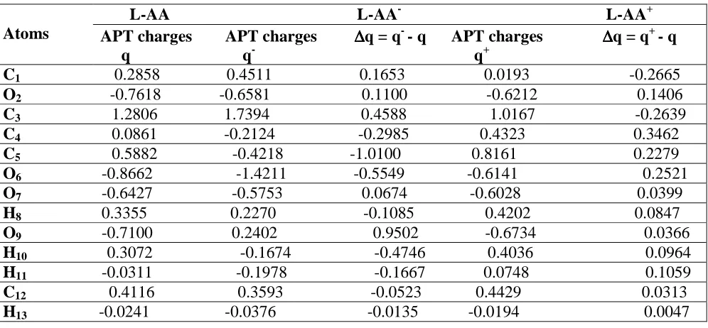
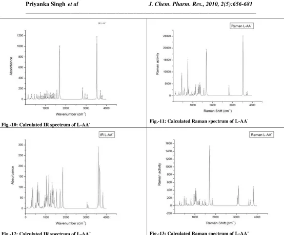
Fig.-10: Calculated IR spectrum of L-AA- Fig.-11: Calculated Raman spectrum of L-AA
[image:23.595.20.574.70.530.2]-
Fig.-12: Calculated IR spectrum of L-AA+ Fig.-13: Calculated Raman spectrum of L-AA+
Figs. 8-13 : The calculated vibrational frequencies, IR intensity and Raman activity of L-AA and its radical ions
Thermodynamics properties
The molar heat capacity (CV) at constant volume, entropy (S), thermal energy (TE) and zero-point vibrational energy (ZPVE), total energy (E) and dipole moment (µ) were obtained for the neutral L-AA molecule and its radical anionic and cationic species and these are collected in the Table-4. The calculated dipole moment decreases in going from the L-AA and L-AA- species. The radical anion is calculated to have lower energies compared to both the neutral and anionic species.
679
S. Species Total Zero-point Dipole Constant Entropy Thermal No. Energy vibrational Moment volume molar (S) Energy (E) energy (µµµµ) heat capacity (TE) (ZPVE) (CV)
1 L-AA -684.993 394852.1 3.325 43.063 105.055 101.622 2 L-AA- -684.998 380384.6 2.329 45.233 110.883 98.743 3 L-AA+ -684.677 393123.5 6.072 42.784 107.543 101.222
r:E & ZPVE are measured in Hartrees & Joules/Mol respectively, CV & S are measured in Cal/Mol-Kelvin, TE is measured in Kcal/Mol.
CONCLUSION
In the L-AA molecule shortening of the r(C3-C4) bond by 0.042 Å as compared to the r(C1-C5) bond is due to attachment of an O atom at the site C3. The carbonyl bond lengths r(C4-O7) and r(C5-O9) for L-AA- are increased whereas for L-AA+ these are found to be decrease as compared to the neutral molecule. The bond angles α(O2-C1-C12), α(H11-C1-C12) and α(O2-C3-O6) for L-AA- increase but L-AA+ decrease as compared to the neutral molecule whereas the bond angle
α(O2-C3-C4) decreases for the both radical ions as compared to the neutral molecule. The dihedral angle H13-C12-C16-H18increases by 12.4° while reverse effect is noticed for the dihedral angles C1-C12-C16-O19 and H13-C12-C16-H17 which decrease by 15.4° and 14.3° respectively, in going from L-AA to L-AA- while the value of the dihedral angles H13-C12-C16-O19 and O14-C12 -C13-O19 increase by ~16° in going from the neutral molecule to anionic species whereas these decrease by 27.8° in going from the anionic to cationic species. The APT charges at the sites O14 and O19 increase in going from the L-AA+ to L-AA- and L-AA+ to L-AA species. In the lactone ring, all the four C atoms possess positive charges but in L-AA-, C4 and C5 are negative as these are hardly affected by bond character. The maximum positive charge is on the atom C3 due to attachment of the two electronegative O atoms at the C3 site. The charges at the sites C12 and C13 decrease by 0.0523 and 0.0261 in going from L-AAto L-AA- but increase by 0.0836 and 0.0187 in going from the L-AA- to L-AA+ species.
680
whereas these increase by 6 and 53 cm-1 in going from the neutral to the cationic species. The magnitudes of the calculated frequencies for the δ(C12-H13) modes ν38 and ν34 decrease by 37 and 27 cm-1 for L-AA- while these increase by 15 and 33 cm-1 for L-AA+ with respect to the neutral molecule. The radicalization of the neutral molecule shifts the magnitude of the frequency of the
ν(C5-OH) mode ν32 by ~30 and 20 cm-1 towards the lower wavenumber side for L-AA- and L-AA+ and the IR intensity for the above mode decreases in going from L-AA to L-AA- to L-AA+. The calculated dipole moment has the highest value for the L-AA+ species and the lowest value for the neutral species. The radical anion is calculated to have lower energies compared to both the neutral and anionic species.
ACKNOWLEDGEMENTS
The author (Priyanka Singh) are thankful to Department of Chemistry, B. H. U., U.P. (India) for giving permission to use the FTIR spectrometer for getting recorded the FTIR spectra.
REFERENCES
[1] M Levine. N. Engl. J. Med., 1986, 314, 892-902. [2] I M. Lee. Proc. Assoc. Am. Phys., 1999, 111, 10-15. [3] A C Carr; B. Frei. Am. J. Clin. Nutr.,1999, 69, 1086-1107.
[4] R E Patterson;, E White; A R Kristal; M L Nuehouser; J D Potter. Cancer Causes Contr., 1997, 8, 786-807.
[5] K. A. Head, Altern. Med. Rev.,1998, 3, 175-186.
[6] K N Prasad; A Kumar; V Kochu Pilla; W C Cole. J. Am. Coll. Nutr., 1999, 18, 13-25.
[7] W. M. Cort, Antioxidant properties of ascorbic acid in foods, in: P.A. Seib, B. M. Tolbest(Eds.), Ascorbic Acid Chemistry, Metabolism and Uses, Adv. Chem. Ser. N. 200 P531, American Chemical Society, Washington, DC, 1982.
[8] M J Barnes; E Kodicek. Vitamins Hormones., 1972, 30, 1-43.
[9] L S Hollis; A R Amudsen; E W Stern. J. Am. Chem. Soc., 1985, 107, 274-276.
[10] C I Rivas; J C Vera; V H Guaiquil; F V Velasquez; O A Borquez-Ojeda; J G Carcamo; I I Concha; D W Golde. J. Biol. Chem., 1997, 272, 5814-5820.
[11] J Hvoslef. Acta Cryst., 1968, B24, 23-35. [12] J Hvoslef. Acta Cryst.1968, B24, 1431-1440. [13] J Hvoslef. Acta Cryst., 1969, B25,2214-2223.
[14] M A Al-Laham; G A Petersson; P Haake. J. Comp. Chem., 1991, 12, 113-118. [15] M A Mora; F J Melendez. J. Mol. Struct., 1998, 454,175-185.
[16] W Lohmann; D Pagel; V Penka. Eur. J. Biochem., 1984, 138, 479-480. [17] J Hvoslef; P K Laeboe. Acta. Chem. Scand., 1971, 25, 3043-3053. [18] Y Dimitrova. Spectrochim. Acta A, 2006, 63, 42-437.
[19] C Y Panicker; H T Vargheseb; D Philip. Spectrochim. Acta A, 2006, 65, 802-804. [20] J T Edsall; E L Sagall. J. Chem. Phys., 1943, 65, 1312-1316.
[21] E G Ferrer; E J Baran. Bio. Tra. Ele. Rese., 2001,83, 111-119. [22] P Singh; N P Singh; R. A. Yadav. Author’s unpublished work.
681
Toyota; R Fukuda; J Hasegawa; M Ishida; T Nakajima; Y Honda; O Kitao; H Nakai; M Klene; X. Li; J E Knox; H P Hratchian; J B Cross; C Adamo; J Jaramillo; R Gomperts; RE Stratmann; O Yazyev; A J Austin; R Cammi; C Pomelli; J W Ochterski; P Y Ayala; K Morokuma; G A Voth; P Salvador; J J Dannenberg; V G Zakrzewski; S Dapprich; A D Daniels; M C Strain; O Farkas; D K Malick; A D Rabuck; K Raghavachari; J B Foresman; J V Ortiz; Q Cui, A G Baboul; S Clifford; J Cioslowski; B B Stefanov; G Liu; A Liashenko; P Piskorz; I Komaromi; R L Martin; D J Fox; T Keith; M A Al-Laham; CY Peng; A Nanayakkara; M Challacombe; P M W Gill; B Johnson; W Chen; M W Wong; C Gonzalez and J A Pople. Gaussian 03, Revision C. 02, Gaussian, Inc., Wallingford, CT, 2004.