Structural Study of Piezo Channel, a Novel
Eukaryotes Mechanosensitive Channel.
Thesis by
Aron Kamajaya
In Partial Fulfillment of the Requirements for the degree of
Doctor of Philosophy
CALIFORNIA INSTITUTE OF TECHNOLOGY Pasadena, California
2017
2017
ACKNOWLEDGEMENTS
First and foremost, I would like to thank Doug Rees for his support and mentorship throughout my graduate school years. I am grateful that you gave me freedom to pursue an independent and unique project, which catalyzes my academic growth to become a scientist. Over the years, you have become my role model both as a scientist and a leader.
I would also express my gratitude for the members of my thesis committee: Henry Lester, Bil Clemons, and David Chan. Your insight, advice, and guidance are critical for my success in this journey. Henry, serving as TA in your class for four terms has ignited my interest in neuroscience. Bil, thank you for being such a cool and approachable person to talk to about my project outside the formal meeting. David, thank you for offering your expertise in cell biology. I love the close interaction between your lab and the Rees lab.
and Pavle Nikolovski. I expect to hear great things from you guys! Allen Lee, Jeffrey Lai, and Phoebe Ray, you have done a wonderful job to keep the lab running smoothly, especially in term of ordering reagents, ensuring the availability of common reagents, scheduling a timely service, resolving occasional conflict, and repair for the malfunction equipment, particularly AKTA and fermenter. I would also like to thank Grant Jensen for organizing the single particle EM sub-group, which helped me during the initial struggle to get started in my single particle EM journey. Thanks to Alasdair McDowall for aiding me with the single particle data collection and to Haoqing Wang for helping me in the EM data processing. Big thanks to Jost Vielmetter for allowing me to use the space at the Protein Expression Center (PEC) facility and providing me with a lot of useful discussion on different options to express my gigantic protein, and to Angela Ho for the assistance on developing the stable cell line.
ABSTRACT
PUBLISHED CONTENT AND CONTRIBUTIONS
Kamajaya, A., Kaiser, J.T., Lee, J., Reid, M., Rees, D.C. (2014) The structure of a conserved piezo channel domain reveals a topologically distinct β sandwich fold. Structure 22(10):1520-7. doi: 10.1016/j.str.2014.08.009.
TABLE OF CONTENTS
Acknowledgements………...iii
Abstract ………..…vi
Published Content and Contributions……….………...vii
Table of Contents………...………viii
Chapter 1: Primer on Mechanosensation and the Piezo
Channel ... 1
Mechanosensation ... 2
Bacterial Mechanosensitive (MS) Channels ... 4
Mechanosensation in Multicellular Eukaryotes ... 5
The Plant MSL channel Family ... 6
Diverse Families of eukaryotic MS Channels ... 7
Piezo Family of Metazoan MS Channel Initial Discovery ... 9
Physiological Role ... 10
Current Puzzles ... 13
The Scope of the Thesis ... 16
Chapter 1 Figure ... 17
Chapter 2: The Structure of a Conserved Piezo Channel Domain
Reveals a Beta Sandwich Fold ... 33
Summary and Highlights ... 34
Introduction ... 35
Results Construct Selection ... 37
Architecture of the C. elegans CTL2 Domain ... 38
Structure of M31R C. elegans PIEZO CTL2 Domain Variant ... 39
Discussion ... 40
Experimental Procedures ... 42
Acknowledgements and Author Contributions ... 46
References ... 47
Chapter 3: The Pursuit Toward Obtaining a Full Length PIEZO
Structure ... 67
Historical Context and the Chapter Outline ... 68
Part 1: Exploring Various Protein Expression Systems Introduction... 71
Bacterial Protein Expression Systems ... 72
Pichia Protein Expression System ... 73
The Journey Begins Mammalian Protein Expression System ... 75
Fluorescence Size Exclusion Chromatography Constructs ... 76
Exploring Other Ways of Expressing Full Lengt PIEZO ... 78
Part 2: Visualizing Full Length Mouse PIEZO1 Using Single Particle Electron Microscope (EM) Transition from X-Ray Crystallography to Single Particle EM ... 83
Initial Negative Stain EM Observation ... 84
Full Length Mouse PIEZO1 is a Trimer ... 86
Chapter Summary and Future Direction ... 88
Methods ... 89
Author Contributions ... 98
References ... 99
Chapter 4: Isolation of a Stable C-terminal Piezo
Fragment ... 126
Background and Motivation ... 127
C-terminal Piezo Constructs Expression and Detergent Extraction Screen ... 130
Isolation of a Stable Fragment of Mouse PIEZO1 ... 133
C-terminal Fragment in the Context of the Full Length Mouse PIEZO1 Structure ... 134
Mouse PIEZO1 C-terminal Fragment can be Expressed and Localized to the Plasma Membrane ... 135
Future Directions ... 138
Methods ... 140
Author Contributions ... 145
Reference ... 146
Chapter 1:
Primer on Mechanosensation and
C h a p t e r 1 : M e c h a n o s e n s a t i o n a n d P i e z o C h a n n e l s
Mechanosensation
Life does not exist in a vacuum. The interactions between cells and their surroundings are crucial for cells’ survival. To persist, cells must be able to detect changes in their environment and execute appropriate responses to adapt to these changes. Living organisms are continually experiencing dynamic mechanical forces, and the ability to detect these forces is essential for many biological processes [1]. Mechanosensation is one sensory modality which allows living organisms to detect mechanical force changes on their surroundings. The mechanistic explanation of how these forces are detected by biological mechanosensors remains incompletely understood. One way cells can convert mechanical cues into intracellular signals is through mechanosensitive (MS) channels. MS channels form permeation pathways upon activation by mechanical stimulation. Typically, ions flow through the permeation pathway, which in turn provides signals that lead to further downstream intracellular signaling cascades.
bioinformatics tools to search for a putative MS channel in the available genomic data, and molecular cloning techniques which enable heterologous expression of a putative MS channel for further biochemical characterization.
Bacterial Mechanosensitive Channels
The two best studied MS channels are the bacterial MS channels: the mechanosensitive channel of large conductance (MscL) and the mechanosensitive channel of small conductance (MscS). These MS channels, first described in 1987 [12] and subsequently cloned [13, 14], are thought to serve as safety valves which protect bacterial cells against osmotic downshock as well as intracellular pressure during cell growth [12, 15-17]. Mycobacterium tuberculosis MscL gene codes for 151 amino acids with molecular weight of 16 kDa, consisting of two transmembrane helices (TM1 and TM2), a short cytoplasmic N-terminal helix, and a C-terminal bundle [18]. In the closed state, TM1 segments from five MscL monomers form a constricted pore likely representing a non-conducting state [18, 19]. The single channel conductance of MscL is measured to be around 3 nS [19], corresponding to a channel pore diameter in the open state estimated to be at least 25 Å wide by Electron Paramagnetic Resonance (EPR) studies [20] as well as molecular sieving experiments using large organic ions [21]. Unexpectedly, the crystal structure of truncated Staphylococcus aureus MscL revealed a homotetrameric arrangement [19], and the variation in oligomeric state between the two MscL structures may reflect unintended consequences of truncation and detergent solubilization [22].
Escherichia coli MscS gene codes for a subunit of 286 amino acids with a molecular weight of almost 31 kDa. The MscS monomer consists of three N-terminal transmembrane segments (TM 1 - 3), a middle-β domain, and a cytoplasmic C-terminal
availability of much biophysical and structural information on these channels, the gating mechanisms remain unclear. In addition to MscL and MscS, there are other bacterial MS channels, including five MscS homologs, such as MscK (potassium-dependent MS channel) and MscM (mini) [25]. The precise role of these MS channels, however, remains an active topic of investigation.
Mechanosensation in Multicellular Eukaryotes
Role of Bacterial MS Channel Homologs in Plants: The MSL Channel Family
In plants, a number of putative mechanosensitive (MS) channel families have been identified. Interestingly, ten homologs of the bacterial MscS channel have been found in the Arabidopsis thaliana genome [34]. They are named the MSL (MscS-Like) family, with the individual genes identified as MSL 1 – 10. The functional properties of these channels have been studied by the Haswell group. Knocking out the MSL2 gene causes aberrant leaf morphology [35]. Both MSL2 and MSL3 are localized to the plastid envelope, and msl2 msl3 double mutants exhibit an abnormal plastid size and shape [36]. It is remarkable that MSL3 is able to rescue MJF456, an E. coli strain which lacks MS channels [36]. Furthermore, MSL9 and MSL10 are found on the plasma membrane of root cells, and are required for mechanosensitivity [37].
Diverse Families of Ion Channel Mediating Mechanosensation in Eukaryotes
Beside plant MSLs, other eukaryotic channel families have been shown to play roles in mechanosensation, including the Degenerin/ Epithelial sodium (DEG/ENaC) channels, two-pore domain potassium (K2P) channels, Transient Receptor Potential (TRP) channels, Trans-Membrane Channel-like (TMC) protein, and Piezo channels.
The Degenerin (deg) genes were first identified in C. elegans mutants that showed constitutively active sodium currents, leading to the degeneration of mechanosensitive neurons [41]. The DEG/ENaC channel family can be activated by multiple stimuli, including mechanical force [42]. Members of this family are proposed to mediate gentle touch, as well as perform other physiological roles, in C. elegans and Drosophila [42, 43]. The structure of an evolutionarily related channel, Acid Sensing Ion Channel (ASIC), has been solved by x-ray crystallography [44]. This structure established that ASIC1 assembles as trimer where each monomer possesses two transmembrane helices connected by extensive extracellular domain.
extracellular helical cap [46]. The channel gating mechanism of K2P family has been proposed based on several crystal structures of TREK-2 and TRAAK [48-51].
The Transient Receptor Potential (TRP) channel family consists of ion channels that respond to a wide variety of stimulants, including chemical, temperature, and mechanical stimuli. Aside from the yeast TRPY, members of this family are classified into seven sub-groups: TRPC, TRPV, TRPA, TRPM, TRPP, TRPN, and TRPML [52]. In general, TRP channels possess six transmembrane segments with varying accessory domains at their cytosolic N- and C-terminal ends [53]. A number of TRP channels are linked to mechanosensation [54]. Through previous biochemical and mutagenesis studies, TRP channels topologies are thought to resemble voltage-gated potassium channels [53, 55]. In fact, the six transmembrane segments of Kv1.2 fit nicely in the low resolution (19 Å) TRPV1 cryo-EM map [56]. Atomic structures of TRP channels have recently been solved by single particle cryo-electron microscopy (cryo-EM) [57-59]. Several members of TRP channel family possess Ankyrin Repeat Domains (ARD) at varying lengths; among them, TRPN and TRPA1 have unusually long ARDs. This domain has been proposed to serve as the gating spring which enables these channels to respond to mechanical cues. In fact, ARD was previously demonstrated to have nanospring behavior using atomic force microscopy [60]. Attaching ARD from NOMPC, a member of TRPN, into voltage-gated channels causes these channels to be mechanosensitive [61]. Previously, TRPA1 was suggested to be the primary mechanotransducer of the auditory system [62]. However, there is no sign of deafness in TRPA1 knock-out mice [63, 64].
are expressed in mice cochlea and vestibular organs during the early postnatal periods [65, 66]. TMC1 becomes a strong candidate for the mechanosensor of the auditory system because a mutation in this gene has been found in patients that have trouble hearing and in the Beethoven mice, which is a mouse model for deafness [65, 67]. There is currently limited structural information on this channel family. TMC1 is predicted to have six transmembrane segments with both N- and C- termini facing the cytosol [68]. Although the predicted topology for TMC1 suggests similarities to the topology of TRP channels, the identity of the putative pore domain is still unclear [4].
Piezo Family of Metazoan Mechanosensitive Channel – Initial Discovery
expression of PIEZO1 or PIEZO2 [69]. Therefore, Piezo satisfies the criteria for a transducer of mechanical signals [2, 70, 71].
Subsequent investigation revealed that Piezo proteins are non-selective cation channel that can be efficiently blocked by 30 – 50 µM of ruthenium red and gadolinium ion [69, 72]
that have been previously identified as general inhibitors of MS channels. Mouse PIEZO1 has a subunit molecular weight of ~250 kDa, while the molecular weight of intact mouse PIEZO1 was estimated as 1.2 MDa, based on native PAGE as detected by western blot using custom polyclonal antibodies raised against a synthetic peptide that corresponds to the conserved region of mouse PIEZO1 [72]. Together with results from photobleaching experiments as well as crosslinking studies, PIEZO1 was proposed to form a tetrameric complex [72]. Mouse PIEZO1 exhibits a single channel conductance of 30 – 50 pS [72] which exceeds the typical conductance of shaker family potassium channel, 18 – 30 pS [73-76], or typical eukaryotic ion channels in general (1 – 30 pS) [77]; it is, nevertheless, about one to two order of magnitude less compared to the bacterial MscS and MscL. Upon reconstitution into lipid bilayers, mouse PIEZO1 exhibited discrete channel opening that could be blocked by ruthenium red. However, the channel conductance of reconstituted mouse PIEZO1 is significantly higher (110 – 120 pS), and does not exhibit mechanosensing activity [72].
Piezo Family of Metazoan Mechanosensitive Channel – Physiological Role
and shows no defects involving either locomotion coordination or the bristle mechanoreceptor; the mutant, however, does exhibit defects in detecting noxious mechanical stimulus at the larval stage [80]. Since the Drosophila mutant lacking Piezo does not exhibit any defects in locomotion, Piezo-dependent detection of noxious mechanical stimuli is thought to function in parallel with PPK26 (pickpocket), a member of DEG/ENac channel family that was previously shown to be essential for the detection of noxious mechanical stimuli [80-83].
In general, PIEZO1 appears to play roles in processes which control physiological homeostasis of multicellular organisms. Before Piezo was established as a MS channel, PIEZO1 (previously known as Fam38a and Mib) was shown to be expressed in astrocytes which are positioned at the proximity of senile plaques in the Alzheimer’s disease mice model; indeed, PIEZO1 expression is significantly increased in rat astrocytes that have been treated with β-amyloid [84]. Also, Piezo is proposed to be involved in sensing the
lines is notably reduced compared to the normal lung epithelial cell line [90]. Human Piezo1 mRNA in the periodontal ligament cells (hPDLC) is upregulated within 0.5 hr in response to the application of static compressive force, and the high mRNA level is maintained for the next 12 hours [93]. hPDLC cells were previously shown to induce differentiation of the progenitor into osteoclasts or osteoblasts in response to mechanical signals [94, 95]. Since GsMTx4, a general chemical inhibitor for MS channels [96], was shown to inhibit osteoclastogenesis, MS channels such as PIEZO1 are thought to direct the cellular differentiation events [93]. Also, live cell extrusion of epithelial cells in response to overcrowding requires PIEZO1; knocking down PIEZO1 in zebrafish prevents live cell extrusion, which leads to formation of epithelial cell aggregates [97]. In addition, PIEZO1 was shown to be essential for the proper vascular development in mice [98, 99] as well as for the maintenance of red blood cell volume in zebrafish [100].
waterfowls, such as ducks and mallards, PIEZO2 is expressed in trigeminal ganglia (TG), and it is thought to be the mechanosensor which enables them to forage for food without using their vision [33]. In addition, both PIEZO1 and PIEZO2 are shown to be the transduction channels for high-strain stimuli on the articular cartilage [114].
In addition, Piezo mutations in human have been associated with multiple clinical disorders [115]. Multiple point mutations found in patients suffering from Dehydrated Hereditary Stomatocytosis (DHS) are mapped onto human PIEZO1 [116-118]. DHS is an autosomal dominant genetic disorder which is characterized by the inability of red blood cells to regulate their volume [119]. Patients suffering from this disorder exhibit clinical hemolytic anemia phenotype; a similar phenotype has also been described in zebrafish [100]. Furthermore, mutation of the human PIEZO2 at a conserved isoleucine residue (I802F) as well as a conserved glutamate residue located at the putative C-terminal cytoplasmic domain (E2727del) are associated with the Gordon syndrome (GS) and Distal Arthrogryposis (DA) type 5 [120, 121]. Both of these disorders are rare autosomal dominant genetic disorders that are characterized by multiple congenital contractures of the hands and feet [122-125].
Piezo Family of Metazoan Mechanosensitive Channel – Current Puzzles
oligomer, potentially a tetramer, with no requirement for direct interaction with other proteins to function [72]. It is unclear whether the Piezo oligomer is assembled to form one permeation pathway or multiple independent pores. Considering that the single channel conductance of Drosophila Piezo is 9-fold less (3.3 pS) than the mouse PIEZOs [72], it is interesting to see whether fly Piezo also assembles in similar oligomeric assembly. Human PIEZO1 is suggested to have a pore diameter of at least 8Å since it is able to permeate organic cations TMA and TEA [128]. Furthermore, efforts to determine the channel topology are limited to bioinformatics predictions with minimal experimental results. Due to differences in the topology prediction algorithms for eukaryotic membrane proteins, different topology prediction algorithms predict that Piezo consists of between 30 to 40 transmembrane segments (Fig 1). Generally, the Piezo protein sequence is conserved toward the C-terminal portion of the protein, especially in the regions which are predicted to be the transmembrane helices [129]; this may highlight the crucial role of Piezo’s C-terminal region in the general function of the channel. In line with this idea, the first two clinically-relevant point mutations of human PIEZO1, M2225R and R2456H, are mapped onto predicted soluble domains at the conserved C-terminal region of the protein [116]. These mutant PIEZOs exhibit delayed inactivation [127, 130]; in fact, all clinically-relevant human PIEZO1 mutants exhibit similar phenotype [117]. In the absence of any structural information, however, it is difficult to propose any mechanistic models to explain how these mutations alter the gating kinetics of the channel.
homolog of stomatin-domain family, MEC-2, which is required for mechanotransduction in C. elegans [132-134]. When overexpressed in HEK293 cells, STOML3 was shown to physically interact with both PIEZO1 and PIEZO2 [131]. The mechanism by which STOML3 binds to Piezo and sensitizes the channel awaits further investigation. Furthermore, wildtype Polycystin2 (PC2), a member of Transient Receptor Potential P (TRPP) subfamily, is shown to inhibit PIEZO1-dependent stretch-activated current in the proximal convoluted tubule cells; a mutant form of Polycystin2 (PC2-740X) is a stronger inhibitor of PIEZO1 compared to the wildtype Polycystin2 [135]. The two proteins were shown to physically interact via the N-terminal domain of Polycystin2 [135]. Polycystin2 is responsible for sensing the deflection of primary cilia of the kidney tubular cells, and mutation of this channel causes polycystic kidney disease (PKD) [136-139]. The nature of the interaction, the interplay between these two channels, and the role of PIEZO1 in kidney pathology are still a mystery.
The Scope of the Thesis
References
1. Peyronnet, R., et al., Mechanosensitive channels: feeling tension in a world under pressure. Front Plant Sci, 2014. 5: p. 558.
2. Arnadottir, J. and M. Chalfie, Eukaryotic mechanosensitive channels. Annu Rev Biophys, 2010. 39: p. 111-37.
3. Kung, C., B. Martinac, and S. Sukharev, Mechanosensitive channels in microbes. Annu Rev Microbiol, 2010. 64: p. 313-29.
4. Ranade, S.S., R. Syeda, and A. Patapoutian, Mechanically Activated Ion Channels. Neuron, 2015. 87(6): p. 1162-79.
5. Kung, C., A possible unifying principle for mechanosensation. Nature, 2005. 436(7051): p. 647-54.
6. Haswell, E.S., R. Phillips, and D.C. Rees, Mechanosensitive channels: what can they do and how do they do it? Structure, 2011. 19(10): p. 1356-69.
7. Sukharev, S., Purification of the small mechanosensitive channel of Escherichia coli (MscS): the subunit structure, conduction, and gating characteristics in liposomes. Biophys J, 2002. 83(1): p. 290-8.
8. Gillespie, P.G. and R.G. Walker, Molecular basis of mechanosensory transduction. Nature, 2001. 413(6852): p. 194-202.
9. Chalfie, M., Neurosensory mechanotransduction. Nat Rev Mol Cell Biol, 2009. 10(1): p. 44-52.
11. Pickles, J.O., S.D. Comis, and M.P. Osborne, Cross-links between stereocilia in the guinea pig organ of Corti, and their possible relation to sensory transduction. Hear Res, 1984. 15(2): p. 103-12.
12. Martinac, B., et al., Pressure-sensitive ion channel in Escherichia coli. Proc Natl Acad Sci U S A, 1987. 84(8): p. 2297-301.
13. Sukharev, S.I., et al., A large-conductance mechanosensitive channel in E. coli encoded by mscL alone. Nature, 1994. 368(6468): p. 265-8.
14. Levina, N., et al., Protection of Escherichia coli cells against extreme turgor by activation of MscS and MscL mechanosensitive channels: identification of genes required for MscS activity. EMBO J, 1999. 18(7): p. 1730-7.
15. Booth, I.R. and P. Louis, Managing hypoosmotic stress: aquaporins and mechanosensitive channels in Escherichia coli. Curr Opin Microbiol, 1999. 2(2): p. 166-9.
16. Nakamaru, Y., et al., Mechanosensitive channel functions to alleviate the cell lysis of marine bacterium, Vibrio alginolyticus, by osmotic downshock. FEBS Lett, 1999. 444(2-3): p. 170-2.
17. Martinac, B., Mechanosensitive ion channels: molecules of mechanotransduction. J Cell Sci, 2004. 117(Pt 12): p. 2449-60.
18. Chang, G., et al., Structure of the MscL homolog from Mycobacterium tuberculosis: a gated mechanosensitive ion channel. Science, 1998. 282(5397): p. 2220-6.
20. Perozo, E., et al., Open channel structure of MscL and the gating mechanism of mechanosensitive channels. Nature, 2002. 418(6901): p. 942-8.
21. Cruickshank, C.C., et al., Estimation of the pore size of the large-conductance mechanosensitive ion channel of Escherichia coli. Biophys J, 1997. 73(4): p. 1925-31.
22. Dorwart, M.R., et al., S. aureus MscL is a pentamer in vivo but of variable stoichiometries in vitro: implications for detergent-solubilized membrane proteins. PLoS Biol, 2010. 8(12): p. e1000555.
23. Bass, R.B., et al., Crystal structure of Escherichia coli MscS, a voltage-modulated and mechanosensitive channel. Science, 2002. 298(5598): p. 1582-7.
24. Lai, J.Y., et al., Open and shut: crystal structures of the dodecylmaltoside solubilized mechanosensitive channel of small conductance from Escherichia coli and Helicobacter pylori at 4.4 A and 4.1 A resolutions. Protein Sci, 2013. 22(4): p. 502-9.
25. Naismith, J.H. and I.R. Booth, Bacterial mechanosensitive channels--MscS: evolution's solution to creating sensitivity in function. Annu Rev Biophys, 2012. 41: p. 157-77.
26. Bastien, R., et al., Unifying model of shoot gravitropism reveals proprioception as a central feature of posture control in plants. Proc Natl Acad Sci U S A, 2013. 110(2): p. 755-60.
28. Sah, R.L., et al., Biosynthetic response of cartilage explants to dynamic compression. J Orthop Res, 1989. 7(5): p. 619-36.
29. O'Conor, C.J., et al., TRPV4-mediated mechanotransduction regulates the metabolic response of chondrocytes to dynamic loading. Proc Natl Acad Sci U S A, 2014. 111(4): p. 1316-21.
30. Yoon, A.R., et al., COX-2 dependent regulation of mechanotransduction in human breast cancer cells. Cancer Biol Ther, 2015. 16(3): p. 430-7.
31. Isnard, S. and W.K. Silk, Moving with climbing plants from Charles Darwin's time into the 21st century. Am J Bot, 2009. 96(7): p. 1205-21.
32. Darwin, C., Insectivorous Plants1875, London: John Murray.
33. Schneider, E.R., et al., Neuronal mechanism for acute mechanosensitivity in tactile-foraging waterfowl. Proc Natl Acad Sci U S A, 2014. 111(41): p. 14941-6.
34. Kloda, A. and B. Martinac, Common evolutionary origins of mechanosensitive ion channels in Archaea, Bacteria and cell-walled Eukarya. Archaea, 2002. 1(1): p. 35-44.
35. Jensen, G.S. and E.S. Haswell, Functional analysis of conserved motifs in the mechanosensitive channel homolog MscS-Like2 from Arabidopsis thaliana. PLoS One, 2012. 7(6): p. e40336.
36. Haswell, E.S. and E.M. Meyerowitz, MscS-like proteins control plastid size and shape in Arabidopsis thaliana. Curr Biol, 2006. 16(1): p. 1-11.
38. Hamilton, E.S., A.M. Schlegel, and E.S. Haswell, United in diversity: mechanosensitive ion channels in plants. Annu Rev Plant Biol, 2015. 66: p. 113-37. 39. Haswell, E.S., MscS‐Like Proteins in Plants. Curr. Top. Membr., 2007. 58: p.
329-59.
40. Maksaev, G. and E.S. Haswell, MscS-Like10 is a stretch-activated ion channel from Arabidopsis thaliana with a preference for anions. Proc Natl Acad Sci U S A, 2012. 109(46): p. 19015-20.
41. Chalfie, M. and J. Sulston, Developmental genetics of the mechanosensory neurons of Caenorhabditis elegans. Dev Biol, 1981. 82(2): p. 358-70.
42. Kellenberger, S. and L. Schild, Epithelial sodium channel/degenerin family of ion channels: a variety of functions for a shared structure. Physiol Rev, 2002. 82(3): p. 735-67.
43. Adams, C.M., et al., Ripped pocket and pickpocket, novel Drosophila DEG/ENaC subunits expressed in early development and in mechanosensory neurons. J Cell Biol, 1998. 140(1): p. 143-52.
44. Jasti, J., et al., Structure of acid-sensing ion channel 1 at 1.9 A resolution and low pH. Nature, 2007. 449(7160): p. 316-23.
45. Ketchum, K.A., et al., A new family of outwardly rectifying potassium channel proteins with two pore domains in tandem. Nature, 1995. 376(6542): p. 690-5. 46. Brohawn, S.G., How ion channels sense mechanical force: insights from
47. Enyedi, P. and G. Czirjak, Molecular background of leak K+ currents: two-pore domain potassium channels. Physiol Rev, 2010. 90(2): p. 559-605.
48. Brohawn, S.G., E.B. Campbell, and R. MacKinnon, Physical mechanism for gating and mechanosensitivity of the human TRAAK K+ channel. Nature, 2014. 516(7529): p. 126-30.
49. Brohawn, S.G., E.B. Campbell, and R. MacKinnon, Domain-swapped chain connectivity and gated membrane access in a Fab-mediated crystal of the human TRAAK K+ channel. Proc Natl Acad Sci U S A, 2013. 110(6): p. 2129-34.
50. Brohawn, S.G., J. del Marmol, and R. MacKinnon, Crystal structure of the human K2P TRAAK, a lipid- and mechano-sensitive K+ ion channel. Science, 2012. 335(6067): p. 436-41.
51. Dong, Y.Y., et al., K2P channel gating mechanisms revealed by structures of TREK-2 and a complex with Prozac. Science, 2015. 347(6227): p. 1256-9.
52. Clapham, D.E., et al., International Union of Pharmacology. XLIX. Nomenclature and structure-function relationships of transient receptor potential channels. Pharmacol Rev, 2005. 57(4): p. 427-50.
53. Montell, C., The TRP superfamily of cation channels. Sci STKE, 2005. 2005(272): p. re3.
54. Eijkelkamp, N., K. Quick, and J.N. Wood, Transient receptor potential channels and mechanosensation. Annu Rev Neurosci, 2013. 36: p. 519-46.
56. Moiseenkova-Bell, V.Y., et al., Structure of TRPV1 channel revealed by electron cryomicroscopy. Proc Natl Acad Sci U S A, 2008. 105(21): p. 7451-5.
57. Cao, E., et al., TRPV1 structures in distinct conformations reveal activation mechanisms. Nature, 2013. 504(7478): p. 113-8.
58. Liao, M., et al., Structure of the TRPV1 ion channel determined by electron cryo-microscopy. Nature, 2013. 504(7478): p. 107-12.
59. Paulsen, C.E., et al., Structure of the TRPA1 ion channel suggests regulatory mechanisms. Nature, 2015. 525(7570): p. 552.
60. Lee, G., et al., Nanospring behaviour of ankyrin repeats. Nature, 2006. 440(7081): p. 246-9.
61. Zhang, W., et al., Ankyrin Repeats Convey Force to Gate the NOMPC Mechanotransduction Channel. Cell, 2015. 162(6): p. 1391-403.
62. Corey, D.P., et al., TRPA1 is a candidate for the mechanosensitive transduction channel of vertebrate hair cells. Nature, 2004. 432(7018): p. 723-30.
63. Kwan, K.Y., et al., TRPA1 contributes to cold, mechanical, and chemical nociception but is not essential for hair-cell transduction. Neuron, 2006. 50(2): p. 277-89.
64. Bautista, D.M., et al., TRPA1 mediates the inflammatory actions of environmental irritants and proalgesic agents. Cell, 2006. 124(6): p. 1269-82.
66. Kawashima, Y., et al., Mechanotransduction in mouse inner ear hair cells requires transmembrane channel-like genes. J Clin Invest, 2011. 121(12): p. 4796-809.
67. Vreugde, S., et al., Beethoven, a mouse model for dominant, progressive hearing loss DFNA36. Nat Genet, 2002. 30(3): p. 257-8.
68. Labay, V., et al., Topology of transmembrane channel-like gene 1 protein. Biochemistry, 2010. 49(39): p. 8592-8.
69. Coste, B., et al., Piezo1 and Piezo2 are essential components of distinct mechanically activated cation channels. Science, 2010. 330(6000): p. 55-60.
70. Ernstrom, G.G. and M. Chalfie, Genetics of sensory mechanotransduction. Annu Rev Genet, 2002. 36: p. 411-53.
71. Delmas, P. and B. Coste, Mechano-gated ion channels in sensory systems. Cell, 2013. 155(2): p. 278-84.
72. Coste, B., et al., Piezo proteins are pore-forming subunits of mechanically activated channels. Nature, 2012. 483(7388): p. 176-81.
73. Heginbotham, L. and R. MacKinnon, Conduction properties of the cloned Shaker K+ channel. Biophys J, 1993. 65(5): p. 2089-96.
74. Conforti, L., et al., O2-sensitive K+ channels: role of the Kv1.2 -subunit in mediating the hypoxic response. J Physiol, 2000. 524 Pt 3: p. 783-93.
76. Grissmer, S., et al., Pharmacological characterization of five cloned voltage-gated K+ channels, types Kv1.1, 1.2, 1.3, 1.5, and 3.1, stably expressed in mammalian cell lines. Mol Pharmacol, 1994. 45(6): p. 1227-34.
77. Hille, B., Ion Channels of Excitable Membranes. Third edition ed2001, Sunderland, CT: Sinauer Associates.
78. Prole, D.L. and C.W. Taylor, Identification and analysis of putative homologues of mechanosensitive channels in pathogenic protozoa. PLoS One, 2013. 8(6): p. e66068.
79. Hedrich, R., Ion channels in plants. Physiol Rev, 2012. 92(4): p. 1777-811.
80. Kim, S.E., et al., The role of Drosophila Piezo in mechanical nociception. Nature, 2012. 483(7388): p. 209-12.
81. Gorczyca, D.A., et al., Identification of Ppk26, a DEG/ENaC Channel Functioning with Ppk1 in a Mutually Dependent Manner to Guide Locomotion Behavior in Drosophila. Cell Rep, 2014. 9(4): p. 1446-58.
82. Guo, Y., et al., The role of PPK26 in Drosophila larval mechanical nociception. Cell Rep, 2014. 9(4): p. 1183-90.
83. Zhong, L., R.Y. Hwang, and W.D. Tracey, Pickpocket is a DEG/ENaC protein required for mechanical nociception in Drosophila larvae. Curr Biol, 2010. 20(5): p. 429-34.
85. Blumenthal, N.R., et al., Stochastic nanoroughness modulates neuron-astrocyte interactions and function via mechanosensing cation channels. Proc Natl Acad Sci U S A, 2014. 111(45): p. 16124-9.
86. Pathak, M.M., et al., Stretch-activated ion channel Piezo1 directs lineage choice in human neural stem cells. Proc Natl Acad Sci U S A, 2014. 111(45): p. 16148-53. 87. Dupont, S., et al., Role of YAP/TAZ in mechanotransduction. Nature, 2011.
474(7350): p. 179-83.
88. Zhao, B., et al., The Hippo-YAP pathway in organ size control and tumorigenesis: an updated version. Genes Dev, 2010. 24(9): p. 862-74.
89. McHugh, B.J., et al., Integrin activation by Fam38A uses a novel mechanism of R-Ras targeting to the endoplasmic reticulum. J Cell Sci, 2010. 123(Pt 1): p. 51-61. 90. McHugh, B.J., et al., Loss of the integrin-activating transmembrane protein
Fam38A (Piezo1) promotes a switch to a reduced integrin-dependent mode of cell migration. PLoS One, 2012. 7(7): p. e40346.
91. Hughes, C.S., L.M. Postovit, and G.A. Lajoie, Matrigel: a complex protein mixture required for optimal growth of cell culture. Proteomics, 2010. 10(9): p. 1886-90. 92. Benton, G., et al., Advancing science and technology via 3D culture on basement
membrane matrix. J Cell Physiol, 2009. 221(1): p. 18-25.
93. Jin, Y., et al., Functional role of mechanosensitive ion channel Piezo1 in human periodontal ligament cells. Angle Orthod, 2015. 85(1): p. 87-94.
up-regulation via prostaglandin E2 synthesis. J Bone Miner Res, 2002. 17(2): p. 210-20.
95. Kim, S.J., et al., Compressive stress induced the up-regulation of M-CSF, RANKL, TNF-alpha expression and the down-regulation of OPG expression in PDL cells via the integrin-FAK pathway. Arch Oral Biol, 2013. 58(6): p. 707-16.
96. Bowman, C.L., et al., Mechanosensitive ion channels and the peptide inhibitor GsMTx-4: history, properties, mechanisms and pharmacology. Toxicon, 2007. 49(2): p. 249-70.
97. Eisenhoffer, G.T., et al., Crowding induces live cell extrusion to maintain homeostatic cell numbers in epithelia. Nature, 2012. 484(7395): p. 546-9.
98. Li, J., et al., Piezo1 integration of vascular architecture with physiological force. Nature, 2014. 515(7526): p. 279-82.
99. Ranade, S.S., et al., Piezo1, a mechanically activated ion channel, is required for vascular development in mice. Proc Natl Acad Sci U S A, 2014. 111(28): p. 10347-52.
100. Faucherre, A., et al., Piezo1 plays a role in erythrocyte volume homeostasis. Haematologica, 2014. 99(1): p. 70-5.
101. Eijkelkamp, N., et al., A role for Piezo2 in EPAC1-dependent mechanical allodynia. Nat Commun, 2013. 4: p. 1682.
102. Hucho, T. and J.D. Levine, Signaling pathways in sensitization: toward a nociceptor cell biology. Neuron, 2007. 55(3): p. 365-76.
104. Dubin, A.E., et al., Inflammatory signals enhance piezo2-mediated mechanosensitive currents. Cell Rep, 2012. 2(3): p. 511-7.
105. Steranka, L.R., et al., Bradykinin as a pain mediator: receptors are localized to sensory neurons, and antagonists have analgesic actions. Proc Natl Acad Sci U S A, 1988. 85(9): p. 3245-9.
106. Leeb-Lundberg, L.M., et al., International union of pharmacology. XLV. Classification of the kinin receptor family: from molecular mechanisms to pathophysiological consequences. Pharmacol Rev, 2005. 57(1): p. 27-77.
107. Mizumura, K., et al., Excitation and sensitization of nociceptors by bradykinin: what do we know? Exp Brain Res, 2009. 196(1): p. 53-65.
108. Ikeda, R., et al., Merkel cells transduce and encode tactile stimuli to drive Abeta-afferent impulses. Cell, 2014. 157(3): p. 664-75.
109. Ikeda, R. and J.G. Gu, Piezo2 channel conductance and localization domains in Merkel cells of rat whisker hair follicles. Neurosci Lett, 2014. 583: p. 210-5.
110. Woo, S.H., et al., Piezo2 is required for Merkel-cell mechanotransduction. Nature, 2014. 509(7502): p. 622-6.
111. Ranade, S.S., et al., Piezo2 is the major transducer of mechanical forces for touch sensation in mice. Nature, 2014. 516(7529): p. 121-5.
112. Schrenk-Siemens, K., et al., PIEZO2 is required for mechanotransduction in human stem cell-derived touch receptors. Nat Neurosci, 2015. 18(1): p. 10-6.
114. Lee, W., et al., Synergy between Piezo1 and Piezo2 channels confers high-strain mechanosensitivity to articular cartilage. Proc Natl Acad Sci U S A, 2014. 111(47): p. E5114-22.
115. Volkers, L., Y. Mechioukhi, and B. Coste, Piezo channels: from structure to function. Pflugers Arch, 2015. 467(1): p. 95-9.
116. Zarychanski, R., et al., Mutations in the mechanotransduction protein PIEZO1 are associated with hereditary xerocytosis. Blood, 2012. 120(9): p. 1908-15.
117. Albuisson, J., et al., Dehydrated hereditary stomatocytosis linked to gain-of-function mutations in mechanically activated PIEZO1 ion channels. Nat Commun, 2013. 4: p. 1884.
118. Andolfo, I., et al., Multiple clinical forms of dehydrated hereditary stomatocytosis arise from mutations in PIEZO1. Blood, 2013. 121(19): p. 3925-35, S1-12.
119. Bruce, L.J., Hereditary stomatocytosis and cation-leaky red cells--recent developments. Blood Cells Mol Dis, 2009. 42(3): p. 216-22.
120. McMillin, M.J., et al., Mutations in PIEZO2 cause Gordon syndrome, Marden-Walker syndrome, and distal arthrogryposis type 5. Am J Hum Genet, 2014. 94(5): p. 734-44.
121. Coste, B., et al., Gain-of-function mutations in the mechanically activated ion channel PIEZO2 cause a subtype of Distal Arthrogryposis. Proc Natl Acad Sci U S A, 2013. 110(12): p. 4667-72.
123. Ioan, D.M., et al., Distal arthrogryposis with autosomal dominant inheritance and reduced penetrance in females: the Gordon syndrome. Clin Genet, 1993. 43(6): p. 300-2.
124. Say, B., et al., The Gordon syndrome. J Med Genet, 1980. 17(5): p. 405.
125. Bamshad, M., A.E. Van Heest, and D. Pleasure, Arthrogryposis: a review and update. J Bone Joint Surg Am, 2009. 91 Suppl 4: p. 40-6.
126. Gottlieb, P.A. and F. Sachs, Piezo1: properties of a cation selective mechanical channel. Channels (Austin), 2012. 6(4): p. 214-9.
127. Bae, C., P.A. Gottlieb, and F. Sachs, Human PIEZO1: removing inactivation. Biophys J, 2013. 105(4): p. 880-6.
128. Gnanasambandam, R., et al., Ionic Selectivity and Permeation Properties of Human PIEZO1 Channels. PLoS One, 2015. 10(5): p. e0125503.
129. Kamajaya, A., et al., The structure of a conserved piezo channel domain reveals a topologically distinct beta sandwich fold. Structure, 2014. 22(10): p. 1520-7.
130. Bae, C., et al., Xerocytosis is caused by mutations that alter the kinetics of the mechanosensitive channel PIEZO1. Proc Natl Acad Sci U S A, 2013. 110(12): p. E1162-8.
131. Poole, K., et al., Tuning Piezo ion channels to detect molecular-scale movements relevant for fine touch. Nat Commun, 2014. 5: p. 3520.
133. O'Hagan, R., M. Chalfie, and M.B. Goodman, The MEC-4 DEG/ENaC channel of Caenorhabditis elegans touch receptor neurons transduces mechanical signals. Nat Neurosci, 2005. 8(1): p. 43-50.
134. Wetzel, C., et al., A stomatin-domain protein essential for touch sensation in the mouse. Nature, 2007. 445(7124): p. 206-9.
135. Peyronnet, R., et al., Piezo1-dependent stretch-activated channels are inhibited by Polycystin-2 in renal tubular epithelial cells. EMBO Rep, 2013. 14(12): p. 1143-8. 136. Delmas, P., Polycystins: from mechanosensation to gene regulation. Cell, 2004.
118(2): p. 145-8.
137. Nauli, S.M. and J. Zhou, Polycystins and mechanosensation in renal and nodal cilia. Bioessays, 2004. 26(8): p. 844-56.
138. Patel, A. and E. Honore, Polycystins and renovascular mechanosensory transduction. Nat Rev Nephrol, 2010. 6(9): p. 530-8.
139. Weinbaum, S., et al., Mechanotransduction in the renal tubule. Am J Physiol Renal Physiol, 2010. 299(6): p. F1220-36.
140. Syeda, R., et al., Chemical activation of the mechanotransduction channel Piezo1. Elife, 2015. 4.
141. Ge, J., et al., Architecture of the mammalian mechanosensitive Piezo1 channel. Nature, 2015.
142. Bernsel, A., et al., TOPCONS: consensus prediction of membrane protein topology. Nucleic Acids Res, 2009. 37(Web Server issue): p. W465-8.
Chapter 2:
The Structure of a Conserved Piezo
Channel Domain Reveals a Novel
Beta Sandwich Fold
C h a p t e r 2 : T h e S t r u c t u r e o f a C o n s e r v e d P i e z o C h a n n e l D o m a i n R e v e a l s a N o v e l B e t a S a n d w i c h
F o l d
Summary
Piezo has recently been identified as a family of eukaryotic mechanosensitive channels composed of subunits containing over 2000 amino acids, without recognizable sequence similarity to other types of channels. Here, we present the crystal structure of a large, conserved extracellular domain located just before the last predicted transmembrane helix of C. elegans PIEZO, which adopts a novel beta sandwich fold. The structure was also determined of a point mutation located in this domain at the equivalent position to the mutation in human PIEZO1 found in the Dehydrated Hereditary Stomatocytosis (DHS) patients (M2225R). While the point mutation does not change the overall domain structure, it does alter the surface electrostatic potential, which may perturb the ability of this domain to interact with a yet-to-be identified ligand or protein. The lack of structural similarity between this domain and any previously characterized fold, including those of eukaryotic and bacterial channels, highlights the distinctive nature of the Piezo family of eukaryotic mechanosensitive channels.
Highlights
• A conserved loop of the Piezo channel is identified that forms a stable domain.
• The structure of this domain reveals a novel beta sandwich fold.
Introduction
The ability to translate environmental cues (chemical, electrical, and mechanical) into intracellular signals is crucial to the functioning of all cells. Although membrane protein receptors for many of these environmental signals have been identified, our understanding of the receptors for mechanical signal transduction remains incomplete. The best characterized models of mechanotransduction are bacterial mechanosensitive (MS) channels that protect cells from osmotic downshock [1]. In multicellular eukaryotes, mechanosensitive channels are proposed to be involved in diverse physiological and developmental processes, such as somatosensory and auditory detection, change in blood osmolarity, muscle stretch, and others [2]. The identity of the MS channels involved in these physiological processes remains contentious, however.
channels are involved in several physiological processes, such as maintaining normal cell density in zebrafish and mammalian epithelial tissue [7], mediating mechanical nociception in Drosophila larvae [8], and light touch in Rohon-Beard neurons in zebrafish [9]. In humans, point mutations in PIEZO1 have been identified in patients suffering from Dehydrated Hereditary Stomatocytosis (DHS), an autosomal dominant hemolytic anemia disease [10-12]. In addition, inactivation of PIEZO1 in lung epithelial cells causes integrin-independent amoeboid cell migration [13].
[15]. We also present the structure of C. elegans PIEZO CTL2 domain carrying a point mutation (M31R) at the equivalent position to the mutation found in DHS patients (M2225R).
Results
Construct Selection
As a starting point for structural analysis of the Piezo channel, we targeted large, well- conserved extramembrane loops. To gain a broad overview of the threading of Piezo protein through the membrane, we used multiple topology prediction webservers: TMHMM2 [16], Phobius [17], TOPCONS [18], TMPred [19], and SMART [20]. Although similar trends were evident, different algorithms predicted different numbers of transmembrane helices (between 30 and 40) for all characterized Piezos and there were differences in the predicted orientation of some of the extramembrane loops (Fig. 1). For our structural study, we selected a Piezo loop that satisfied the following criteria:
1. it is consistently predicted by all the topology-prediction algorithms used, 2. it is larger than 100 amino acid residues,
3. it is highly conserved across the Piezo family.
molecular weight between 24 – 32 kDa, making CTL2 the largest conserved loop among the predicted loops of the Piezo channel. In addition, the Phyre2 webserver [22] predicts that CTL2 has well-defined secondary structure (Fig S2). Therefore, we selected CTL2 for crystallographic analysis.
Architecture of the C. elegans CTL2 Domain
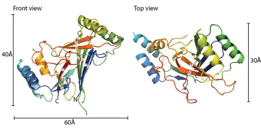
Following a screen of various Piezo orthologues (see Materials and Methods), the C. elegans CTL2 domain was found to be amenable for crystallographic analysis. The structure of wild type C. elegans CTL2 establishes that this domain is organized around a core of three beta sheets flanked by two pairs of helices with overall dimensions of ~60 Å x 40 Å x 30 Å (Fig 2). The secondary structure observed in the crystal structure generally matches that predicted by the Phyre2 webserver (Fig S2). The three beta sheets are arranged such that sheet 1 (strands 1, 2, 5, 6, with strands numbered from the N-terminus) is nearly parallel to sheet 2 (strands 3, 4, 11, 8), and perpendicular to sheet 3 (strands 7, 9, 10). Interestingly, analysis of residue conservation done with Profunc [23, 24] and Consurf [25] reveals that CTL2 possess conserved patch on one side but not the other (Fig 3). Analysis done by Profunc is more automated, such that residue conservation is calculated using 50 of the closest C. elegans CTL2 homologs as obtained by BLAST search against Uniprot database. Consequently, Profunc shows a higher residue conservation at this patch compared to calculation done by Consurf.
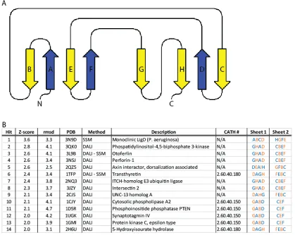
z-score: 2.8). Most of these candidate proteins possess a beta sandwich fold composed of two beta sheets; this is different than our Piezo soluble domain structure that consists of three beta sheets. However, if we consider sheet 1 and sheet 2 of CTL2 as the beta sandwich core with the rest of the protein as connectors, we obtain a two beta sheet sandwich core with a 2D connectivity diagram illustrated in (Fig S3a). A comparison between the Piezo loop structure and the identified proteins did not reveal any significant connectivity similarity (Fig S3b). Subsequently, we explored the structural classification database CATH [28] for two-sheet beta sandwich containing proteins (CATH ID: 2.60). Examination of the strand order in the beta sheets of these proteins failed to identify any close matches between CTL2 and previously solved structures. Therefore, we conclude that Piezo C-terminal soluble domain exhibits a novel beta sandwich fold.
Structure of M31R C. elegans PIEZO CTL2 Domain Variant
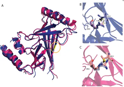
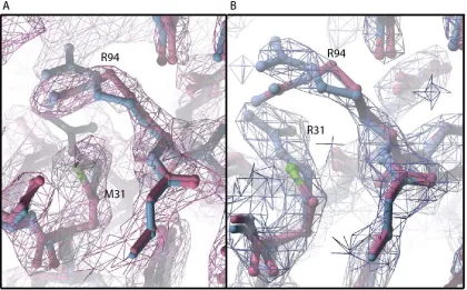
neighboring arginine residue, R94 (Fig. 4b). The most significant change in the M31R mutant CTL2 structure is the shift of the R94 sidechain ~3 Å away from R31, presumably due to electrostatic repulsion (Fig. 4b,c, Fig S5). However, R94 in C. elegans PIEZO loop is not conserved in the multiple sequence alignment (Fig. S4). R94 aligns to a glutamate residue that is primarily conserved in the vertebrate Piezo1 and Piezo2. Even though the C. elegans PIEZO CTL2 does not have a glutamate at this position, there are three glutamates in proximity to M31: E30, E32, and E98. This results in a net acidic local environment surrounding M31 in the C. elegans PIEZO.
Discussion
The model of CTL2 establishes that the region forms a compact domain containing a beta sandwich with three sheets. The CTL2 fold is apparently unrelated to any other family of proteins, including the β sheet domains present in other eukaryotic channels, specifically the extracellular domains of the two eukaryotic ion channels of known structure [29, 30], ASIC (CATH ID: 2.60.470.10) and P2X4 (CATH ID: 2.60.490.10). There is no evidence from either structure of the wild type or M31R mutant that this domain forms a tetramer that might help stabilize the oligomeric state of the intact channel. Mutating the conserved methionine residue into arginine (M31R), corresponding to a human DHS mutation (M2225R), does not cause a significant conformational change in C. elegans PIEZO CTL2 domain, suggesting that the effect of this mutation may be to more subtly perturb conformational energetics through changes in electrostatics, or perhaps to interfere with the ability to interact with other domains by changing the interface region.
investigation. The lack of structural similarity between this domain and any known protein fold, including those of eukaryotic and bacterial channels, reinforces the lack of recognizable sequence homology between Piezo and other channels, and highlights the distinctive nature of this enigmatic family of eukaryotic mechanosensitive channels.
Experimental Procedures Protein cloning and expression
The CTL2 regions from the mouse PIEZO1, mouse PIEZO2, human PIEZO1, Drosophila PIEZO and C. elegans PIEZO orthologues were cloned into a pET21 vector between the NdeI and XhoI restriction sites, generating a construct with a C-terminal 6xHis tag. The plasmid DNA was transformed into BL-21 gold E. coli cells (Stratagene). Cells were grown in Terrific Broth medium until they reached an OD600 of 1.0, when 1 mM IPTG was added. Protein induction was carried on overnight at 20 oC. The CTL2 of C. elegans PIEZO (GenBank ID: 392900032) was found to be best suited for structural analysis; the protein and nucleotide sequences of the coding region for this construct are: MSLLNQIGTISMPEKVTLRISIEGYPPLYEMEAQGSNHDNAELGMIKPDQLASLNQ ALTDSYTTRDTNSILRSRMSVSYLKGYTYEDILIVRFRPESEIYWPISQDSRNAMID KLSRNTSVNFEVSLEFTRPYDPNENAALKHSKSWLVPISLDMTIRAKIQSALRGDP GHPILIPQSIPAFIQVPNQGELTLPTSIGNTIINDGNPRINTTGMEKSDEARAWFDSLT LNLEQGKSQNEKMWIATSEHPGDQNAKLWIKTANTTYSGRPYLQVVGFIDRAFPS LEHHHHHH
Protein purification
Cells were homogenized in lysis buffer (20 mM Tris-HCl pH 8.0, 50 mM NaCl, lysozyme, DNAse, PMSF) at a ratio of 1 g cells / 10 mL of buffer. Cells were lysed using a microfluidizer (Microfluidics, Model M-110L), and the lysate was centrifuged to remove insoluble material. The protein was purified in three steps. First, lysate was run through a NiNTA column, washed with 40 mM imidazole for 15 column volumes, and eluted with 250 mM imidazole. Next, the eluate from the NiNTA column was run through a Superdex-200 16/60 column (GE Healthcare), and the corresponding peak fraction was collected. Finally, the resulting protein fraction was run through an anion exchange column, HiTrap Q Sepharose FF (GE Healthcare), to obtain the final protein preparation. The identities of the purified wild type (WT) and M31R variant proteins were confirmed by mass spectrometry.
Crystallization & model refinement
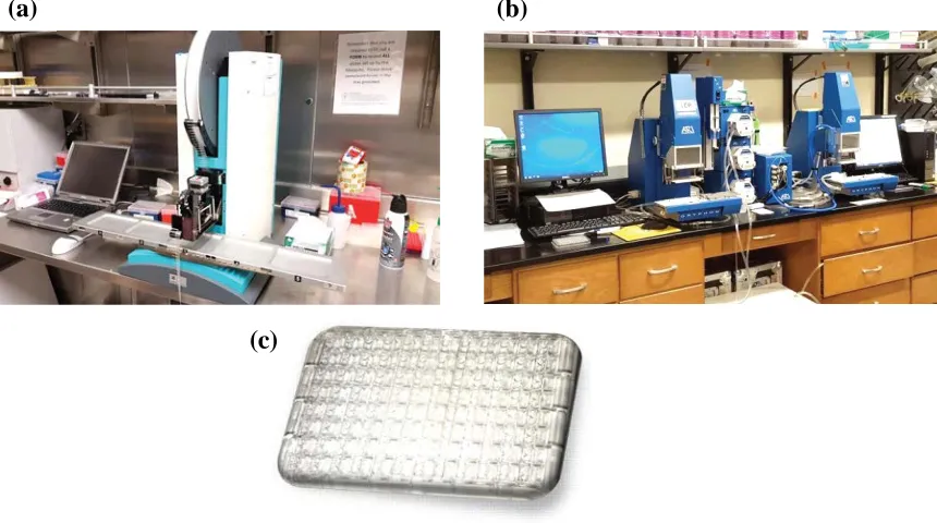
WT CTL2 was crystallized by the sitting drop vapor diffusion method against a reservoir containing 0.1 M phosphate-citrate buffer pH 4.0, 26% PEG1000, and 0.2 M Li2SO4 at 20 oC in a 96-well plate, at a ratio 0.2 μL protein to 0.2 μL reservoir drop, using intelli sitting drop crystallization plates (ArtRobbins). The protein concentration was 10-12 mg/mL. WT CTL2 crystals appeared after 18 hrs and grew to maturation after 3 days to approximate dimensions of 50 µm x 50 µm x 150 µm. M31R CTL2 was crystallized against a reservoir
Acknowledgments:
We thank A. Patapoutian for providing the C. elegans PIEZO construct, S. Hess from the Proteome Exploration Laboratory at the Beckman Institute at the California Institute of Technology for mass spectrometry analysis of the protein crystals, and P. Nikolovski from the Molecular Observatory at the Beckman Institute at the California Institute of Technology. This research was supported by National Institute of Health (NIH GM84211). We acknowledge the Gordon and Betty Moore Foundation, the Beckman Institute, and the Sanofi-Aventis Bioengineering Research Program and the Beckman Institute at Caltech for their generous support of the Molecular Observatory at Caltech. Operations at SSRL are supported by the US DOE and NIH.
Authors Contributions
References:
1. Levina, N., et al., Protection of Escherichia coli cells against extreme turgor by activation of MscS and MscL mechanosensitive channels: identification of genes required for MscS activity. EMBO J, 1999. 18(7): p. 1730-7.
2. Chalfie, M., Neurosensory mechanotransduction. Nat Rev Mol Cell Biol, 2009. 10(1): p. 44-52.
3. Coste, B., et al., Piezo proteins are pore-forming subunits of mechanically activated channels. Nature, 2012. 483(7388): p. 176-81.
4. Coste, B., et al., Piezo1 and Piezo2 are essential components of distinct mechanically activated cation channels. Science, 2010. 330(6000): p. 55-60. 5. Prole, D.L. and C.W. Taylor, Identification and analysis of putative homologues
of mechanosensitive channels in pathogenic protozoa. PLoS One, 2013. 8(6): p. e66068.
6. Bae, C., F. Sachs, and P.A. Gottlieb, The mechanosensitive ion channel Piezo1 is inhibited by the peptide GsMTx4. Biochemistry, 2011. 50(29): p. 6295-300. 7. Eisenhoffer, G.T., et al., Crowding induces live cell extrusion to maintain
homeostatic cell numbers in epithelia. Nature, 2012. 484(7395): p. 546-9.
8. Kim, S.E., et al., The role of Drosophila Piezo in mechanical nociception. Nature, 2012. 483(7388): p. 209-12.
10. Zarychanski, R., et al., Mutations in the mechanotransduction protein PIEZO1 are associated with hereditary xerocytosis. Blood, 2012. 120(9): p. 1908-15. 11. Albuisson, J., et al., Dehydrated hereditary stomatocytosis linked to
gain-of-function mutations in mechanically activated PIEZO1 ion channels. Nat Commun, 2013. 4: p. 1884.
12. Andolfo, I., et al., Multiple clinical forms of dehydrated hereditary stomatocytosis arise from mutations in PIEZO1. Blood, 2013. 121(19): p. 3925-35, S1-12. 13. McHugh, B.J., et al., Loss of the integrin-activating transmembrane protein
Fam38A (Piezo1) promotes a switch to a reduced integrin-dependent mode of cell migration. PLoS One, 2012. 7(7): p. e40346.
14. Bae, C., et al., Xerocytosis is caused by mutations that alter the kinetics of the mechanosensitive channel PIEZO1. Proc Natl Acad Sci U S A, 2013. 110(12): p. E1162-8.
15. Bae, C., P.A. Gottlieb, and F. Sachs, Human PIEZO1: removing inactivation. Biophys J, 2013. 105(4): p. 880-6.
16. Sonnhammer, E.L., G. von Heijne, and A. Krogh, A hidden Markov model for predicting transmembrane helices in protein sequences. Proc Int Conf Intell Syst Mol Biol, 1998. 6: p. 175-82.
17. Kall, L., A. Krogh, and E.L. Sonnhammer, Advantages of combined
transmembrane topology and signal peptide prediction--the Phobius web server. Nucleic Acids Res, 2007. 35(Web Server issue): p. W429-32.
19. Hofmann, K. and W. Stoffel, TMBASE - A database of membrane spanning protein segments. Biol. Chem. Hoppe-Seyler, 1993. 374: p. 166.
20. Schultz, J., et al., SMART, a simple modular architecture research tool:
identification of signaling domains. Proc Natl Acad Sci U S A, 1998. 95(11): p. 5857-64.
21. Katoh, K., et al., MAFFT: a novel method for rapid multiple sequence alignment based on fast Fourier transform. Nucleic Acids Res, 2002. 30(14): p. 3059-66. 22. Kelley, L.A. and M.J. Sternberg, Protein structure prediction on the Web: a case
study using the Phyre server. Nat Protoc, 2009. 4(3): p. 363-71.
23. Laskowski, R.A., J.D. Watson, and J.M. Thornton, Protein function prediction using local 3D templates. J Mol Biol, 2005. 351(3): p. 614-26.
24. Laskowski, R.A., J.D. Watson, and J.M. Thornton, ProFunc: a server for
predicting protein function from 3D structure. Nucleic Acids Res, 2005. 33(Web Server issue): p. W89-93.
25. Ashkenazy, H., et al., ConSurf 2010: calculating evolutionary conservation in sequence and structure of proteins and nucleic acids. Nucleic Acids Res, 2010. 38(Web Server issue): p. W529-33.
26. Krissinel, E. and K. Henrick, Secondary-structure matching (SSM), a new tool for fast protein structure alignment in three dimensions. Acta Crystallogr D Biol Crystallogr, 2004. 60(Pt 12 Pt 1): p. 2256-68.
28. Sillitoe, I., et al., New functional families (FunFams) in CATH to improve the mapping of conserved functional sites to 3D structures. Nucleic Acids Res, 2013. 41(Database issue): p. D490-8.
29. Jasti, J., et al., Structure of acid-sensing ion channel 1 at 1.9 A resolution and low pH. Nature, 2007. 449(7160): p. 316-23.
30. Kawate, T., et al., Crystal structure of the ATP-gated P2X(4) ion channel in the closed state. Nature, 2009. 460(7255): p. 592-8.
31. Battye, T.G., et al., iMOSFLM: a new graphical interface for diffraction-image processing with MOSFLM. Acta Crystallogr D Biol Crystallogr, 2011. 67(Pt 4): p. 271-81.
32. Potterton, E., et al., A graphical user interface to the CCP4 program suite. Acta Crystallogr D Biol Crystallogr, 2003. 59(Pt 7): p. 1131-7.
33. Kabsch, W., Integration, scaling, space-group assignment and post-refinement. Acta Crystallogr D Biol Crystallogr, 2010. 66(Pt 2): p. 133-44.
34. Evans, P., Scaling and assessment of data quality. Acta Crystallogr D Biol Crystallogr, 2006. 62(Pt 1): p. 72-82.
35. Terwilliger, T.C., et al., Decision-making in structure solution using Bayesian estimates of map quality: the PHENIX AutoSol wizard. Acta Crystallogr D Biol Crystallogr, 2009. 65(Pt 6): p. 582-601.
36. Zwart, P.H., et al., Automated structure solution with the PHENIX suite. Methods Mol Biol, 2008. 426: p. 419-35.
38. Emsley, P. and K. Cowtan, Coot: model-building tools for molecular graphics. Acta Crystallogr D Biol Crystallogr, 2004. 60(Pt 12 Pt 1): p. 2126-32.
39. Murshudov, G.N., et al., REFMAC5 for the refinement of macromolecular crystal structures. Acta Crystallogr D Biol Crystallogr, 2011. 67(Pt 4): p. 355-67.
Figure 1. Membrane topology of mouse PIEZO1 as predicted by various membrane
topology prediction algorithms. Depending on the membrane topology prediction
algorithm, mouse PIEZO1 is predicted to have 30 to 40 transmembrane segments. The
numbers of amino acids (aa) for the larger predicted loops are indicated. The loop of
interest, CTL2 (red box), is predicted to be an extracellular loop by 3 of the 4 topology
prediction algorithms. Since mouse PIEZO1 is the most extensively characterized Piezo
channel, it is used to illustrate the topology predictions; Diagrams are drawn using TOPO2
Figure 2. Structure of the wild type C. elegans CTL2 domain. The structure of the wild
type Piezo CTL2 is colored in a rainbow scheme, progressing from the N-terminus (blue)
to C-terminus (red). Front view (left): The loop is oriented so that the connections to the
putative transmembrane helices are positioned toward the bottom. The β-strands are
numbered sequentially from the N-terminus. Top view (right): the front view, rotated 90˚
Figure 3. Residue conservation analysis of CTL2
(A) Surface representation of the WT CTL2 domain (marine blue) is oriented in similar
fashion to the one displayed in Figure 2 (inside the black square). N and C terminus are
colored in light gray. (B) The top panel displays surface conservation representation from
the closest Piezo homologs as calculated by ProFunc using the pdb coordinate of the WT
CTL2 domain structure, whereas the bottom panel display surface conservation from
representative Piezo homologs taken from broad branches of eukaryotic kingdom as
calculated by ConSurf. Left panel shows the surface of CTL2 upon 90 degree rotation
along the y-axis, showing the right-side view of CTL2. Right panel shows the surface of
CTL2 upon -90 degree rotation, showing the left-side view of CTL2. Surface conservation
is colored in a rainbow gradient scheme in such a way that the least conserved region is
colored in blue and the most conserved region is colored in red. M31R is located at the
Figure 4. Superposition of wild type and M31R substituted CTL2 domains. (A)
Superposition of the wild type (pink) and M31R substituted (blue) CTL2 structures. The
position of residue 31 is highlighted in yellow (circled). (B, C). The M31R mutant structure
is displayed in blue and the WT structure is displayed in pink; this substitution is
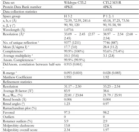
Table 1. Data collection and refinement statistics of the wild type and mutant
a Values in parentheses represents the highest resolution shell
Data set Wildtype CTL2 CTL2 M31R
Protein Data Bank number 4PKE 4PKX
Data collection statistics
Space group H 3 2 P 1 21 1
a, b, c (Å) 72.59, 72.59, 241.6 45.18, 37.25, 73.56
α, β, γ (o) 90, 90, 120 90, 91.58, 90
Wavelength (Å) 1.0716 1.0332
Resolution (Å)a 33.09 – 2.45 (2.57 –
2.45) 38.97 – 2.54 (2.68 – 2.54)
No. of unique reflection a 9377 (1211) 7746 (887)
Mean (I/sigma I) a 17.7 (3.0) 28.4 (11.2)
Completeness a 99.9% (100%) 93.6% (75.4%)
Average multiplicity a 10.1 (10.6) 3.1 (2.8)
Anom. Completeness a 99.9% (99.9%)
DelAnom. correlation between half-sets
a 0.915 (0.061)
R-merge a 0.093 (0.810) 0.028 (0.085)
Matthew Coefficient 1.951 1.92
Refinement statistics
Resolution 31.17 – 2.50 33.23 – 2.54
Average B factor (Å2) 83.9 38.6
Rwork/Rfree (%) 22.81 / 23.84 21.79 / 25.91
Rmsd bonds (Å) 0.006 0.004
Rmsd angles (o) 1.23 0.97
Ramachandran plot (%)
Favored 97.5 98.2
Outliers 0 0
Rotamer outliers (%) 5.9 2.9
Molprobity clashscore 13.8 12.8
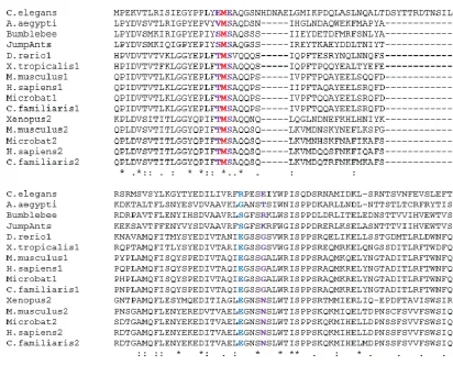
Figure S1. Multiple sequence alignment of Piezo homologs. Multiple sequence alignment was performed using representative Piezo homologs (listed below) from vertebrate Piezo1, vertebrate Piezo2, invertebrate Piezo, plant Piezo, and unicellular eukaryote Piezo. Protein sequence conservation is represented by the shading and height of the bar corresponding to each residue, with the tall yellow bar, short brown bar, and no bar representing high levels, low levels, and poor sequence conservation, respectively. The green bars highlight the transmembrane (TM) segment for mouse PIEZO1 as predicted by Topcons [18]. CTL2 (shaded in light blue) is the largest conserved Piezo soluble domain. The Piezo sequence conservation is relatively higher in the C-terminal region. The diagram was generated using the MAFFT multiple sequence alignment program [21], available at the EMBL-EBI web page.
List of Piezo homologs used:
Chapter 3:
The Pursuit Toward Obtaining a Full
C h a p t e r 3 : T h e P u r s u i t T o w a r d O b t a i n i n g a F u l l L e n g t h P i e z o S t r u c t u r e
Historical Context and The Chapter Outline
The notion of “a picture is worth a thousand words” is certainly true in the field of molecular biology. In the absence of images, it is difficult to deduce a detailed mechanism on how a particular molecular machine works. Consequently, there have been major efforts dedicated to elucidating the structure of biological macromolecules. However, these molecules are much too small for direct visualization by regular light microscopy techniques. Therefore, we have to utilize specialized imaging techniques which employ electromagnetic radiation of much smaller wavelength than visible light to obtain snapshots of these molecules at atomic resolution.
formulation of alpha helical model of protein secondary structure [4]. Then, another structural biology milestone took place when James Watson and Francis Crick deduced the structure of the DNA double helix from x-ray diffraction images obtained from DNA fibers by Rosalind Franklin [5].
In a typical x-ray crystallography experiment, data is collected as the intensities of x-ray diffraction reflections, containing the amplitudes but missing the phase information. This is commonly referred to as the ‘phase problem’. In 1953, Max Perutz invented a way to solve this problem by utilizing the diffraction pattern difference between the native protein crystal and its heavy metal derivatized isomorphic crystal, a method that is known as isomorphous replacement [6]. Using this method, we can now obtain atomic structures of any well-diffracting protein crystals from which we can deduce detailed mechanisms on a variety of molecular processes; this gave rise to a new biological science field known as the Structural Biology.
With the advancement of recombinant DNA technology in the late 1970s, proteins that are not naturally abundant could finally be produced in cells that were engineered to overexpress the protein of interest [15]. Although progress in solving the structures of soluble proteins experienced a significant boost with the development of over-expression methods, membrane protein structure determination has still trailed behind (Figure 1); the first structure of a membrane protein did not come out until 1985 [16]. This is partly because many membrane proteins failed to be expressed in sufficient amounts using commonly used recombinant systems; this is especially true for many eukaryotic membrane proteins [17].
Furthermore, solubilization of membrane proteins by amphipathic reagents, such as detergents, can be problematic. For this reason, significant efforts have been dedicated to optimize the expression of membrane proteins using different expression systems and different purification conditions. Therefore, in the first part of this chapter, I will describe our efforts in exploring several different protein expression systems and detergent-buffer conditions to improve the yield of full length Piezo protein for x-ray crystallographic study.