Open Access
Study protocol
Comparison of embedded and added motor imagery training in
patients after stroke: study protocol of a randomised controlled
pilot trial using a mixed methods approach
Corina Schuster*
1,2, Jenny Butler
2, Brian Andrews
3, Udo Kischka
1,4and
Thierry Ettlin
1,5,6Address: 1Reha Rheinfelden, Rheinfelden, Switzerland, 2School of Health and Social Care, Oxford Brookes University, Oxford, UK, 3School of
Technology, Oxford Brookes University, Oxford, UK, 4Oxford Centre for Enablement, Oxford, UK, 5Department of Neurology, Medical faculty,
University of Basel, Basel, Switzerland and 6Department of Education, University of Applied Science Northwestern Switzerland, Basel, Switzerland
Email: Corina Schuster* - [email protected]; Jenny Butler - [email protected]; Brian Andrews - [email protected]; Udo Kischka - [email protected]; Thierry Ettlin - [email protected]
* Corresponding author
Abstract
Background: Two different approaches have been adopted when applying motor imagery (MI) to stroke patients. MI can be conducted either added to conventional physiotherapy or integrated within therapy sessions. The proposed study aims to compare the efficacy of embedded MI to an added MI intervention. Evidence from pilot studies reported in the literature suggests that both approaches can improve performance of a complex motor skill involving whole body movements, however, it remains to be demonstrated, which is the more effective one.
Methods/Design: A single blinded, randomised controlled trial (RCT) with a pre-post intervention design will be carried out. The study design includes two experimental groups and a control group (CG). Both experimental groups (EG1, EG2) will receive physical practice of a clinical relevant motor task ('Going down, laying on the floor, and getting up again') over a two week intervention period: EG1 with embedded MI training, EG2 with MI training added after physiotherapy. The CG will receive standard physiotherapy intervention and an additional control intervention not related to MI.
The primary study outcome is the time difference to perform the task from pre to post-intervention. Secondary outcomes include level of help needed, stages of motor task completion, degree of motor impairment, balance ability, fear of falling measure, motivation score, and motor imagery ability score. Four data collection points are proposed: twice during baseline phase, once following the intervention period, and once after a two week follow up. A nested qualitative part should add an important insight into patients' experience and attitudes towards MI. Semi-structured interviews of six to ten patients, who participate in the RCT, will be conducted to investigate patients' previous experience with MI and their expectations towards the MI intervention in the study. Patients will be interviewed prior and after the intervention period.
Discussion: Results will determine whether embedded MI is superior to added MI. Findings of the semi-structured interviews will help to integrate patient's expectations of MI interventions in the design of research studies to improve practical applicability using MI as an adjunct therapy technique.
Trial registration: ClinicalTrials.gov NCT00858910
Published: 22 October 2009
Trials 2009, 10:97 doi:10.1186/1745-6215-10-97
Received: 20 March 2009 Accepted: 22 October 2009
This article is available from: http://www.trialsjournal.com/content/10/1/97
© 2009 Schuster et al; licensee BioMed Central Ltd.
Background
Regardless of the decreasing trend in long-term statistics stroke is still one of the three leading causes of death in the US [1] and in Europe. The incidence for individuals older than 55 years range from 4.2 to 11.7 per 1000 [2]. Stroke has a sudden onset resulting from cerebral haemor-rhage or ischemia in the brain. In the US, 600'000 first-ever strokes occurred in 2005 with an estimated cost to the community of $ 65 billion in both direct and indirect costs in 2008 [1,3]. In Europe the total costs of stroke are aggregate to 38 € billions in 2006: 49% arise from direct costs, 23% from productivity loss, and 29% from the for-mal care [4]. Only 25% of the affected individuals recover with minor problems, 45% sustain moderate to severe impairments that necessitate special attention and life-long care [5].
Several rehabilitation methods address patient recovery, are based on motor learning or neuro-developmental approaches [6-11]. Physiotherapy focuses on regaining motor function, postural alignment and independence in activities of daily living (ADL). Regaining functional abil-ity with a focus on mobilabil-ity is one of the most frequently targeted short-term rehabilitation goals in patients [12]. New rehabilitation approaches have been reported, e.g. robot-aided [13], virtual reality rehabilitation [14], and MI [15,15]. MI does not require expensive technology, equipment, instrumented locations, and it does not phys-ically exhaust the individual [16]. After initial learning, the MI technique can be practiced by the patient inde-pendent from the therapist, location, and time of the day.
The literature on MI techniques has increased tremen-dously since the late 1990s and there have been several lit-erature reviews related to the application of stroke [17-19]. All found a beneficial value for the recovery of stroke patients when MI was added after physiotherapy or occu-pational therapy sessions. The analysed randomised or clinical controlled trials used different MI methodologies and compared MI versus an equivalent amount of atten-tion time, where patients listened to informaatten-tion about stroke and relaxation exercises.
MI has its origin in the sports psychology and behavioural psychology in the end of the 19th century [20]. It involves rehearsing a known motor act without any visible muscle contraction or motor output [21]. Several theories are pro-posed to explain the neuro-physiological mechanisms of MI. The 'psycho neuromuscular theory' was proposed by Jacobson in the early 1930s based on the detection of myoelectrical changes related to the imagined movement [22]. Another theory is based on co-location of brain acti-vation during imagined and real movements in healthy individuals [23-25] as well as in stroke patients [26,27]. Functional magnetic resonance imaging (fMRI) showed
activation in frontal, parietal cortical, and sub-cortical areas that are involved in action planning, execution and modulation [25,28]. Recently, the first fMRI study was published that investigated brain activation during imag-ination of whole body movements [29], supporting the findings from many MI intervention studies in sport psy-chology.
To date intervention studies in stroke rehabilitation have focused on simple movements, such as reaching and grasping [15,30], whereas investigations in sports psy-chology used more complex motor skills [31]. Complex motor activities are relevant in rehabilitation as well, since patients seek to regain independence in ADL. Previous stroke research has compared MI to a different mental practice method, e.g. relaxation, or listening to informa-tion about stroke. Some researchers have added MI to conventional physiotherapy or occupational sessions [17]. More recently, study proposals were published that will investigate the effect of embedded MI in more com-plex tasks of daily living [32,33]. Holmes and Collins developed the PETTLEP model to embed MI in training for sports psychology in 2001 [34] based on a combina-tion of seven real-life condicombina-tions: physical, environment, task, timing, learning, emotion, and perspective. The PET-TLEP model has improved outcome when applied in a MI intervention [35,36]. MI training may be embedded into physiotherapy sessions offering the prospect of more effective and systematic MI training compared to added MI approaches. However, to the authors' knowledge, the PETTLEP model has not been adapted and tested in a stroke population.
Aside the quantitative assessment of MI it is essential to determine patients' experiences with this training method. In sports research the athlete's usage of imagery was assessed with semi-structured interviews, e.g. during rehabilitation of an injury [37] as well as during and after training periods [38]. Driediger et al. [37] reported posi-tive effects of imagery regarding cogniposi-tive, motivational, and healing intentions in those athletes. Imagery was used frequently during the day, during rest, e.g. before sleeping, and various other activities such as car driving. Positive and exact images were used to set goals for the rehabilita-tion process, to handle pain intensity, and to keep a posi-tive stance. These studies found that interviewed athletes were experienced and knowledgeable about imagery and made frequent use of different imagery types, e.g. kinaes-thetic, visual, and auditory imagery. Research is needed to transfer these qualitative findings from sports to a stroke population.
Study aims and research questions
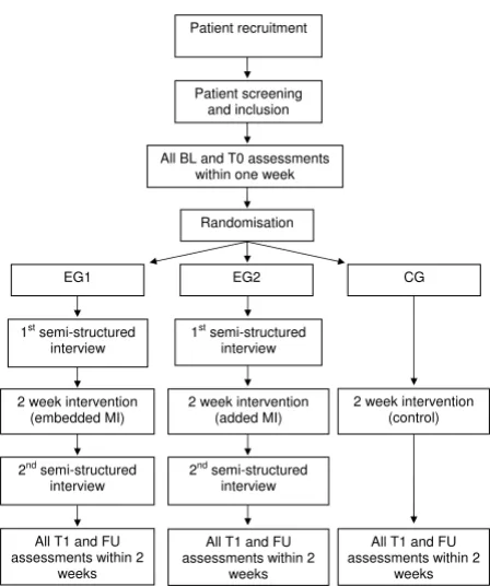
interven-tion with a randomised controlled trial (RCT) study design. The RCT includes three study groups to evaluate the effect MI training embedded into physiotherapy ver-sus MI training added to physiotherapy. In the qualitative part semi-structured interviews of six to ten patients who participate in the RCT will be interviewed to gain knowl-edge about the patient's experiences and expectations regarding MI. These study parts are compatible to each other and the results are expected to interact. Conse-quently, the overall study design has been described as a 'mixed methods approach'. Figure 1 illustrates the planned project.
The following sections describe the research protocol of both parts in detail.
Aim of the quantitative study part
The aim of the study part is to compare the effect of embedded MI training in physiotherapy (EG1, PETTLEP approach) to added MI (EG2) after regular physiotherapy sessions, regarding improvements of time needed to per-form a motor task ('Going down, laying on the floor, and getting up again'). A third patient group with no MI will serve as a control group (CG) to provide an overall base-line for this intervention.
(EG1= experimental group 1, EG2 = experimental group 2)
Research question of the quantitative study part
Do patients in the embedded MI training group (EG1) require less time to perform the motor task compared to patients in the added MI training group (EG2)?
Aim of the qualitative study part
This qualitative part of the study should add an important insight into patients' experience and attitude to imagery, especially MI. With the help of semi-structured interviews the following aspects will be investigated:
- Before MI intervention:
ⴰ Patient's previous experience with MI.
ⴰ Patient's usage of MI regarding the 'W-questions' in imagery research (When, Where, What, Why) [39].
ⴰ Patient's expectations towards the MI
interven-tion in the RCT.
- After MI intervention:
ⴰ Patient's attitudes towards the MI after the inter-vention.
ⴰ Patient's tentative changes in MI usage.
Research question of the qualitative study part
What are the thoughts, feelings, and experience of people with stroke who are participating in MI training? Does it change after a two week MI intervention?
Methods
Methodology of the quantitative study part
Quantitative study design and setting
In this single-centred randomised controlled trial with a pre-post intervention design, embedded and added MI techniques will be compared to improve a learned motor task ('Going down, laying on the floor, and getting up again'; for a detailed description please see Section 'Motor task and fear of falling on page 13). The study design includes two experimental groups and one control group. Both experimental groups (EG1, EG2) will receive physi-otherapy for the motor task: EG1 with an embedded MI training into physiotherapy sessions (PETTLEP approach), EG2 with an added MI training after physio-therapy. The third group is the control group (CG) with physiotherapy and an added control intervention not related to MI. All groups receive the same intervention time of 45 minutes. The investigation will have an inter-vention period of two weeks and will be carried out in the rehabilitation centre Reha Rheinfelden in Switzerland.
The study has been approved by the responsible cantonal review board of the health department Aarau (Switzer-land) and the ethics committee of the School of Health and Social Care at Oxford Brookes University (United Kingdom).
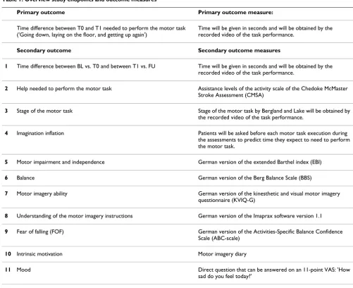
Primary and secondary outcome measures
Table 1 provides an overview on all study outcome meas-ures. All outcome measures will be assessed at four times: twice during the baseline phase (BL, T0), after the inter-vention (T1), and after the two week follow-up phase (FU). Changes over time will be calculated by the differ-ences between BL vs. T0, T0 vs. T1 and T1 vs. FU.
Primary outcome measure
The primary study outcome has been defined as changes in time needed to perform the motor task due to the study intervention. This primary outcome will be assessed by the difference between measurement points T0 and T1. For performance and validity reasons all assessment ses-sions will be videotaped. Time needed to perform the task in seconds will be derived from the video recordings.
Secondary outcome measures
Motor task related profile
1. Time difference to perform the motor task between BL and T0 as well as between T1 and FU.
2. Help needed to perform the motor task will be eval-uated using classification levels of the activity scale of the Chedoke McMaster Stroke Assessment (CMSA) [40,41]. The CMSA is a validated and very reliable per-formance-based assessment [42-44]. The activity scale belongs to the gross function index and is scored with one (patient needs total assistance or the task is not tested for safety reasons) to seven (patient can perform the task completely independent without any assist-ance or devices, in a reasonable time) on an ordinal scale [45].
3. Stage of the motor task will be evaluated using the classification of Bergland and Laake [46] (please see section 'Stages of the motor task').
4. 'Imagination inflation': Undergraduate college stu-dents overestimated their motor performance after MI
training. This effect is called 'Imagination inflation' and was detected by Landau et al [47]. To monitor the inflation in this investigation all patients will be asked before each motor task execution during the assess-ments to predict time they will need to perform the task before each motor task execution.
Motor impairment and balance profile
The motor impairment and balance profile will be evalu-ated with the German versions of the extended Barthel index (EBI) and the Berg Balance Scale (BBS). Both assess-ments will take about 15 to 20 minutes each to complete.
5. The EBI is a 16-item performance-based measure-ment. Activities of daily living (ADL), e.g. personal hygiene, dressing, feeding, and cognitive aspects, e.g. communication and problem solving, will be scored on a five-point Likert scale (0 = cannot perform the task, 4 = independent) [42,48]. The total score will be used to evaluate trends. Validity and reliability were examined with patients after stroke and multiple scle-rosis [49-52]. A validated German version is available [50].
6. The BBS consists of 14 items to assess two dimen-sions of balance impairments in (older) individuals and patients undergoing rehabilitation. The ability to maintain an upright posture in different positions and the ability to adjust posture when reducing support surface will be scored on a 5-point ordinal scale (0 to 4). High scores are interpreted as a sufficient ability to perform a task or to perform it within a given time frame [42,53]. The total score will be used to evaluate trends. Validity, reliability and sensitivity to change were evaluated [53-56]. A validated German version is available [57].
Motor imagery profile
The motor imagery profile will be assessed with the Kines-thetic and Visual Imagery Questionnaire (KVIQ) and the Imaprax software, version 1.1. The completion of both assessments will take 30 to 40 minutes.
7. The KVIQ includes a visual and a kinaesthetic imagery scale and was developed based on the revised movement imagery questionnaire (MIQ) [58]. The new measure was specifically developed to assess motor imagery ability for individuals with motor impairments in 2007 by Malouin et al. [59]. The ques-tionnaire is available in a short (10 items) and a long version (20 items). The latter will be used in this inves-tigation. All items will be evaluated in a standardised sequence while sitting for visual and kinaesthetic scales. The assessment requires performing a sequence of simple movements once, imagine the movement
[image:4.612.64.288.94.362.2]Study overview Figure 1
Study overview. MI = motor imagery, BL, T0 = 1st and 2nd
baseline measurement event, EG1, EG2 = experimental group 1 and 2, CG = control group, T1 = measurement event after the 2 week intervention period, FU = measure-ment event after a 2 week follow-up period.
Randomisation Patient recruitment
Patient screening and inclusion
CG EG2
EG1
2 week intervention (embedded MI)
1st semi-structured interview 1st semi-structured
interview
2 week intervention (control) 2 week intervention
(added MI) All BL and T0 assessments
within one week
All T1 and FU assessments within 2
weeks 2nd semi-structured
interview
2nd semi-structured interview
All T1 and FU assessments within 2
weeks
All T1 and FU assessments within 2
once and score the vividness of the 'inner picture' on a 5 point Likert scale (1 - 'image as clear as actually see-ing it' to 5 'no image') as well as the feelsee-ing of the imagined movements (1 - 'as intense as making the movement' to 5 - 'no sensation'). The total score of each subscale will be used to evaluate trends.
8. The Imaprax 1.1 software was developed to evaluate understanding of MI, vividness of movement imagery, and imagery perspective in patients with apraxia fol-lowing stroke. It is based on software that was used with skydivers [60,61]. Six gestures or activities will be evaluated in a three step standardised procedure: select the correct gesture or activity from three pro-posed ones, evaluate the vividness of the 'inner pic-ture', and determine the internal or external perspective of your 'inner picture'. The total score of MI vividness will be used to evaluate trends.
KVIQ and Imaprax assessment tools were published in other languages than German. Following a cross-cultural translation and adaptation based on guidelines for self-report questionnaires by Beaton et al. [62] both assess-ments were forward translated into German. To check for consistency both tools were backward translated into their original publication languages and checked by the origi-nal authors. We obtained the permission from those authors to use the translated assessments in our study.
Psychological profile
[image:5.612.52.553.91.499.2]The psychological effect of MI is not yet proven. Neverthe-less, authors of published MI studies proposed to include assessments of selected psychological factors, e.g. atten-tion and working memory [63]. The psychological profile includes the evaluation of patient's fear of falling (FOF) using the Activities-Specific Balance Confidence Scale (ABC-scale).
Table 1: Overview study endpoints and outcome measures
Primary outcome Primary outcome measure:
Time difference between T0 and T1 needed to perform the motor task ('Going down, laying on the floor, and getting up again')
Time will be given in seconds and will be obtained by the recorded video of the task performance.
Secondary outcome Secondary outcome measures
1 Time difference between BL vs. T0 and between T1 vs. FU Time will be given in seconds and will be obtained by the
recorded video of the task performance.
2 Help needed to perform the motor task Assistance levels of the activity scale of the Chedoke McMaster
Stroke Assessment (CMSA)
3 Stage of the motor task Stage of the motor task by Bergland and Lake will be obtained by
the recorded video of the task performance.
4 Imagination inflation Patients will be asked before each motor task execution during
the assessments to predict time they expect to need to perform the motor task.
5 Motor impairment and independence German version of the extended Barthel index (EBI)
6 Balance German version of the Berg Balance Scale (BBS)
7 Motor imagery ability German version of the kinesthetic and visual motor imagery
questionnaire (KVIQ-G)
8 Understanding of the motor imagery instructions German version of the Imaprax software version 1.1
9 Fear of falling (FOF) German version of the Activities-Specific Balance Confidence
Scale (ABC-scale)
10 Intrinsic motivation Motor imagery diary
11 Mood Direct question that can be answered on an 11-point VAS: 'How
sad do you feel today?'
9. The ABC-scale is 16-item questionnaire that evalu-ates the self-confidence of a person based on Ban-dura's theory of efficacy [42,64]. It can be self-administered or completed during a face to face inter-view. Patients determine their self-perceived confi-dence to remain in balance on a visual analogue scale (VAS) ranging from zero to 100 percent (10 cm). The mean value will be used to evaluate trends. The scale is a valid and reliable assessment that is available in German language [65,66].
10. Furthermore, intrinsic motivation will be evalu-ated from the patient's MI diary. Using details on fre-quency of independent MI practice reported in the patient's diary motivation to practice and the compli-ance with the training can be determined [67]. Other investigations showed that patients regained self-con-trol about their recovery process. They felt more skil-ful, and patients as well as athletes expressed their satisfaction and belief in the MI training, which helped to improve their performance [68,69].
11. Patient's mood will be enquired by a direct ques-tion: 'How do you feel today?'. This will be scored on an 11-point VAS ranging from zero (very good) to ten (very bad).
Further evaluations and assessments
To further describe the included study population the fol-lowing descriptive data will be obtained from each partic-ipant: age, gender, weight, disease, affected side, time since stroke onset, actual medication, cognitive function, handedness, rehabilitation history (treatments until study start), patient's sports history, and patient's history of falls since the stroke onset (over the last six to twelve months). Data will be obtained by direct questions to patients, from medical records, and from a relative or proxy.
Cognitive function will be assessed with the Mini-Mental State Examination (MMSE). MMSE is a short screening tool for dementia symptoms [70]. MMSE includes items to evaluate spatial and temporal orientation, short-term memory, attention and calculation abilities, language, thinking, and action planning. The total score is 30 points. MMSE will be performed once at a baseline assess-ment and will take about 15 to 20 minutes.
Patient's handedness will be determined with the Edin-burgh Handedness Inventory (EHI) once. The EHI is a valid and reliable short 10-item questionnaire. Patients will be asked what is the preferred hand to carry out activ-ities of daily living (ADL), e. G. Using toothbrush, cut something with a scissor, or use a spoon [71]. The EHI will take about five to ten minutes to complete.
Further important information that may influence the effect of MI are:
- patient's type of learning (visual or kinaesthetic),
- patient's belief in the therapy [72].
Both will be assessed by direct questions during baseline measurement events.
Recruitment process and patient selection criteria
Patients will be recruited according to the following pro-cedure:
1. A procedure common in retrospective research will be used. Former inpatients of the rehabilitation centre with a cerebrovascular ischaemia or a hemorrhagic bleeding will receive an information letter including a prepared answer sheet and a paid envelope. When interested in receiving more information patients may return the envelope with the answer sheet or contact the project leader by telephone or email.
2. Upon receiving answer sheets back in the rehabilita-tion centre, patients will be sent the patient informa-tion sheet to inform them about the planned study.
3. Three to ten days after sending the patient informa-tion sheet, patients will be contacted by phone to ask, if they have received the patient letter and if they have any questions they would like to have answered. If the patient is interested, a first appointment will be arranged to provide detailed information about the study procedure and check for study eligibility. This appointment will be arranged at rehabilitation centre, the patient's home or institution.
Additionally, patients will be recruited as leaving inpa-tients or from the outpatient therapy department of the rehabilitation centre by their treating therapist, through an advertisement at the centre's homepage, and through flyers at several locations of the centre. Interested patients can contact the project leader through their therapist, by phone, email, or mail. Patients will be selected based on the criteria in Table 2.
Study procedure and measurement events
CG. Each group will receive three therapy sessions per week for two weeks, hence six sessions in total. Each ses-sion lasts 45 minutes. Based on the main study hypothe-sis, therapy for EG1 will embed MI training during physiotherapy, while EG2 will receive MI training after the physiotherapy part. For a detailed description please see Section 'Study intervention' on page 14. Baseline meas-urements will be repeated directly after the therapy ses-sion weeks to evaluate short-term effects of the MI intervention (T1), as well as after a follow up period of two weeks (FU). All intervention and assessment sessions will take place at the rehabilitation centre's physiotherapy department.
Randomisation and allocation concealment
Patients will be randomly allocated to the study groups (EG1, EG2, CG) to ensure internal validity of results. Group allocation to one of the three study groups will be performed after the second baseline assessment (T0) using a randomisation list. The list has been generated with MATLAB 2007b (Mathworks Inc., USA) by a researcher not involved in the study. The generated list has been sent to the clinic's pharmacist, who is not involved in the study and will have no contact with study patients. The pharmacist will prepare sealed envelopes with the patient allocation based on the randomisation list. Patients will be given the sealed envelope by the treating therapist and they will open the envelop themselves. After opening, envelopes will be stored with the patient's per-sonal documents in a locked cabinet. Only the treating therapist and the research assistant will have access to the documents. The independent examiner will not have access to the documents. Patients will be told not to talk to the examiner about the group allocation or therapy content during the post-intervention assessments. Ran-domisation will be concealed to the independent exam-iner until the last follow-up assessment of the patient has been performed.
Blinding and study group interaction
Blinding of study personnel in research projects is a main quality criterion of a study [73]. Nevertheless, blinding of therapists in an intervention study who perform
experi-mental treatment in patients is not always feasible, espe-cially if experimental intervention shall be integrated in the therapy. Consequently, hiding the patient's allocation in this setting will not be possible. Therefore, neither ther-apists nor patients will be blinded in this study. Neverthe-less, the assessor of post-intervention and follow-up assessments and the independent statistician who will control statistical analyses will be blinded to patient's group allocation until all assessments have been per-formed. Interaction of patients with other study partici-pants will be minimised by the following preventive measures:
- The patients will be asked to not speak about their study and therapy content as well as imagery sessions outside their families, and neither to talk to the asses-sor about the study and session content.
- All examiners will be instructed not to ask patients about their study content.
- All patients are outpatients, coming to the rehabilita-tion centre for the study intervenrehabilita-tion only. They do not stay together in one clinic ward.
- Group sessions will be avoided.
Motor task and fear of falling
The motor task 'Going down, laying on the floor, and get-ting up again' is clinically relevant, in particular for older people who have problems with ambulation and balance. This task is an important skill to live independently and maintain activities of daily living (ADL) [46,66]. A fall is defined as an involuntary position alteration that results in laying on the floor or ground. It can cause injuries such as fractures, soft tissue injury or joint dislocations [46,74]. In particular, lacking the ability to get up from the floor is related to fear of falling (FOF) in the elderly [46]. FOF is a psychological construct that helps to estimate an individ-ual's ability to perform ADL without falling [66]. Fried-man and colleagues found that FOF can emerge from an experienced fall and it can exist within non-fallers [75]. Patients after stroke have a 2.3 times higher risk of falling
Table 2: Patient inclusion and exclusion criteria
Inclusion criteria Exclusion criteria
- Patients after a first-ever ischemic or hemorrhagic stroke - Outpatients or inpatients 3 months after stroke
- Ability to stand with or without a cane for at least 30 sec on a normal hard floor
- Ability to walk for 20 metres with or without a cane or an orthosis - MMSE with at least 20 points
- Age older than 18 years - Signed written informed consent
- Joint replacements (knee, hip, shoulder) - Limiting pain in the upper or lower body (evaluated with an eleven point VAS rating scale)
- Limiting range of motion (ROM) in the hip, knee, ankle joint or toes - Body weight more than 90 kilograms
than the age-matched normal population [76]. This risk can increase up to 3.4 times if more than one fall already occurred. It is known that a majority of falls happen dur-ing day time, indoors as well as outdoors, while walkdur-ing, and while changing the body position, e.g. from sitting to standing [76]. These findings confirm the relevance of the selected motor task in this intervention study. Hence, it is expected that MI training and physiotherapy sessions will reduce FOF and improve patient's self-confidence in walk-ing.
Stages of the motor task
The stages of the motor task are a modified version from the task stages of Bergland and Laake [46]. They proposed a movement sequence with 13 stages that is helpful for training with elderly individuals having FOF. The patient stands facing a mat on the floor. After the instruction 'Get down to the floor mat' the patient moves down to the mat in a standardised procedure to lie straight on the mat with legs extended. After the command 'Stand up' the patient stands up, moving from the laying position on the floor to an upright position by repeating all stages in reverse order. Two stages were modified: the starting position (stride standing) will be included as the first stage because this stride standing is already challenging for patients after stroke. The original stage 5 (to prone kneeling and up) will be left out because only a small portion of patients after stroke are able to bear the trunk weight at the affected hand, arm, and shoulder while maintaining the arm in an extended position.
Motor task stages considered in this study
- Stage 0 - Standing
- Stage 1 - Stride standing
- Stage 2 - To half-kneeling on to a large foam wedge and up
- Stage 3 - To half-kneeling on to a small wedge and up
- Stage 4 - To half-kneeling on a mat and up
- Stage 5 - To high-kneeling on a mat and up
- Stage 6 - To half-sitting on two pillows and up
- Stage 7 - To half-sitting on one pillow and up
- Stage 8 - To half-sitting on a mat and up
- Stage 9 - Side laying on a large wedge and up
- Stage 10 - Side laying on a small wedge and up
- Stage 11 - Side laying on a mat and up
- Stage 12 - Supine laying on a mat and up
The motor task from Bergland and Lake [46] for elderly people contains thirteen stages and the highest level rep-resents the highest level of motor task completion. This classification will be maintained for the modified motor task in this study with stroke patients.
Study intervention
Therapy session and physiotherapy
All groups receive physiotherapy treatment based on a mixed neuro-physiological and motor learning approach three times a week for two weeks [77]. Patients will be treated by an experienced physiotherapist with a working experience of at least two years in neurological rehabilita-tion. Each session will include activities while sitting and walking depending on the motor impairment level of the patient. The main content of the sessions will focus on exercises and activities to improve postural control in dif-ferent starting positions, preferable positions (or surfaces) with small support to bear body weight (e.g. sitting, stand-ing). The motor task 'Going down, laying on the floor, and getting up again' will be practised once during physi-otherapy in all study groups. To maintain an equivalent practice level, patients will be asked not to practice the motor task at home during the intervention period.
In the therapy sessions it is not allowed to:
- practice the motor task more than once,
- practice the motor task in a different order,
- practise parts of the motor task on a treatment bench.
Embedded motor imagery training (EG1)
The MI intervention will be embedded into physiotherapy of the six therapy sessions which last for 45 minutes each. For EG1 the complete motor task will be divided into its thirteen stages. Each stage will be mentally rehearsed before and after it is once physically practised.
The embedded MI procedure is based on the PETTLEP approach [34] that can be summarised as follows:
- Timing: Duration of the motor task should not exceed the real performance duration.
- Environment: Using (personalised) multisensory environmental cues.
- Task/Learning/Perspective: Depending on the patient's learning type and its familiarisation with the task, external or internal perspective is chosen.
Patients will be encouraged to rehearse the motor task mentally as often as possible between therapy sessions. They will be asked to keep a diary of their individual men-tal rehearsals to measure rehearsal frequency.
Added motor imagery training (EG2)
After 30 minutes of physiotherapy in each therapy session the participants of EG2 will receive an added MI training. This training will be based on the studies of Page et al. [30,78,79]. Patients will listen to a tape that consists of three parts: part one is a brief relaxation period up to three minutes, afterwards in part two, patients listen to the description of each motor task step that should be imag-ined, and finally, in part three, patients will have a short period to refocus on the room and the situation (two min-utes). Patients will listen to the tape in a separate quiet room in a supine laying position on a treatment bench. As in EG1, participants of EG2 will be encouraged to rehearse the motor task mentally as often as possible between ther-apy sessions. They will be asked to keep a diary of their individual mental rehearsals.
Control group (CG)
Besides receiving physiotherapy during a 30 minutes ses-sion, participants in the CG will listen to a tape lasting for 15 minutes. The rationale for the tape is to provide in CG participants with the same therapeutic attention as applied in EG1 and EG2. It is important that the partici-pants do not imagine movements. Therefore, the tape will start with a short relaxation period up to three minutes. Afterwards patients will listen to information about stroke: its cause, its consequences for different body func-tions and its recovery phase, therapy opfunc-tions, prevention of potential complications, self-help groups and their offers. This control protocol has been used in other MI studies without negative effect reported by authors [30,80]. All tapes will have an encouraging character and patients will be asked how they liked the information on the tape. Patients will listen to the tape in a separate quiet room in a supine laying position on a treatment bench.
The CG is a very vital aspect of this pilot study to show a treatment effect of both experimental groups versus a CG [81-83]. Since the PETTLEP approach has been
investi-gated in athletes, its efficacy in a stroke population is not known. Therefore, a CG helps to determine the benefit of embedded MI following the PETTLEP approach in a stroke population. Furthermore, if no difference between both experimental groups can be detected a comparison with the CG will show the overall effect of the MI inter-vention. Without this option no conclusion could be drawn from the experimental intervention and the scien-tific value of the investigation would be diminished.
Embedded MI (as in EG1) is the novel MI approach inves-tigated in this study. Added MI (as in EG2) is the current MI therapy standard against EG1 will be benchmarked. The investigated methodologies result in different thera-pist contact times for all study groups. To nevertheless compare all groups in this study, a methodology was implemented to minimise the effect of different therapist contact times. Our approach is to embed the tape listening (EG2, CG) into times of patient-therapist contact and cue the patient involvement. In particular, the following measures have been taken:
- The tape used in EG2 and the CG has been recorded with the research therapist guiding the patient. Hence patients hear the same voice from the tape as they are used in the physiotherapy training.
- While listening to the instructions from the tape, patients in EG2 will be asked to imagine the complete motor task and to report the number of motor task imaginations to the research therapist afterwards. That request has been included to remain concentrated and attentive during the added MI session.
- Before and after listening to the tape (EG2, CG), the research therapist will be present in the room, to help patients in sitting up and put on/off cloths. The thera-pist will ask patients how they liked the tape content.
Statistical analyses
All necessary statistical analyses will be performed with the Statistical Package for the Social Sciences (SPSS) ver-sion 16. The significance level will be set at p ≤ 0.05. For all outcome measures the final analysis method will be determined after data collection tests of normal distribu-tion and homogeneity of variances. Before data analyses collected patient data will be anonymised.
Sample size calculation
As the hypothesis of this investigation has not been addressed before, an a priori sample size cannot be esti-mated.
Group comparison
Group equality will be determined at baseline regarding all descriptive variables: age, gender, disease, affected brain hemisphere, time since stroke onset, cognitive func-tion, handedness, rehabilitation history, patient's sports history, and patient's history of falls since stroke onset (over the last six to twelve months). Parametric and non-parametric tests will be used depending on statistical data level and data distribution. Furthermore, inter-group comparisons between BL and T0, T0 and T1, as well as T1 and FU will be calculated to analyse changes among all three groups over time. Additionally, intra-group compar-isons for each measurement event (pre- and post-inter-vention; T0 vs. T1 vs. FU) will be calculated for all three groups; means, standard deviations and confidence inter-vals, will be given where feasible. An intention to treat analysis will be performed for drop outs. Missing values will be replaced by the average trend of all participants of the respective group.
Presentation of results
The patient recruitment process, the total number of included and excluded patients, as well as the dropout rate will be summarised in a CONSORT flow diagram [84]. All data describing the study population (general profile) will be presented in an overview table. The changes between assessment events and differences between study groups will be displayed in graphs and tables. Significant changes will be marked. Standard devi-ations and confidence intervals will be used to describe dispersion characteristics.
Methodology of the qualitative study part
Our exploratory approach applied in the qualitative part evaluates patient's prior experience and usage of MI as well experience they have gained during the study inter-vention. Current literature did not consider stroke patient's experience obtained with MI. In consequence it is not clear how patients can use MI. As the MI techniques are not yes standardized, no assessment on user experi-ence of MI exists. We have chosen semi-structured inter-views to obtain an important insight into patient's experience and attitudes to MI. Additionally, new hypoth-eses for further investigations can be derived. Embedding MI into physiotherapy is a new therapy approach. The patient's awareness and positive or negative experience during the embedded intervention could be explored in detail during the semi-structured interviews. The qualita-tive approach allows to discover potentially vital aspects promoting or inhibiting the MI execution. Insight gained from the interviews can help to adapt the MI approach to patient's needs. This may have a critical impact on the
fur-ther development of MI techniques, in particular, on fol-low up studies after this pilot trial.
Semi-structured interviews will be conducted twice for this nested qualitative research study. Patients will be interviewed after randomisation to one of the two experi-mental groups of the RCT and after the last MI interven-tion session.
Sampling and comparison
The inclusion and exclusion criteria of the RCT (please see Table2) describe main characteristics of the patients. For the qualitative part, six to ten patients will be recruited from the whole study sample. Regarding the diversity and heterogeneity of the sample, the following additional cri-teria will be considered:
- Ability to speak and express thoughts and feelings.
- Inclusion of both genders.
- Inclusion of different ages (younger and older patients).
- Inclusion of patients at different stages of the recov-ery process (sub-acute or chronic stage).
- Inclusion of patients with different levels of motor impairment.
- Inclusion of different kinds of professions.
- Inclusion of different amount/level of sports before stroke.
- Willingness to participate in the interviews and signed informed consent.
At least three statements of the interviewees will be com-pared among each other. If possible, a group comparison analysis will be included based on the patient's time since stroke onset.
Data collection
the interviewer directly after each interview. Field notes will include information on interview situation; the patient's acting during the interview, her/his facial expres-sions, gestures, mood, feelings, and course of the inter-view. Further information about unexpected events or statements, feelings, and impression of the interviewer will be included as well. Depending on fatigue and con-centration ability of the patient, interviews will last between 30 minutes up to one hour.
Interview guide and questions
The interview can be seen as an active inter-action of two people. Regardless of the prepared interview questions, the interviewer will react on the patient's statements. The interviewer has to formulate spontaneous questions to follow-up answers into more detail.
The interview is divided into three parts: introduction, main and final parts. During the introduction patients will be familiarised with the interview procedure. After starting the recording participants will be asked if she/he has any open questions regarding the information sheet or the informed consent. Additionally, the interviewer will point out that the recording or the whole interview can be stopped at any time if the participant wishes to do so. The introductory part includes broad start questions regarding the stroke event, recovery process, and rehabili-tation phase up to this point. Furthermore, interviewees will be asked about previously occurred falls, their fear of falling, and their coping strategies. The participants will be encouraged to talk about their impressions and feelings.
The main part of the interview will focus on the patient's experience with MI. Interviewees will be asked
- what individual experiences they have in learning and using MI,
- what MI means to them,
- what exactly do they imagine,
- when do they use MI,
- why they use MI and
- where do they perform MI.
Additionally, to check for patient's understanding, all par-ticipants have to perform and imagine the task 'Standing up from the chair and sitting down'. After performing the task, interviewees will be asked: what they have imagined, how did it feel to imagine, how did they like it, what they think about MI therapy, what they expect from the study intervention, and how MI could help them.
Moreover, time needed for the physical and imagined activity performance will be compared to check for con-gruence of activity duration. To assess activity duration participants will time physical and imagined activity themselves with a stopwatch. In the last part of the inter-view the interinter-viewer will briefly summarise patient's state-ments. Participants will be asked if they would like to make additional comments to anything they have said. Furthermore, interviewees can talk about their impres-sions from the interview (situation, questions). They can make suggestions, express critique or encouragement. All important statements of patients after stopping recordings will be noted by the interviewer during or after the inter-view and will be included in field notes.
Semi-structured interviews after the two week interven-tion period will mainly focus on the experience of patients during this time and, if their usage of MI and their attitude towards MI have changed.
All interview questions were developed based on the find-ings from Munroe et al. [39] and the interview guide from MacIntyre et al. [38].
Transcribing verbal data
All recorded data will be verbatim transcribed by a research assistant with good typewriting skills and with the help of the f4 software for digital transcriptions [85]. As there exists no defined standard to transcribe verbal data, the research assistant will include pauses and repeti-tions during the interview and will follow the transcrip-tion suggestranscrip-tions of Kvale [86] and Bortz [87]. The text will be checked for congruity with the audio data by the research assistant who did the transcription and double-checked by the interviewer. The transcription documents will be complemented by detailed field notes of the inter-viewer and notes of the analysis process. In combination, these documents will provide a picture of the interview situation and will provide the basis for data analysis.
Data analysis
suitable quotations. The main analyses of the interview will be done with the Nvivo software [88]. Data collection and data analysis will be based on a phenomenological approach to build a structure from which themes will emerge (thematic analysis). The interview material will be categorized and coded using both, software and personal immersion in the data following Gibbs' recommended approach [89].
Quality issues
Transparency of the interview and analysis process is one major opportunity to increase quality in qualitative research and allows a stepwise replication of the research process. Transparency is achieved in this study by docu-menting 'second questions' during the interviews and maintaining a process for categorisation and coding of transcribed interviews.
An essential approach to increase research quality is to plan and conduct investigations based on trustworthi-ness, which includes credibility and transferability. Credi-bility will be addressed in this study by member validation and peer validation [86]. Member validation aims to decrease misinterpretation of interviewees and provides them with the opportunity to comment on the data interpretation. All interviewed participants will be given the opportunity to read the themed analysis to con-firm the resource of the themes with their own experi-ences that have emerged to confirm the validity of the data. Peer examination helps the interviewer to discuss data interpretation with other colleagues and get feedback on the analysis. A third approach to improve quality aspects is the mixed methods design of the research pro-posal. Quantitative data, e.g. of the KVIQ, can support the qualitative analysis. Triangulation of both methods will provide complementary information and an in-depth understanding of the patient's attitude towards MI and benefits from the intervention study.
Presentation of analysis
The analysis will include the common structure of a jour-nal article: introduction/background, methods, results, discussion, and conclusion, adapted to the journal's instructions for authors. Extracts from the transcribed material will be included in the article and will be embed-ded into quotation marks. The categorisation scheme and the category definitions will be included in the appendix of the publication.
Discussion
This mixed methods protocol describes an intervention pilot study proposal aiming to compare two different MI techniques in patients after stroke: the sports psychology approach of an embedded MI training into physiotherapy (based on the PETTLEP model) and added MI training
after physiotherapy. The third group serves as a control to evaluate the effect of MI vs. A control intervention. A qual-itative was integrated into the protocol to gain in-depth knowledge of participant's attitude towards MI and mod-ifications in MI usage during the intervention study.
Findings from fMRI studies provide the neuro-physiolog-ical basis of current MI training interventions. Brain areas activated during MI and real movements show a strong congruity for single arm movements as well as complex whole body movements in stroke patients [26,29]. Simi-lar findings were made for other neurological disorders e.g. Mb. Parkinson and amyotrophic lateral sclerosis [90,91]. Intervention studies confirmed a beneficial effect of MI in patients after stroke. Moreover, these results were confirmed in further patient groups, including traumatic brain injury, multiple sclerosis, and Mb. Parkinson [92-94].
The PETTLEP approach was developed for performance improvement of athletes and describes seven important conditions that should be considered during MI training. To this end, the PETTLEP model provides a systematic and embedded approach to MI training. In contrast to PET-TLEP, the approach established in stroke rehabilitation adds MI to therapy sessions by including a relaxation phase between physiotherapy and MI training. This added MI training was neither performed in the actual physical training environment nor in the task-specific body posi-tion, as is requested for PETTLEP [34]. Based on a prelim-inary analysis and the current study design we expect that the PETTLEP model can be transferred to stroke rehabili-tation.
The perspective that is taken by participants during MI interventions is a controversially discussed issue [34]. Which perspective is the right one? External, hence the participant watching herself/himself or another person performing a task in front of her/his inner eye, or internal, where the participant watching her/his arm or leg from own eye perspective? Do all patients choose the same per-spective at the beginning? Does the perper-spective selection depend on the level of experience with a particular motor task? By determining the learning type and the preferred self-selected perspective the authors hope to contribute towards resolving this debate.
similarly complex motor tasks have not been investigated in stroke rehabilitation before.
Three outcome measures (time and help needed to per-form the task as well as level of task completion) will be supplemented by psychological assessment of FOF and motor imagery understanding and ability assessment. Taken together, all assessments will draw a comprehen-sive picture of a patient's capabilities and subsequent changes during the intervention and follow up period. The motor task assessments will be video recorded. From analysis of assessment videos, it is expected to obtain a more detailed description of the motor task stages, quality of movements, and impact of MI training on the motor task. Furthermore, time ratio of imagination and perform-ance of the motor task will be calculated from patient pre-diction and actual scores to evaluate the 'Imagination inflation' [47]. These results will provide information about the patient's therapy coherence and acceptance of MI.
Duration of the intervention period was defined after con-sidering MI studies in stroke rehabilitation as well as in sports psychology. In sports psychology, very short MI treatment durations were chosen in comparison to reha-bilitation [95]. The duration in this study design is shorter than that of added MI interventions, since the approach using MI embedded in standard physiotherapy requires less time. However, it is expected that the embedding approach will compensate for the reduced duration.
A comparison of embedded and added approaches to MI training in a stroke population sample will contribute to a broader understanding and more focused design of MI interventions in stroke patients. Results will help to answer the question on which MI training approach is more beneficial for patients after stroke and whether a sport psychology model can be transferred directly to rehabilitation practice. Findings from the semi-structured interviews will help to integrate patient's expectations on MI interventions in the design of research studies to improve practical applicability.
Abbreviations
ABC-Scale: Activities-Specific Balance Confidence Scale; ADL: Activities of daily living; BL - 1st Baseline measure-ment event; BBS: Berg Balance Scale; CG: Control group; CMSA: Chedoke-McMaster Stroke Assessment; EBI: Extended Barthel Index; EG1: Experimental group 1; EG2: Experimental group 2; EHI: Edinburgh Handedness Inventory; fMRI: Functional magnetic resonance imaging; FOF: Fear of falling; FU: Follow up; KVIQ-G: Kinesthetic and Visual Imagery Questionnaire - German version; MI: Motor imagery; MIQ: Revised Movement Imagery
Ques-tionnaire; RCT: Randomised controlled trial; ROM: Range of motion; SPSS: Statistical package for social sciences; T0:
2nd baseline measurement event; T1: Post-intervention
measurement event; VAS: Visual analogue scale.
Competing interests
The authors declare that they have no competing interests.
Authors' contributions
CS, JB, BA and UK participated in the design of the study and helped to draft the manuscript. CS and TE are the leading researchers at the trial site. All authors helped to draft the manuscript and approved the final version of the manuscript.
Acknowledgements
The authors would like to thank Derick Wade (Oxford Centre for Enable-ment), Helen Dawes and Thamar Bovend'Eerdt (Oxford Brookes Univer-sity) for their support and discussion of the study design.
References
1. Kung H-C, Hoyert DL, Xu J, Murphy SL: Deaths: Final Data for 2005. In National Vital Statistics ReportVolume 56. services USdohah; 2008.
2. Feigin VL, Lawes CM, Bennett DA, Anderson CS: Stroke epidemi-ology: a review of population-based studies of incidence, prevalence, and case-fatality in the late 20th century. Lancet neurology 2003, 2(1):43-53.
3. Sosulski MR, Lawrence C: Mixing Methods for Full-Strength Results: Two Welfare Studies. Journal of Mixed Methods Research 2008, 2(2):121-148.
4. Allender S, Scarborough P, Peto V, Rayner M, Leal J, Luengo-Fernan-dez R, Gray A: European cardiovascular disease statistics. third edition. European Heart Network; 2008.
5. Rehabiliation Therapy [http://www.stroke.org/site/PageS erver?pagename=REHABT]
6. Tyson SF, Selley AB: The effect of perceived adherence to the Bobath concept on physiotherapists' choice of intervention used to treat postural control after stroke. Disabil Rehabil 2007,
29(5):395-401.
7. Taub E, Uswatte G, King DK, Morris D, Crago JE, Chatterjee A: A placebo-controlled trial of constraint-induced movement therapy for upper extremity after stroke. Stroke 2006,
37(4):1045-1049.
8. Khadilkar A, Phillips K, Jean N, Lamothe C, Milne S, Sarnecka J:
Ottawa panel evidence-based clinical practice guidelines for post-stroke rehabilitation. Top Stroke Rehabil 2006, 13(2):1-269. 9. Ada L, Dorsch S, Canning CG: Strengthening interventions increase strength and improve activity after stroke: a sys-tematic review. Aust J Physiother 2006, 52(4):241-248.
10. Woldag H: Modern therapeutic approaches in the rehabilita-tion of walking ability after stroke. Stroke 2005, 36(5):932. author reply 932-933.
11. van Vliet PM, Lincoln NB, Foxall A: Comparison of Bobath based and movement science based treatment for stroke: a ran-domised controlled trial. J Neurol Neurosurg Psychiatry 2005,
76(4):503-508.
12. Geschwindner HM, Rettke H, Heuvel WJ van den, Halfens RJ, Dassen T: Rehabilitation in acute stroke patients in German-speak-ing Switzerland. Swiss Med Wkly 2007, 137(13-14):205-211. 13. Mayr A, Kofler M, Quirbach E, Matzak H, Frohlich K, Saltuari L:
Pro-spective, blinded, randomized crossover study of gait reha-bilitation in stroke patients using the Lokomat gait orthosis.
Neurorehabil Neural Repair 2007, 21(4):307-314.
15. Liu KP, Chan CC, Lee TM, Hui-Chan CW: Mental imagery for promoting relearning for people after stroke: a randomized controlled trial. Arch Phys Med Rehabil 2004, 85(9):1403-1408. 16. Jackson PL, Doyon J, Richards CL, Malouin F: The efficacy of
com-bined physical and mental practice in the learning of a foot-sequence task after stroke: a case report. Neurorehabil Neural Repair 2004, 18(2):106-111.
17. Zimmermann-Schlatter A, Schuster C, Puhan M, Siekierka E, Steurer J: Efficacy of Motor Imagery in post-stroke rehabilitation: a systematic review. J NeuroEng Rehabil 2008, 5:8.
18. Braun SM, Beurskens AJ, Borm PJ, Schack T, Wade DT: The effects of mental practice in stroke rehabilitation: a systematic review. Arch Phys Med Rehabil 2006, 87(6):842-852.
19. Sharma N, Pomeroy VM, Baron JC: Motor imagery - A backdoor to the motor system after stroke? Stroke 2006,
37(7):1941-1952.
20. Jastrow JA: Study of involuntary movements. Am J Psychol 1892,
4:398-407.
21. Decety J, Grezes J: Neural mechanisms subserving the percep-tion of human acpercep-tions. Trends Cogn Sci 1999, 3(5):172-178. 22. Jacobson E: Electrical measurements of neuromuscular states
during mental activities; 5. Variation of specific muscles con-tracting during imagination. Am J Physiol 1931, 96:115-121. 23. Kuhtz-Buschbeck JP, Mahnkopf C, Holzknecht C, Siebner H, Ulmer S,
Jansen O: Effector-independent representations of simple and complex imagined finger movements: a combined fMRI and TMS study. Eur J Neurosci 2003, 18(12):3375-3387.
24. Abbruzzese G, Assini A, Buccolieri A, Marchese R, Trompetto C:
Changes of introcortical inhibition during motor imagery in human subjects. Neuroscience Letters 1999:113-116.
25. Naito E, Kochiyama T, Kitada R, Nakamura S, Matsumura M, Yonekura Y, Sadato N: Internally simulated movement sensa-tions during motor imagery activate cortical motor areas and the cerebellum. J Neurosci 2002, 22(9):3683-3691. 26. Weiss T, Hansen E, Beyer L, Conradi ML, Merten F, Nichelmann C,
Rost R, Zippel C: Activation processes during mental practice in stroke patients. Int J Psychophysiol 1994, 17(1):91-100. 27. Shackell EM, Standing LG: Mind Over Matter: Mental Training
Increases Physical Strength. N Am J Psychol 2007, 9(1):189-200. 28. Chainay H, Krainik A, Tanguy ML, Gerardin E, Le Bihan D, Lehericy S:
Foot, face and hand representation in the human supple-mentary motor area. Neuroreport 2004, 15(5):765-769. 29. Szameitat AJ, Shen S, Sterr A: Motor imagery of complex
every-day movements. An fMRI study. Neuroimage 2007,
34(2):702-713.
30. Page SJ, Levine P, Leonard A: Mental practice in chronic stroke -Results of a randomized, placebo-controlled trial. Stroke 2007, 38(4):1293-1297.
31. Isaac AR: Mental practice: Does it work in the field? SO: Sport-Psychologist 1992, 6(2):192-198.
32. Verbunt JA, Seelen HA, Ramos FPR, Michielsen BH, Wetzelaer WL, Moennekens M: Mental practice-based rehabilitation training to improve arm function and daily activity performance in stroke patients: a randomized clinical trial. BMC Neurology 2008, 8:7.
33. Braun SM, Beurskens AJ, van Kroonenburgh SM, Demarteau J, Schols JM, Wade DT: Effects of mental practice embedded in daily therapy compared to therapy as usual in adult stroke patients in Dutch nursing homes: design of a randomised controlled trial. BMC Neurology 2007 2007, 7:34.
34. Holmes PS, Collins DJ: The PETTLEP approach to motor imagery: A functional equivalence model for sport psycholo-gists. J Appl Sport Psychol 2001, 13(1):60-83.
35. Smith D, Wright C, Allsopp A, Westhead H: It's All in the Mind: PETTLEP-Based Imagery and Sports Performance. J Appl Sport Psychol 2007, 19(1):80-92.
36. Wright CJ, Smith DK: The Effect of a Short-term PETTLEP Imagery Intervention on a Cognitive Task. JIRSPA 2007, 2(1):. Article 1
37. Driediger M, Hall C, Callow N: Imagery use by injured athletes: a qualitative analysis. J Sports Sci 2006, 24(3):261-271.
38. MacIntyre TE, Moran AP: A qualitative investigation of imagery use and meta-imagery processes among elite canoe-slalom competitors. Journal of Imagery Research in Sport and Physical Activity 2007, 2(1):1-23.
39. Munroe KJ, Giacobbi PR, Hall C, Weinberg R: The four Ws of imagery use: Where, when, why, and what. Sport Psychol 2000,
14(2):119-137.
40. Gowland C, Stratford P, Ward M, Moreland J, Torresin W, Van Hul-lenaar S, Sanford J, Barreca S, Vanspall B, Plews N: Measuring phys-ical impairment and disability with the Chedoke-McMaster Stroke Assessment. Stroke 1993, 24(1):58-63.
41. Moreland J: Theoretical basis of the Chedoke-McMaster Stroke Assessment. Physiother Can 1993, 45(4):231-238. 42. Finch E, Brooks D, Stratford PW, Mayo NE: Physical Rehabilitation
Out-come Measures - A Guide to Enhanced Clinical Decision Making Second edition. Hamilton BC Decker Inc; 2002.
43. Huijbregts MPJ, Gowland C, Gruber RA: Measuring clinically-important change with the activity inventory of the chedoke mcmaster stroke assessment. Physiotherapy Canada 2000:295-304.
44. Valach L, Signer S, Hartmeier A, Hofer K, Steck GC: Chedoke-McMaster stroke assessment and modified Barthel Index self-assessment in patients with vascular brain damage. Int J Rehabil Res 2003, 26(2):93-99.
45. Gowland C, VanHullenaar S, Torresin W, et al.: Chedoke-McMaster Stroke Assessment: development, validation, and administration manual Hamilton (ON): School of Rehabilitation Science, McMaster Univer-sity; 1995.
46. Bergland A, Laake K: Concurrent and predictive validity of "get-ting up from lying on the floor". Aging Clin Exp Res 2005,
17(3):181-185.
47. Landau JD, Leynes PA, Libkuman TM: Mental simulation increases physical performance estimates but not physical perform-ance. SO: Journal-of-Mental-Imagery 2001, 25(3-4):93-105.
48. Schädler S, Kool J, Lüthi H, Marks D, Oesch P, Pfeffer A, Wirz M: Assessments in der Neurorehabilitation Bern: Hans Huber; 2006. 49. Marolf M, Vaney C, Prosiegel M, König N: Evaluation of disability
in multiple sclerosis patients: a comparative study of the functional independence measure, the extended barthel index and the expanded disability status scale. Clin Rehabil 1996:309-313.
50. Prosiegel M, Böttger S, Schenk T, König N, Marolf M, Vaney C, Gar-ner C, Yassouridis A: Der Erweiterte Barthel-Index (EBI) - eine neue Skala zur Erfassung von Fähigkeitsstörungen bei neu-rologischen Patienten. Neurol Rehabil 1996:7-13.
51. Weimar C, Kurth T, Kraywinkel K, Wagner M, Busse O, Haberl RL, Diener HC: Assessment of functioning and disability after ischemic stroke. Stroke 2002, 33(8):2053-2059.
52. Jansa J, Pogacnik T, Gompertz P: An evaluation of the Extended Barthel Index with acute ischemic stroke patients. Neuroreha-bil Neural Repair 2004, 18(1):37-41.
53. Berg KO, Wood-Dauphinee SL, Williams JI, Maki B: Measuring bal-ance in the elderly: validation of an instrument. Can J Public Health 1992, 83(Suppl 2):S7-11.
54. Berg K, Wood-Dauphinee S, Williams JI: The Balance Scale: relia-bility assessment with elderly residents and patients with an acute stroke. Scandinavian journal of rehabilitation medicine 1995,
27(1):27-36.
55. Stevenson TJ: Detecting change in patients with stroke using the Berg Balance Scale. Aust J Physiother 2001, 47(1):29-38. 56. Conradsson M, Lundin-Olsson L, Lindelof N, Littbrand H, Malmqvist
L, Gustafson Y, Rosendahl E: Berg Balance Scale: Intrarater Test-Retest Reliability Among Older People Dependent in Activities of Daily Living and Living in Residential Care Facil-ities. PHYS THER 2007, 87(9):1155-1163.
57. Scherfer E, Bohls C, Freiberger E, Heise KF, Hogan D: Berg-Bal-ance-Scale - German Version - Translation of a Standardized Instrument for the Assessment of Balance and Risk of Fall-ing. physioscience 2006, 2:59-66.
58. Hall C-R, Martin K-A: Measuring movement imagery abilities: A revision of the Movement Imagery Questionnaire. Journal of Mental Imagery 1997, 21:143-54.
59. Malouin F, Richards C, Jackson P, Lafleur M, Durand A, Doyon J: The Kinesthetic and Visual Imagery Questionnaire (KVIQ) for Assessing Motor Imagery in Persons with Physical Disabili-ties: A Reliability and Construct Validity Study. J Neurol Phys Ther 2007, 31(1):20-29.
60. Fournier JF: IMAGIX: Multimedia Software For Evaluating the Vividness of Movement-Imagery. Percep Motor Skills 2000,