www.impactjournals.com/oncotarget/ Oncotarget, September, Vol.2, No 9
Human amniotic fluid stem cells as a model for functional studies
of genes involved in human genetic diseases or oncogenesis
Margit Rosner, Helmut Dolznig, Katharina Schipany, Mario Mikula, Oliver Brandau, and Markus Hengstschläger1 Medical Genetics, Medical University of Vienna, Währinger Strasse 10, 1090 Vienna, Austria
Correspondence to: : Markus Hengstschläger, PhD, Prof, email: [email protected]
Keywords: stem cells, human genetics, oncogenesis, amniocentesis, RNA interference, mTOR
Received: September 12, 2011, Accepted: September 14, 2011, Published: September 14, 2011
Copyright: © Rosner et al. This is an open-access article distributed under the terms of the Creative Commons Attribution License, which permits unrestricted use, distribution, and reproduction in any medium, provided the original author and source are credited.
ABSTRACT:
Besides their putative usage for therapies, stem cells are a promising tool for functional studies of genes involved in human genetic diseases or oncogenesis. For this purpose induced pluripotent stem (iPS) cells can be derived from patients harbouring specific mutations. In contrast to adult stem cells, iPS cells are pluripotent and can efficiently be grown in culture. However, iPS cells are modulated due to the ectopic induction of pluripotency, harbour other somatic mutations accumulated during the life span of the source cells, exhibit only imperfectly cleared epigenetic memory of the source cell, and are often genomically instable. In addition, iPS cells from patients only allow the investigation of mutations, which are not prenatally lethal. Embryonic stem (ES) cells have a high proliferation and differentiation potential, but raise ethical issues. Human embryos, which are not transferred in the course of in vitro fertilization, because of preimplantation genetic diagnosis of a genetic defect, are still rarely donated for the establishment of ES cell lines. In addition, their usage for studies on gene functions for oncogenesis is hampered by the fact the ES cells are already
tumorigenic per se. In 2003 amniotic fluid stem (AFS) cells have been discovered, which meanwhile have been demonstrated to harbour the potential to differentiate into cells of all three germ layers. Monoclonal human AFS cell lines derived from amniocenteses have a high proliferative potential, are genomically stable and are not associated with ethical controversies. Worldwide amniocenteses are performed for routine human genetic diagnosis. We here discuss how generation and banking of monoclonal human AFS cell lines with specific chromosomal aberrations or monogenic disease mutations would allow to study the functional consequences of disease causing mutations. In addition, recently a protocol for efficient and highly reproducible siRNA-mediated long-term knockdown of endogenous gene functions in AFS cells was established. Since AFS cells are not tumorigenic, gene modulations not only allow to investigate the role of endogenous genes involved in human genetic diseases but also may help to reveal putative oncogenic gene functions in different biological models, both in vitro and in vivo. This concept is discussed and a “proof of principle”, already obtained via modulating genes involved in the mammalian target of rapamycin (mTOR) pathway in AFS cells, is presented.
INTRODUCTION
In medical genetics the future development of new prophylactic and therapeutic strategies directly depends on a better understanding of the mechanisms by which
genetic variation contributes to disease. It must be a major goal of human genetics to use optimal biological models, that allow the investigation of the consequences of a
specific genetic aberration. On the one hand, in the past a
biological differences many phenotypes of human genetic
diseases fail to be successfully replicated in mice. On the
other hand, human clinical studies can per se also only provide limited information: 1) It is almost impossible
to obtain the specific tissue of interest (e.g. neuronal or
cardiac cells), given it is not blood. 2) For more detailed biochemical and cell biological investigations a distinct quantity of material must be available. Unfortunately, long expansion of human biopsy material is often accompanied by the induction of mutations in cell culture. Accordingly, today, human stem cells are in the focus of interest of medical genetics [1, 2].
To be useful in medical genetics human stem cells
must fulfil certain criteria. They should be available with specific natural occurring genetic aberrations, which are
of relevance for certain human pathological phenotypes. In addition, they should harbour high proliferative activities and pluripotency, the potential to differentiate in cells of all three germ layers. Human ES cells are derived from the inner cell mass of the blastocyst. They can be grown in culture in an undifferentiated state and retain their pluripotent differentiation capacity for an unlimited period of time. Since over twenty years human embryos are diagnosed via preimplantation genetic diagnosis
in the course of in vitro fertilization. This is offered to couples, whose potential offsprings are at risk of single gene disorders or structural and numerical chromosome aberrations [3]. As a consequence, embryos with all kinds of numerical chromosomal abnormalities and unbalanced
chromosomal translocations, as well as with specific
monogenic disease mutations are excluded from transfer into the uterus and could be used to generate human ES cell lines. The top ten of monogenic diseases diagnosed has been relatively constant over the years, including cystic
fibrosis, β-thalassaemia, spinal muscular atrophy, sickle cell disease, Huntington´s disease, myotonic dystrophy, Charcot-Marie-Tooth disease, fragile X syndrome, Duchenne muscular dystrophy and haemophilia [3, 4].
[image:2.612.59.554.351.678.2]In countries, where it is legal to destroy human embryos for research, ES cell lines carrying certain inherited defects have been generated [see e.g. 5, 6]. The fact, that ES cell lines harbouring natural occurring mutations have a great potential in the research on the pathophysiology of these diseases, as well as for the development of new therapies [7], has initiated the creation of different registries for human ES cells [8-10; see also http://www.hescreg.eu]. However, ES cells have certain disadvantages. Firstly, destroying a human
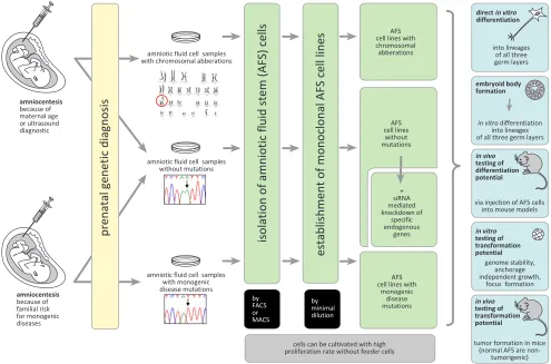
Figure 1: Human AFS cells as a biological model to study gene functions. Monoclonal human AFS cell lines can be established from amniocentesis performed because of different medical indications. These stem cell lines can be used for in vitro and in vivo investigations of the consequences of the functional loss of specific endogenous genes for differentiation and oncogenic processes. For
details see the text.
amniocentesis because of maternal age or ultrasound diagnostic
amniocentesis because of familial risk for monogenic diseases
amniotic fluid cell samples with chromosomal abberations
amniotic fluid cell samples without mutations
amniotic fluid cell samples with monogenic disease mutations
prenatal genetic diagnosis
isolation of amniotic fluid stem (AFS) cells
establishment of monoclonal AFS cell lines
by FACS or MACS
by minimal dilution
cells can be cultivated with high proliferation rate without feeder cells
AFS cell lines with chromosomal abberations
AFS cell lines without mutations
AFS cell lines with
monogenic disease mutations
+ siRNA mediated knockdown of
specific endogenous
genes
directin vitro differentiation
embryoid body formation
in vivo testing of differentiation potential
in vitro testing of transformation potential
in vivo testing of transformation potential
into lineages of all three germ layers
in vitro differentiation into lineages of all three germ layers
via injection of AFS cells into mouse models
genome stability, anchorage independent growth,
focus formation
tumor formation in mice (normal AFS are
embryo for research raises ethical issues, regarding when human life begins and the moral status of a few-days-old embryos. In addition, the spectrum of ES cells carrying an inherited defect is limited due to the fact that in vitro fertilization with preimplantation genetic diagnosis is only rarely applied [11-14]. And last but not least, ES
cells cannot be used to investigate the role of specific
mutations in tumorigenesis in in vivo models, since when ES cells are transplanted in mice, they are already per se tumorigenic [15].
A second stem cell type, which fulfils the criterias
described above, are iPS cells. In 2006, Yamanaka´s
group demonstrated for the first time that adult mouse fibroblasts can be reprogrammed to pluripotent stem
cells via ectopic overexpression of the genes Pou5f1, Sox2, Klf4 and cMyc, which are known to be expressed in ES cells [16]. Such iPS cells can be grown in culture maintaining their pluripotency to form all three embryonic tissue types. In 2008, the generation of iPS
cells from patients with specific genetic diseases was first described [17,18]. Inbetween, a variety of iPS lines
from single-gene disorders, chromosome syndromes and complex diseases, including e.g. amyotrophic lateral sclerosis, diabetes mellitus, Parkinson´s diseases, Down syndrome, epidermolysis bullosa, or Rett syndrome, have been generated [1, 2, 17-20]. Still, iPS cells also harbour relevant disadvantages. Using cells from adult patients has the advantage that the detailed clinical history of the patient is known, but one must take into account that such cells might have already accumulated other mutations without relevance for the disease of interest. And clearly, such an approach only allows to study mutations, which are not prenatally lethal. Importantly, the process of ectopic induction of pluripotency might also negatively affect the usefulness of these cells as models for human diseases [1, 2, 19, 20]. In addition, recently it was demonstrated that the epigenetic memory of the original differentiated state is not perfectly erased during reprogramming [21,
22]. And finally, it seems to be inevitable that iPS cells
accumulate karyotypic abnormalities and gene mutations during propagation in culture [23-26]. We here discuss that human AFS cells might be a powerful alternative for disease modelling.
HUMAN AFS CELLS TO STUDY
NATURAL OCCURRING DISEASE
CAUSING MUTATIONS
Ultrasound investigations and maternal serum screening are routinely used in prenatal diagnosis.
Specific ultrasound signs and/or maternal age are classical
indications for invasive genetic prenatal diagnosis, mostly to detect fetal numerical chromosomal aberrations. Invasive prenatal diagnosis is also recommended to detect unbalanced segregation of e.g. balanced parental chromosome translocations. In addition, prenatal
chromosome testing also leads to the detection of a major category of “unexpected” chromosomal abnormalities, including numerical aneuploidies other than trisomies 13, 18, and 21, and the wide variety of possible de novo structural chromosomal aberrations. Since the Human
Genome Project was officially launched the knowledge
on single-gene disorders has grown dramatically. Today, a wide variety of mutations, causatively involved in monogenic diseases, can be prenatally diagnosed. Predominantly such prenatal diagnoses are performed as
a consequence of a specific family history (Figure 1)
[27-29]. Besides other invasive approaches, amniocentesis is a widespreadly accepted standard procedure of care since the 1970s. It is almost unpredictable how many amniocenteses are worldwide performed per year. An older study reported that more than 200.000 amniocentesis procedures were performed already 20 years ago, only in
one year (1990) and only in the United States [30, 31].
Despite this wide and well established usage of
human amniotic fluid cells in routine prenatal diagnosis, the knowledge about the cells contained in amniotic fluid
remained rather elusive [32, 33]. New interest in these cells was initiated in 2001, when Fauza´s group reported
that amniotic fluid cells could be used in tissue engineering
approaches for the surgical repair of congenital anomalies in the perinatal period [34]. This was also about the time
when the first suggestion of human amniotic fluid as
a new putative source for stem cells was reported [35]. In 2003, a highly proliferative cell type, expressing the
pluripotent stem cell marker Oct4, was discovered to exist in human amniotic fluid, providing the first evidence for AFS cells [36]. The confirmation of Oct4-positive cells in amniotic fluid by different other groups then initiated a fast growing stem cell research field [37-45]. In the last
years, it became evident that AFS cells can differentiate into cells of all three embryonic tissue types. AFS cell differentiation e.g. upon hematopoietic, neurogenic, osteogenic, chondrogenic, adipogenic, renal and hepatic lineages has been demonstrated [37-63]. Descending
from one single Oct4- and NANOG-positive AFS cell,
it was possible to induce adipogenic, osteogenic and neurogenic differentiation [47]. In another study, isolation of monoclonal AFS cells, which expressed the markers
Oct4 and CD117 (c-kit), allowed to demonstrate that
adipogenic, osteogenic, myogenic, endothelial, neurogenic and hepatic cell differentiation could be induced [42]. Monoclonal human AFS cells can form embryoid bodies
(EBs) when cultured without anti-differentiation factors
under conditions in which they are unable to attach to the surface of culture dishes and without contact to feeder cells. The formation of such three-dimensional multicellular aggregates is accompanied by a decrease of stem cell marker expression and by the induction of differentiation into different lineages [64].
Taken together, these findings prove that human AFS
and to differentiate into cell types of all three germ layers. These data were obtained using monoclonal human AFS cell lines generated via magnetic cell sorting and minimal dilution approaches from amniocentesis samples. So established monoclonal lines can be expanded as immature stem cells with high proliferation rate in culture without the need of feeder cells [42, 60, 61, 64]. We here suggest the generation and banking of normal human AFS cell lines, of AFS cell lines with chromosomal aberrations, as
well as of AFS cell lines with specific monogenic disease mutations for research purposes (Figure 1). Such lines
carrying natural occurring mutations could provide an optimal tool for disease modelling with the aim to obtain more detailed insights into the functions of the involved chromosomal regions or genes. As in the past already performed with normal monoclonal human AFS cell lines [42, 58, 60, 61, 63, 64], lines harbouring mutations could be investigated in in vitro differentiation experiments
(including e.g. organotypic reaggregation assays, see
below), in embryoid body formation assays, and could be investigated in mouse models in vivo (Figure 1). Adult
stem cells exhibit lower differentiation potential than AFS cells and cannot be grown with high proliferative activity. Compared to ES cells, AFS cells do not raise ethical issues and the spectrum of samples with pathogenic mutations should be larger. Compared to iPS cells, there is no need for ectopic induction of pluripotency in AFS cells. They are genomically stable, they do neither harbour the epigenetic memory nor somatic mutations of already differentiated source cells, and mutations, which allow implantation but are not compatible with a prenatal development until birth, could also be included [1, 12, 19, 20, 42-45, 55-57, 60, 63-65]. Accordingly, AFS cells could provide an optimal tool for basic research and disease modelling in
medical genetics (Figure 1).
HUMAN AFS CELLS TO INVESTIGATE
GENES INVOLVED IN ONCOGENESIS
Stem cells could also be a powerful tool to study the consequences of genetic modulations for transformation processes, both, in vitro e.g. in colony formation assays and in vivo in mouse models (Figure 1). Such approaches
are hampered when the chosen stem cell type, in its undifferentiated form, is tumorigenic per se. This holds true for ES cells [1, 13-15]. Similarly, in the study
describing the first generation of iPS cells the authors
reported that subcutaneous transplantation of these stem cells into nude mice also resulted in tumor development [16]. Recently, it became clear that the tumor-forming propensities of the many different types of iPS cells vary
significantly. This is due to the different methods used for reprogramming (the genes or proteins, which have been
modulated), the different iPS cells´ tissues of origin, the
different genetic background of the donor (including the
somatic mutations accumulated during the life span of the
source cell), and very likely also to the propensity of iPS cells to accumulate mutations during cultivation [1, 19, 66, 67]. Accordingly, although the question whether ES
or iPS cells, after differentiation along specific lineages,
could be used for studies on transformation processes still needs to be further investigated, it is obvious that such experimental approaches would have to face many
difficulties and limitations, which might negatively affect
the level of interpretation.
AFS cells are primary cells of a very early stage of human development. Accordingly, they very likely did not have time to accumulate many somatic mutations yet [44, 45, 55-57]. In addition, the investigations performed so far revealed that monoclonal human AFS cell lines maintain genome stability during expansion [42, 64]. And most importantly, AFS cells, unlike ES and iPS cells, do not induce tumor formation in severe combined
immunodeficient mice [42]. Accordingly, we here want to
suggest the usage of different AFS cell lines to investigate
the role of specific genetic modulations for transformation processes and tumor development (Figure 1). Although
possible, we would not favour the ectopic expression of oncogenes in AFS cells, because of the risk that the so obtained high ectopic levels of expression would not necessarily mimick a natural occurring situation. We rather want to suggest the usage of siRNA-mediated
knockdown of endogenous tumor suppressor genes (for
examples see below). Monoclonal human AFS cells with so downregulated endogenous gene functions could then be investigated in in vitro and in vivo transformation assays as indicated in Figure 1. For this purpose we have recently
established a protocol for efficient siRNA-mediated
prolonged gene silencing in AFS cells. This protocol allows a 96-98% downregulation of the endogenous expression of tumor suppressors, such as the TSC2 gene product tuberin or the cyclin-dependent kinase inhibitor p27, over a time period of about 14 days [68].
MODULATION OF MTOR CASCADE
COMPONENTS IN HUMAN AFS CELLS
Very recently we already made use of the approach discussed above to functionally investigate the role of
components of the mTOR signalling cascade in human AFS cells. mTOR is the key component of the insulin
signalling cascade, which is involved in a wide variety of different processes such as cell growth, proliferation, metabolism, transcription, translation, survival, autophagy, aging, differentiation and oncogenesis. An important
upstream regulator of mTOR is the oncogenic kinase
Akt, which itself is activated via the enzyme cascade
of PI3K (phosphatidylinositol-3-kinase) and PDK1 (phosphoinositide-dependent kinase-1). Akt-triggered
phosphorylation of the tumor suppressor protein tuberin
mammalian cells, two mTOR containing complexes are described, mTORC1 (consisting of mTOR, raptor and mLST8) and mTORC2 (containing mTOR, mLST8, rictor and sin1). mTORC1 is involved in the control of mRNA
translation e.g. via phosphorylating the p70S6Kinase,
whereas mTORC2 phosphorylates and activates Akt [69-72]. Deregulation of upstream regulators of mTOR, such
as e.g. Wnt, Ras, TNF-α, PI3K, or Akt is a hallmark in
many human cancers. Mutations in the mTOR pathway
component genes TSC1, TSC2, LKB1, PTEN, VHL, NF1 and PKD1 trigger the development of the human genetic syndromes Tuberous sclerosis, Peutz-Jeghers syndrome, Cowden syndrome, Bannayan-Riley-Ruvalcaba syndrome, Lhermitte-Duclos disease, Proteus syndrome,
von Hippel-Lindau disease, Neurofibromatosis type 1,
and Polycystic kidney disease. Besides a variety of single
gene disorders and tumorigenesis, the mTOR pathway has
also been shown to be of relevance for the development of complex diseases, such as cardiac hypertrophy, obesity or type 2 diabetes [73-75].
Studies performed in hematopoietic stem cells
provided first evidence that mTOR might also play a role
in stem cell physiology [76]. A major characteristic of pluripotent stem cells is their potential to spontaneously form EBs. EB formation is a commonly used in vitro approach to recapitulate and investigate the three-dimensional and tissue level contexts of the cell differentiation phenomena mimicking early mammalian embryogenesis [77, 78]. Since it was already earlier shown
that the mTOR cascade is fully active in human AFS cells
[79], we induced EB formation of monoclonal human
AFS cell lines to investigate the role of mTOR.
siRNA-mediated knockdown of raptor or rictor demonstrated that
EB formation depends on both, mTORC1 and mTORC2
[64]. Together with similar results in human ES cells [80],
these findings were the first demonstration of the role of mTOR for EB formation of pluripotent stem cells.
We have further shown that human monoclonal AFS cells are able to contribute to the formation process of renal tissues. For this purpose, murine embryonic kidneys were dissociated into a single-cell suspension and then reaggregated to form organotypic renal structures. Using this approach it was possible to form chimeric renal structures via mixing murine embryonic kidney cells with monoclonal human AFS cells selected from amniocentesis
samples. Monoclonal, pluripotent Oct4- and
CD117-positive AFS cells were able to participate in the formation process of different renal tissues accompanied by the induction of the expression of well known renal markers [60]. In this study the highly efficient and reproducible
protocol for prolonged siRNA-mediated gene silencing in human monoclonal AFS cells [68], which was used for the EB study described above, also allowed to demonstrate
the pivotal role of mTOR component genes for renal
tissue formation [60].
Taken together the approach of siRNA-mediated
knockdown of mTOR cascade components in monoclonal
human AFS cell lines allowed to study the consequences on EB formation and on the potential to differentiate along renal lineages. These investigations already provided a “proof of principle” for the here discussed usage of human
AFS cells as a model for functional studies of specific
genes.
REFERENCES
1. Zhu H, Lensch WM, Cahan P, Daley GQ. Investigating monogenic and complex diseases with pluripotent stem cells. Nat Rev Genet. 2011; 12:266-275.
2. Stadtfeld M, Hochedlinger K. Induced pluripotency: history, mechanisms, and applications. Genes Dev. 2010; 24:2239-2263.
3. Geraedts JPM, DeWert GMWR. Preimplantation genetic diagnosis. Clin Genet. 2009; 76:315-325.
4. Simpson JL. Preimplantation genetic diagnosis at 20 years. Prenat Diag 2010; 30:682-695.
5. Verlinsky Y, Strelchenko N, Kukharenko V, Rechitsky S,
Verlinsky O, Galat V, Kuliev A. Human embryonic stem cell lines with genetic disorders. Reprod Biochem Online.
2004; 10:105-110.
6. Pickering S, Minger S, Patel M, Taylor H, Black C, Burns C, Ekonomou A, Braude P. Generation of a human embryonic
stem cell line encoding the cystic fibrosis mutation F508,
using preimplantation genetic diagnosis. Reprod Biomed
Online. 2005; 10:390-397.
7. Ben-Yosef D, Malcov M, Eiges R. PGD-derived human embryonic stem cell lines as a powerful tool for the study of human genetic disorders. Mol Cell Endocrinol. 2008; 282:153-158.
8. Sermon KD, Simon C, Braude P, Viville S, Borstlap J, Veiga A. Creation of a registry for human embryonic stem cells carrying an inherited defect: joint collaboration between ESHRE and hESCreg. Hum Reprod. 2009; 24:1556-1560. 9. Taymor K, Scott CT. The practical consequences of a
national human embryonic stem cell registry. Stem Cell Rev and Rep. 2009; 5:315-318.
10. Isasi RM, Knoppers BM. Governing stem cell banks and registries: emerging issues. Stem Cell Res. 2009; 3:96-105. 11. Rosenthal N. Prometheus´s vulture and the stem cell
promise. New Engl J Med. 2003; 349:267-274.
12. Wood A. Ethics and embryonic stem cell research. Stem Cell Reviews 2005; 1:317-324.
13. Schulman A. The search for alternative sources of human pluripotent stem cells. Stem Cell Rev. 2005; 1:291-292. 14. Kamm FM. Ethical issues in using and not using embryonic
stem cells. Stem Cell Reviews. 2005; 1:325-330.
16. Takahashi K, Yamanaka S. Induction of pluripotent stem
cells from mouse embryonic and adult fibroblast cultures by defined factors. Cell. 2006; 126:663-676.
17. Dimos JT, Rodolfa KT, Niakan KK, Weisenthal LM, Mitsumoto H, Chung W, Croft GF, Saphier G, Leibel R, Goland R, Wichterle H, Henderson CE, Eggan K. Induced pluripotent stem cells generated from patients with ALS can be differentiated into motor neurons. Science. 2008; 321:1218-21.
18. Park IH, Arora N, Huo H, Maherali N, Ahfeldt T, Shimamura A, Lensch MW, Cowan C, Hochedlinger K,
Daley GQ. Disease-specific induced pluripotent stem cells.
Cell. 2008; 134:877-86.
19. Mattis V, Svendsen CN. Induced pluripotent stem cells: a new revolution for clinical neurology? Lancet Neurol. 2011; 10:383-394.
20. Wu SM, Hochedlinger K. Harnessing the potential of induced pluripotent stem cells for regenerative medicine. Nat Cell Biol. 2011; 13:497-505.
21. Kim K, Doi A, Wen B, Ng K, Zhao R, Cahan P, Kim J, Aryee MJ, Ji H, Ehrlich LI, Yabuuchi A, Takeuchi A, Cunniff KC, Hongguang H, McKinney-Freeman S,
Naveiras O, Yoon TJ, Irizarry RA, Jung N, Seita J, Hanna J, Murakami P, Jaenisch R, Weissleder R, Orkin SH,
Weissman IL, Feinberg AP, Daley GQ. Epigenetic memory in induced pluripotent stem cells. Nature. 2010; 467:285-90.
22. Stadtfeld M, Apostolou E, Akutsu H, Fukuda A, Follett P, Natesan S, Kono T, Shioda T, Hochedlinger K. Aberrant silencing of imprinted genes on chromosome 12qF1 in mouse induced pluripotent stem cells. Nature. 2010; 465:175-81.
23. Laurent LC, Ulitsky I, Slavin I, Tran H, Schork A, Morey R, Lynch C, Harness JV, Lee S, Barrero MJ, Ku S, Martynova M, Semechkin R, Galat V, Gottesfeld J, Izpisua Belmonte JC, Murry C, Keirstead HS, Park HS, Schmidt U, Laslett AL, Muller FJ, Nievergelt CM, Shamir R, Loring JF. Dynamic changes in the copy number of pluripotency and cell proliferation genes in human ESCs and iPSCs during reprogramming and time in culture. Cell Stem Cell. 2011; 8:106-18.
24. Mayshar Y, Ben-David U, Lavon N, Biancotti JC, Yakir B,
Clark AT, Plath K, Lowry WE, Benvenisty N. Identification and classification of chromosomal aberrations in human
induced pluripotent stem cells. Cell Stem Cell. 2010; 7:521-31.
25. Gore A, Li Z, Fung HL, Young JE, Agarwal S, Antosiewicz-Bourget J, Canto I, Giorgetti A, Israel MA, Kiskinis E, Lee JH, Loh YH, Manos PD, Montserrat N, Panopoulos AD, Ruiz S, Wilbert ML, Yu J, Kirkness EF, Izpisua Belmonte JC, Rossi DJ, Thomson JA, Eggan K, Daley GQ, Goldstein LS, Zhang K. Somatic coding mutations in human induced pluripotent stem cells. Nature. 2011;471:63-7.
26. Pasi CE, Dereli-Öz A, Negrini S, Friedli M, Fragola G, Lombardo A, Van Houwe G, Naldini L, Casola S, Testa G,
Trono D, Pelicci PG, Halazonetis TD. Genomic instability in induced stem cells. Cell Death Differ. 2011; 18:745-53. 27. Hengstschläger M. Fetal magnetic resonance imaging and
human genetics. Europ J Radiol. 2006; 57:312-315. 28. Raymond FL, Whittaker J, Jenkins L, Lench N., Chitty
LS. Molecular prenatal diagnosis: the impact of modern technologies. Prenat Diagn 2010; 30:674-681.
29. Nicolaides KH. Turning the pyramid of prenatal care. Fetal Diagn Ther 2011; 29:183-196.
30. Meaney FJ, Riggle SM, Cunningham GC, Stern KS, David JG. Prenatal genetic services: toward a national data base.
Clin Obstet Gynecol. 1993; 36:510-520.
31. Nizard J. Amniocentesis: technique and education. Curr Op Obstet Gynecol. 2010; 22: 152-154.
32. Hoehn H, Salk D. Morphological and biochemical
heterogeneity of amniotic fluid cells in culture. Meth Cell
Biol. 1982; 26:11-34.
33. Gosden CM. Amniotic fluid cell types and culture. Brit
Med Bull. 1983; 39:348-354.
34. Kaviani A, Perry TE, Dzakovic A, Jennings RW, Ziegler
MM, Fauza DO. The amniotic fluid as a source of cells for
fetal tissue engineering. J Pediat Surg. 2001; 36:1662-1665.
35. Prusa A, Hengstschläger M. Amniotic fluid cells and
human stem cell research – a new connection. Med Sci Monit. 2002; 8:253-7.
36. Prusa A, Marton E, Rosner M, Bernaschek G, Hengstschläger
M. Oct-4 expressing cells in human amniotic fluid: a new
source for stem cell research? Hum Reprod. 2003; 18:1489-93.
37. in t`Anker PS, Scherjon SA, Kleijburg-van der Keur C, Noort WA, Claas FHJ, Willemze R, Fibbe WE, Kanhai
HHH. Amniotic fluid as a novel source of mesenchymal
stem cells for therapeutic transplantation. Blood. 2003; 102:1548-9.
38. Tsai M-S, Lee J-L, Chang Y-J, Hwang S-M. Isolation of human multipotent mesenchymal stem cells from
second-trimester amniotic fluid using a novel two-stage culture
protocol. Hum Reprod. 2004; 19:1450-6.
39. Karlmark KR, Freilinger A, Marton E, Rosner M, Lubec
G, Hengstschläger M. Activation of Oct-4 and Rex-1 promoters in human amniotic fluid cells. Int J Mol Med.
2005; 16:987-92.
40. Bossolasco P, Montemurro T, Cova L, Zangrossi S, Calzarossa C, Miatiotis S, Soligo D, Bosari S, Silani V, Deliliers GL, Rebulla P, Lazzari L. Molecular and
phenotypic characterization of human amniotic fluid cells
and their differentiation potential. Cell Res. 2006; 16: 329-36.
41. Kim J, Lee Y, Kim H, Hwang KJ, Kwon HC, Kim SK, Cho
DJ, Kang SG, You J. Human amniotic fluid-derived stem
cells have characteristics of multipotent stem cells. Cell Prolif. 2007; 40:75-90.
Furth ME, Soker S, Atala A. Isolation of amniotic stem cell lines with potential for therapy. Nature Biotech. 2007; 25:100-6.
43. Siegel N, Rosner M, Hanneder M, Freilinger A,
Hengstschläger M. Human amniotic fluid stem cells: a new
perspective. Amino Acids. 2008; 35:291-3.
44. Pappa KI, Anagnou NP. Novel sources of fetal stem cells:
where do they fit on the developmental continuum? Regen
Med. 2009; 4:423-33.
45. Dobreva P, Pereira PNG, Deprest J, Zwijsen A. On the
origin of amniotic stem cells: of mice and men. Int J Dev Biol. 2010; 54:761-77.
46. Prusa AR, Marton E, Rosner M, Bettelheim D, Lubec G, Pollak A, Bernaschek G, hengstschläger M. Neurogenic
cells in human amniotic fluid. Am J Obstet Gynecol. 2004;
191:309-14.
47. Tsai M-S, Hwang S-M, Tsai Y-L, Cheng F-C, Lee J-L.
Chang Y-J. Clonal amniotic fluid-derived stem cells
express characteristics of both mesenchymal and neural stem cells. Biol Reprod. 2006; 74:545-51.
48. Rehni AK, Singh N, Jaggi AS, Singh M. Amniotic fluid
derived stem cells ameliorate focal cerebral
ischaemia-reperfusion injury induced behavioural deficits in mice.
Behav Brain Res. 2007; 183:95-100.
49. Kolambkar YM, Peister A, Soker S, Atala A, Guldberg
RE. Chondrogenic differentiation of amniotic fluid-derived
stem cells. J Mol Hist. 2007; 38:405-13.
50. Orciani M, Emanuelli M, Martino C, Pugnaloni A, Tranquilli
AL, DiPrimio R. Potential role of culture mediums for successful isolation and neuronal differentiation of
amniotic fluid stem cells. Int J Immunopathol Pharmacol.
2008; 21:595-602.
51. Ditadi A, de Coppi P, Picone O, Gautreau L, Smati R,
Six E, Bonhomme D, Ezine S, Frydman R, Cavazzano-Calvo M, Andre-Schmutz I. Human and murine amniotic c-Kit+Lin- cells display hematopoietic activity. Blood, 2009; 113:3953-60.
52. Jezierski A, Gruslin A, Tremblay R, Ly D, Smith C, Turksen K, Skorska M, Bani-Yaghoub M. Probing stemness and
neural commitment in human amniotic fluid cells. Stem
Cell Rev Rep. 2010; 6:199-214.
53. Mareschi K, Rustichelli D, Comunanza V, DeFazio R, Cravero C, Morterra G, Martinoglio B, Medico E, Carbone E, Benedetto C, Fagioli F. Multipotent mesenchymal stem
cells from amniotic fluid originate neural precursors with
functional voltage-gated sodium channels. Cytotherapy. 2009; 11:534-8.
54. Pfeiffer S, McLaughlin D. In vitro differentiation of
human amniotic fluid-derived cells: augmentation towards
a neuronal dopaminergic phenotype. Cell Biol Int .2010; 34:959-67.
55. Pozzobon M, Ghionzoli M, DeCoppi P. ES, iPS, MSC and AFS cells. Stem cells exploitation for pediatric surgery: current research and perspective. Pediatr Surg Int. 2010;
26:3-10.
56. Abdulrazzak H, Moschidou D, Jones G, Guillot PV. Biological characteristics of stem cells from foetal, cord blood and extraembryonic tissues. J R Soc Interface. 2010; 7:S689-S706.
57. Antonucci I, Stuppia L, Kaneko Y, Yu S, Tajiri N, Bae EC, Chheda SH, Weinbren NL, Borlongan CV.. Amniotic
fluid as rich source of mesenchymal stromal cells for transplantation therapy. Cell Transplant. 2011; DOI:
10.3727/096368910X539074
58. Perin L, Giuliani S, Jin D, Sedrakyan S, Carraro G, Habibian R, Warburton D, Atala A, DeFilippo RE. Renal
differentiation of amniotic fluid stem cells. Cell Prolif.
2007; 40:936-48.
59. Siegel N, Valli A, Fuchs C, Rosner M, Hengstschläger M. Induction of mesenchymal/epithelial marker expression
in human amniotic fluid stem cells. RBM Online. 2009;
19:838-46.
60. Siegel N, Rosner M, Unbekandt M, Fuchs C, Slabina N, Dolznig H, Davies JA, Lubec G, Hengstschläger M.
Contribution of human amniotic fluid stem cells to renal tissue formation depends on mTOR. Hum Mol Genet.
2010; 19:3320-31.
61. Perin L, Sedrakyan S, Giuliani S, DaSacco S, Carraro G, Shiri L, Lemley KV, Rosol M, Wu S, Atala A, Warburton
D, DeFilippo RE. Protective effect of human amniotic fluid stem cells in an immunodeficient mouse model of acute tubular necrosis. Plos One. 2010; 5:1-16
62. Hauser PV, DeFazio R, Bruno S, Sdei S, Grange C, Bussolati B, Benedetto C, Camussi G. Stem cells derived
from human amniotic fluid contribute to acute kidney inury
recovery. Am J Pathol. 2010; 177:2011-21.
63. DaSacco S, Sedrakyan S, Boldrin F, Giuliani S, Parnigotto PP, Habibian R, Warburton D, DeFilippo RE, Peril L.
Human amniotic fluid as a potential new source of organ specific precursor cells for future regenerative medicine
applications. J Urol. 2010; 183:1193-200.
64. Valli A, Rosner M, Fuchs C, Siegel N, Bishop CE, Dolznig H, Mädel U, Feichtinger W, Atala A, Hengstschläger M.
Embryoid body formation of human amniotic fluid stem cells depends on mTOR. Oncogene. 2010: 29:966-77.
65. Prusa AR, Marton E, Rosner M, Freilinger A, Bernachek G, Hengstschläger M. Stem cell marker expression in human
trisomy 21 amniotic fluid cells and trophoblasts. J Neural
Transm. 2003; 67:235-42.
66. Miura K, Okada Y, Aoi T, Okada A, Takahashi K, Okita K, Nakagawa M, Koyanagi M, Tanabe K, Ohnuki M, Ogawa D, Ikeda E, Okano H, Yamanaka S. Variation in the safety
of induced pluripotent stem cell lines. Nat Biotechnol. 2009; 27:743-5.
67. Yamashita T, Kawai H, Tian F, Ohta Y, Abe K. Tumorigenic
development of induced pluripotent stem cells in ischemic mouse brain. Cell Transplant. 2011; 20:883-91.
Hengstschläger M. Efficient siRNA-mediated prolonged gene silencing in human amniotic fluid stem cells. Nature
Protocols. 2010; 5:1081-95.
69. Polak P, Hall MN. mTOR and the control of whole body metabolism. Curr Opin Cell Biol. 2009; 21:209-18.
70. Sengupta S, Peterson TR, Sabatini DM. Regulation of the
mTOR complex 1 pathway by nutrients, growth factors,
and stress. Mol Cell. 2010; 40:310-22.
71. Ma XM, Blenis J. Molecular mechanisms of
mTOR-mediated translational control. Nat Rev Mol Cell Biol. 2009; 10:307-18.
72. Blagosklonny MV. Revisiting the antagonistic pleiotropy
theory of aging: TOR-driven program and quasi-program.
Cell Cycle. 2010; 9:3151-6.
73. Chappell WH, Steelman LS, Long JM, Kempf RC, Abrams SL, Franklin RA, Bäsecke J, Stivala F, Donia M, Fagone P, Malaponte G, Mazzarino MC, Nicoletti F, Libra M, Maksimovic-Ivanic D, Mijatovic S, Montalto G, Cervello M, Laidler P, Milella M, Tafuri A, Bonati A, Evangelisti C, Cocco L, Martelli AM, McCubrey JA. Ras/Raf/MEK/
ERK and PI3K/PTEN/Akt/mTOR inhibitors: rationale and
importance to inhibiting these pathways in human health.
Oncotarget. 2011; 2:135-64.
74. Wong KK, Engelman JA, Cantley LC. Targeting the PI3K
signaling pathway in cancer. Curr Opin Genet Dev. 2010;
20:87-90.
75. Rosner M, Hanneder M, Siegel N, Valli A, Fuchs C,
Hengstschläger M. The mTOR pathway and its role in
human genetic diseases. Mutat Res Rev Mutat Res. 2008; 659:284-92.
76. Russel RC, Fang C, Guan K-L. An emerging role for TOR
signaling in mammalian tissue and stem cell physiology. Development. 2011; 138:3343-3356.
77. Koike M, Sakaki S, Amano Y, Kurosawa H. Characterization of embryoid bodies of mouse embryonic stem cells formed under various culture conditions and estimation of differentiation status of such bodies. J Biosi Bioeng. 2007; 104: 294-299.
78. Ungrin MD, Joshi C, Nica A, Bauwens C, Zandstra PW. Reproducible, ultra high-troughput formation of multicellular organization from single cell suspension-derived human embryonic stem cell aggregates. PLoS
ONE. 2008; 3:e1565.
79. Siegel N, Valli A, Fuchs C, Rosner M, Hengstschläger M.
Expression of mTOR pathway proteins in human amniotic fluid stem cells. Int J Mol Med. 2009; 23:779-784.
80. Zhou J, Su P, Wang L, Chen J, Zimmermann M, Genbacev
O, Afonja O, Horne MC, Tanaka T, Duan E, Fisher SJ, Liao J, Chen J, Wang F. mTOR supports long-term