A decade of molecular pathogenomic analysis
of group A Streptococcus
James M. Musser, Samuel A. Shelburne III
J Clin Invest.
2009;
119(9)
:2455-2463.
https://doi.org/10.1172/JCI38095
.
Molecular pathogenomic analysis of the human bacterial pathogen group A
Streptococcus
has been conducted for a decade. Much has been learned as a consequence of the
confluence of low-cost DNA sequencing, microarray technology, high-throughput
proteomics, and enhanced bioinformatics. These technical advances, coupled with the
availability of unique bacterial strain collections, have facilitated a systems biology
investigative strategy designed to enhance and accelerate our understanding of disease
processes. Here, we provide examples of the progress made by exploiting an integrated
genome-wide research platform to gain new insight into molecular pathogenesis. The
studies have provided many new avenues for basic and translational research.
Review Series
Find the latest version:
A decade of molecular pathogenomic analysis
of group A
Streptococcus
James M. Musser1 and Samuel A. Shelburne III2
1Center for Molecular and Translational Human Infectious Diseases Research, The Methodist Hospital Research Institute, and Department of Pathology,
The Methodist Hospital, Houston, Texas, USA. 2Department of Infectious Diseases, Division of Internal Medicine,
MD Anderson Cancer Center, Houston, Texas, USA.
Molecular pathogenomic analysis of the human bacterial pathogen group A
Streptococcus
has been conducted for a
decade. Much has been learned as a consequence of the confluence of low-cost DNA sequencing, microarray
tech-nology, high-throughput proteomics, and enhanced bioinformatics. These technical advances, coupled with the
availability of unique bacterial strain collections, have facilitated a systems biology investigative strategy designed
to enhance and accelerate our understanding of disease processes. Here, we provide examples of the progress made
by exploiting an integrated genome-wide research platform to gain new insight into molecular pathogenesis. The
studies have provided many new avenues for basic and translational research.
Introduction
Group A Streptococcus (GAS) is a Gram-positive bacterium that causes several diseases in humans, including pharyngitis and/or tonsillitis, skin infections (including impetigo, erysipelas, and other forms of pyoderma), acute rheumatic fever (ARF), scar-let fever, poststreptococcal glomerulonephritis (PSGN), a toxic shock–like syndrome, and necrotizing fasciitis (NF) (1–3). The organism is a human-adapted pathogen, that is, humans are the natural host and there is no animal or environmental reservoir that contributes to the life cycle of GAS. On a global basis, ARF and the rheumatic heart disease that follows are the most com-mon cause of preventable pediatric heart disease worldwide (4). There are 25–35 million cases of GAS pharyngitis per year in the United States, which are responsible for about 1–2 billion dollars per year in direct health care costs, and 600 million cases globally each year (Table 1) (2, 3). The continued high morbidity and mor- tality caused by GAS in developing nations, the substantial finan-cial burden attributable to GAS-related health care costs in the United States, the development and spread of antibiotic resistance in this pathogen (5–7), and the lack of a licensed human vaccine (8, 9) highlight the need for a fuller understanding of the molecu- lar pathogenesis of GAS. Especially vital is the need for informa- tion bearing on the molecular events contributing to pathogen-host interaction, clone emergence, and epidemics.
In recent years, a paradigm shift has occurred in the manner in which microbial pathogenesis problems are studied. The conflu-ence of data derived from genome-sequencing projects and the development of microarray technology permit a genome-wide strategy to be used for pathogenesis research, now commonly referred to as molecular pathogenomics investigation (10). Com- parative genome sequencing and other types of analyses of mul-tiple isolates of the same pathogenic bacterial species have revealed unexpectedly large amounts of intraspecies variation in gene content and uncovered strategies used in genome diversification (11–19). Strains of some bacterial species differ in gene content by up to 25% and have extensive allelic variation (11, 13, 15). This striking magnitude of genetic differentiation provides tremendous adaptive flexibility and influences the spectrum of clinical disease
caused by distinct clones of the species. For example, the genome of E. coli serotype O157:H7, which is responsible for hemolytic uremic syndrome, is approximately 25% larger (i.e., approximately 1 Mb of DNA larger) than that of many other strains of E. coli that do not cause significant disease in humans (13).
A molecular pathogenomics approach has been applied to GAS for nearly a decade and has yielded new information about the genetic basis of GAS pathogenesis, clone emergence, and strain genotype–disease phenotype relationships. Herein we highlight key findings that illustrate that molecular pathogenomics can be used to greatly accelerate the rate of obtaining new information about long-standing and previously intractable infectious disease ques-tions. Only by applying hypothesis-driven research and using new technologies will the elucidation of molecular events underlying infectious disease processes proceed with maximum efficiency.
GAS genome sequences
General concepts. The full-genome sequences that are now available for 13 strains of GAS make it one of the more deeply sequenced species of human pathogens (15, 20–28). GAS strains can be clas-sified by either M protein or T protein serotype, depending on the composition of bacterial antigens expressed on their cell surface, and the 13 genome sequences represent serotypes responsible for more than 70% of M protein serotypes that commonly cause GAS pharyngitis and invasive infections in the Western Hemisphere (29–31). The strains selected for sequencing were chosen mainly because they represent abundantly occurring M protein sero-types or because they had a distinct clinical phenotype of special interest, such as high virulence, antimicrobial agent resistance, or association with a particular disease (15, 20–28). Several common themes have emerged from comparison of the genome sequences (15). Each genome is approximately 1.9 Mb in size, with approxi-mately 10% of the overall gene content encoded on variably present exogenous genetic elements such as prophages and integrated con-jugative elements (15), with the former accounting for most of the variably present gene content (15, 20–28). Importantly, the marked heterogeneity of gene content is even observed among GAS strains of the same M protein serotype. This extensive variation in gene content stands in stark contrast to humans and many other higher eukaryotic species, which have less than 0.1% variation in gene con-tent among different members of a species. The 13 GAS genome
Conflict of interest: J.M. Musser received research support from Novartis Vaccines.
review series
sequences provide a critical resource for initiating investigation into how variation in gene content influences pathogenesis.
Discovery of new virulence factors through whole-genome sequence analy-sis. As recently as a decade ago, only relatively few GAS virulence factors had been identified. These included M protein, hyaluronic acid capsule, and the extracellular cysteine protease streptococcal pyrogenic exotoxin B (SpeB). Within a few years of the first GAS genome sequence becoming available, no fewer than 13 new pro-teins that contribute to pathogenesis had been described (32–45). Novel pathways of GAS host-pathogen interaction also had been elucidated. For example, Edwards et al. demonstrated that super- natants derived from GAS cleaved IL-8, resulting in reduced neu-trophil activation and migration (37). Using the GAS genome data, the IL-8–degrading activity was shown to be encoded by a previously uninvestigated gene (spy0416, using the serotype M1 numbering system for strain SF370), and the proteinase encoded by this gene was subsequently termed “Streptococcus pyogenes cell envelope proteinase” (SpyCEP) (46, 47). In a short time, it has been learned that SpyCEP-mediated cleavage of IL-8 decreases neutro- phil endothelial transmigration and that degradation of addition-al chemokines by SpyCEP retards neutrophil activation (46–48). Together, these discoveries indicate a key role for SpyCEP in GAS host-pathogen interaction (46–50). Similar rapid progress, facili-tated by the availability of the GAS genome sequences, has been made in identifying and characterizing the function of novel GAS immunoglobulin-degrading enzymes (39, 40, 51), collagen-like proteins (41, 44, 52), and several superantigens (53–55). Given that numerous open reading frames in the GAS genome that encode putative cell surface proteins have yet to be investigated, it seems highly likely to us that heretofore unidentified GAS virulence fac-tors will continue to be discovered in the coming years.
Expanded understanding of virulence factor regulation pathways
As noted above, the GAS genome encodes a broad range of viru-lence factors that are critical to the diverse array of infections that the bacterium causes (1). One important impact of GAS genome-wide investigations has been to provide an enhanced molecular
understanding of how the pathogen coor-dinates virulence factor production (56). There are 13 conserved two-component regulatory systems (TCSs) in the complet-ed GAS genomes, only one of which had been studied prior to the availability of the first GAS genome sequence in 2001 (57). TCSs regulate multiple unlinked chro-mosomal genes and control coordinated expression of genes encoding virulence fac-tors, such as toxins, degradative enzymes, and immune-modulating molecules, in response to environmental stimuli. Since 2001, each of the 13 TCSs has been studied by genome-wide analyses to some degree, many in detail (58–62). Similarly, there are more than 100 putative stand-alone transcriptional regulators encoded within the GAS genome, only a few of which had been identified before the availability of a genome sequence. Of the 100 putative stand-alone transcriptional regulators, 12 have now been investigated in detail, and the transcriptomes that eight of these regulate in vitro have been determined (17, 63–68).
Genome-wide investigations of GAS regulatory pathways have transformed our understanding of pathogenesis. A key finding has been that global gene transcription varies highly depending on the environment, growth conditions, and stage of growth of the bacterium (69–72). For example, Voyich et al. (73) have shown that the GAS transcriptome is substantially altered in response to phagocytosis by human polymorphonuclear leukocytes (PMNs), a key step in invasive GAS disease. Similarly, interaction with pharyngeal epithelial cells and human saliva was found to induce marked alterations in the GAS transcriptome, thereby providing new information about potential strategies used for infection of and persistence in the oropharynx (69, 74). Longitudinal analysis of changes in the GAS transcriptome over time in nonhuman primates has revealed that the temporal pattern of GAS gene transcription in pharyngitis is very closely linked to three dis-tinct phases of infection, namely colonization, acute infection, and asymptomatic carriage (75). During the colonization phase, when GAS CFUs were low, the expression of genes involved in carbohydrate metabolism was greatly increased, suggesting that carbon source acquisition is a key step in initial GAS growth and establishment of infection. Expression of genes encoding GAS virulence factors with known roles in GAS survival, dissemina-tion, inhibition of PMN recruitment, and induction of host cytokines were highly expressed in the acute phase of infection, concomitant with an increase in GAS CFUs and host inflamma-tion. Thus, the in vivo infection data are in strong alignment with ex vivo results (i.e., those obtained using saliva, epithelial cells, and PMNs).
A key finding of repeated GAS transcriptome analyses has been the elucidation of previously unappreciated connections between distinct gene categories. For example, recent work has shown that transcript levels of genes encoding proteins involved in carbohy-drate catabolism and genes encoding virulence factors change in concert in response to environmental stimuli (74, 75). Such changes are at least partially due to a regulatory circuit controlled
by catabolite control protein A (CcpA) (Figure 1) (59, 74–78). Simi-Table 1
Minimum summary estimates of the global burden of GAS diseases
Existing cases Annual new cases Annual deaths Severe GAS disease
RHD 15.6 million 282,000 233,000
History of ARF without carditis, 1.88 million 188,000 — requiring secondary prophylaxis
RHD-related infective endocarditis — 34,000 8,000 RHD-related stroke 642,000 144,000 108,000 Acute poststreptococcal — 472,000 5,000 glomerulonephritis
Invasive GAS diseases — 663,000 163,000 Total severe cases 18.1 million 1.78 million 517,000 Superficial GAS disease
Pyoderma 111 million — —
Pharyngitis — 616 million —
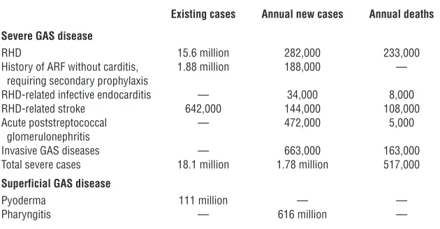
[image:3.585.53.368.112.276.2]larly, a genome-wide investigation of SpeB regulation led to the discovery that a lactose catabolism enzyme (LacD.1) has evolved to coordinate alterations in GAS virulence factor production as a result of changes in carbon source availability (Figure 1) (79). Anal-ysis of completed GAS genomes led to the discovery that one of the two GAS lactose operons has retained a catabolic role, whereas the other has evolved a regulatory function (Figure 1) (80).
In addition to discovering ties between central metabolic pro-cesses and pathogenesis, GAS genome-wide studies have also revealed interactions between metal regulation, oxidative stress, and pathogenesis. Analysis of the completed genomes indicates that GAS encodes two highly conserved metalloregulators, MtsR and PerR, that regulate proteins involved in iron and manganese uptake (81, 82). Animal studies have found that PerR is needed for full GAS virulence in skin and soft tissue infection and in oro-pharynx infection (65, 82, 83). Surprisingly, the PerR regulon was found to include numerous carbohydrate utilization genes, sug-gesting links among central metabolic processes, oxidative stress
response, and virulence (65). The key role for MtsR in the develop-ment of NF is discussed below in “Genome-wide dissection of the molecular events underlying GAS epidemics.”
Insights into the molecular basis of GAS disease specificity
[image:4.585.47.544.82.447.2]Investigations into the molecular basis of GAS host site predilection. The two major sites of GAS infection are the human throat and skin (84). It has long been recognized that particular M protein sero-types mainly cause pharyngitis, whereas others predominate in skin infection, leading to the idea of skin-specialist and throat-specialist GAS strains (85, 86). However, the molecular basis for these observations has been unclear (86). Bioinformatic study of GAS genomes led to the identification of an area of the genome that is highly variable between M protein serotypes, referred to as the fibronectin-binding, collagen-binding T antigen (FCT) region (85). Lately, evidence has accumulated that genetic heterogeneity within the FCT region may be a major factor determining why par-ticular GAS strains colonize and infect distinct host regions (87). For example, the FCT region contains genes encoding the recently
Figure 1
review series
discovered cell surface pili that are critical to GAS epithelial cell adhesion (32, 33, 36). Thus, strain-to-strain differences in pili com-position may contribute to the predisposition of particular GAS M protein serotypes for certain host sites in an analogous fashion to that observed for E. coli (88). Elucidation of the molecular basis for why particular GAS strains colonize and infect particular host environments holds the promise for developing novel preventive and therapeutic targets.
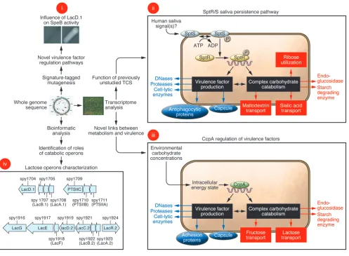
New insights into relationships between mucosal and invasive GAS dis-ease. GAS thrives at human mucosal sites and also causes devas-tating invasive infections. Strains isolated from mucosal sites are genetically indistinguishable from invasive strains by standard assays, such as M protein serotyping and multi-locus sequence typing (89, 90). However, these techniques index only a very small part of the genome, which means that they greatly underestimate the amount of genetic variation present. Using newly developed genome resequencing techniques, Sumby et al. (91) analyzed the
complete genomes of GAS isolates recovered from the spleen of mice that had been infected subcutaneously. Compared with the strain used to inoculate the mouse skin, the invasive GAS isolates (i.e., those obtained from the spleen after skin inoculation) had mutated forms of the control of virulence (CovR/S) TCS (91). Mutation of this TCS resulted in derepression of numerous viru- lence factors critical for combating the host immune system com-ponents encountered during bloodstream infection (Figure 2) (91). For example, GAS secretes a potent DNase that is involved in escape from neutrophil extracellular traps (NETs), a host immune defense mechanism generated by dying PMNs (92). The DNase is upregulated in CovR/S mutants, thereby contributing to the devel-opment of invasive disease. The clinical relevance of these findings was confirmed by the discovery that many GAS strains causing invasive infections in humans often have function-altering muta-tions in the genes that encode the components of the CovR/S TCS (91, 93). Thus, it is currently thought that GAS mucosal isolates have an intact CovR/S system that limits GAS virulence factor pro-duction, whereas interaction with the host immune system and/or deep tissues selects for strains with CovR/S mutations, leading to a hypervirulent phenotype and the serious manifestations of inva-sive GAS disease (Figure 2).
Establishment of the key role of lateral gene transfer in the emergence of unusually virulent clones
[image:5.585.50.280.77.529.2]Several types of molecular events contribute to the evolution and emergence of bacterial strains with enhanced virulence. The most well-understood and by far the most studied process is horizon-tal gene transfer (HGT), which involves bacterial viruses known as bacteriophages (transduction), plasmids (conjugation), and genomic DNA (transformation) (16, 94). HGT events create new strain genotypes by moving blocs of genetic material — sometimes large pieces of DNA that exceed 40–60 kb in size — between strains. Thus, HGT events represent a quantum evolutionary leap that can increase bacterial fitness by enhancing antimicrobial agent resistance, immune avoidance, and capacity to colonize or infect a new ecological niche. As in eukaryotes, bacterial evolution also occurs by more subtle processes, including accumulation of point mutations and small genomic changes such as those created by slipped-strand mispairing. Several of these molecular processes have contributed to the recent emergence and intercontinental dissemination of a new clone of serotype M1 GAS with distinct virulence properties (22, 95–97). This understanding was revealed by several lines of work, including comparative genome charac-terization conducted in several laboratories in a span of almost
Figure 2
20 years (22, 46, 95, 96, 98, 99). Comparative pathogenomic analy-sis resulted in two particularly important findings (22). First, low- and high-virulence serotype M1 strains differ in bacteriophage content and chromosomal integration site (22). Second, it was unexpectedly also discovered that another HGT event, involving reciprocal recombination of a 36-kb chromosomal region encod-ing the secreted toxins streptolysin O and NAD+-glycohydrolase,
was a critical evolutionary event that shaped the genome of con- temporary virulent M1 strains (22). The likely mechanism under-lying this event was generalized transduction, a process involving inadvertent packaging of a random chromosomal segment from a donor strain into a bacteriophage capsid head, followed by transfer to a new recipient bacterial strain. Importantly, contemporary vir-ulent M1 strains produce high levels of these two toxins compared with older, less virulent M1 strains, but the underlying molecular mechanism of increased expression is not yet understood.
Host susceptibility to invasive GAS disease
Like other infectious diseases, the clinical manifestations of GAS infection reflect interaction between the bacterium and the host. In contrast to situations in which infection with the pathogen is the prime determinant of disease, such as occurs for Bacillus anthra-cis or ebola virus, most humans are repeatedly exposed to and colo-nized by GAS without developing clinical symptoms. Moreover, GAS isolates that cause superficial infections such as pharyngitis and impetigo can be genetically closely related to those of the same M protein serotype that cause lethal infections such as NF and
toxic shock syndrome (90, 100). Therefore, it is highly probable that the development of severe clinical manifestations following GAS infection has a strong host susceptibility component. Most investigations of host susceptibility to severe GAS infection have focused on the role of HLA polymorphisms (101, 102). The inter- action of GAS superantigens with HLA class II molecules on anti-gen-presenting cells can result in the activation of up to 25% of all T cells at a given time, although the number of T cells activated by a particular GAS superantigen varies substantially from per-son to person (103, 104). The protective and deleterious roles of particular HLA alleles in the development of streptococcal toxic shock syndrome were observed in an epidemiologic investigation and confirmed using transgenic mice (101, 102, 105).
[image:6.585.48.539.85.374.2]In addition to focused research on HLA polymorphisms, recent studies have also begun to dissect the molecular basis of host susceptibility to serious GAS infection in mice using a genome-wide approach (106, 107). Researchers have exploited differences in susceptibility to GAS infection among strains of genetically defined mice to begin to localize protective and deleterious host genetic polymorphisms (106, 107). For example, a heightened inflammatory response to GAS was associated with worse out-comes in a mouse model of infection and linked to alterations in the expression of genes involved in apoptosis, macrophage activation, and prostaglandin synthesis (106). A genome-wide transcriptome analysis of murine macrophages also identified genes encoding proteins involved in prostaglandin synthesis as being upregulated during interaction with GAS, and the use of
Figure 3
review series
inhibitors of prostaglandin synthesis has been associated with severe GAS infection in humans (108, 109). There are significant experimental design and execution barriers to extrapolating the genome-wide approach to enhance understanding of human sus-ceptibility to GAS infections. However, the key point is that the increasing availability of tools such as genome-wide SNP analysis holds significant promise for increasing our understanding of human host determinants of GAS infection.
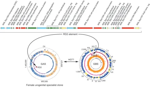
Serotype M28 and puerperal sepsis: pathogenesis consequences of a chimeric genome
Although group B Streptococcus (GBS) is a well-known cause of seri-ous neonatal or maternal infections, GAS can also be responsible for these infections. Genome sequencing and pathogenesis stud-ies have provided unexpected clues as to why serotype M28 GAS strains are repeatedly overrepresented in puerperal sepsis (child-bed fever) cases (23).
It was hypothesized (23) that analysis of the genome sequence of a serotype M28 GAS strain causing puerperal sepsis would iden- tify novel genetic elements that contributed to the overrepresenta-tion of strains of this serotype in this infection type. Analysis of the genome sequence of a serotype M28 strain has borne this out, providing the highly unexpected discovery that these GAS strains have a genome that is a chimera, composed of largely GAS genetic material onto which has been molecularly grafted a large piece of foreign DNA shared with GBS strains (23) (Figure 3). The foreign DNA is 37.4 kb in size, was acquired by HGT, and encodes seven secreted proteins that are produced in human infections (35). With very few exceptions, this genetic element is not present in other GAS strains. Importantly, one of the secreted proteins medi-ates attachment of M28 strains to human urogenital epithelium; another binds to GP340, a large human glycoprotein abundantly present in vaginal and oral secretions (35, 110). Thus, the M28 genome has been shaped by acquisition of a foreign genetic ele-ment that assisted in creating a disease-specialist GAS strain. As with other GAS molecular pathogenomics studies, these discover-ies have served to catalyze downstream pathogenesis experiments. Studies are ongoing that are designed to provide a deeper under-standing of the precise molecular processes involved in this niche adaptation and subsequent infection.
Genome-wide dissection of the molecular events underlying GAS epidemics
GAS has been used as a model system to study the molecular pro- cesses contributing to epidemics. Since 1992, more than 350 sero- type M3 strains have been recovered in a prospective population-based surveillance study of GAS invasive infections being conducted in Ontario, Canada (27, 111, 112). These strains have caused two temporally distinct epidemic waves, centered in 1995 and 2000 (19). A molecular pathogenomics approach allowed the identification of key contributors to the episodic behavior of the GAS invasive iso- lates. First, the distinct epidemics were shown to be caused by a het-erogeneous array of serotype M3 subclones, rather than recycling of a single clone (19). These distinct clones were characterized by the acquisition or loss of specific prophages that encode known GAS virulence factors. Second, host selective pressure appears to have resulted in a highly successful clone that rose to dominance in the second epidemic wave. DNA sequence analysis, coupled with immu-nologic studies, identified a four–amino acid duplication in the amino terminus of M protein in the new subclone responsible for
many of the invasive cases in the 2000 epidemic (19). This duplica-tion resulted in alterations in linear B cell epitopes, which produced substantial differences in the ability of human PMNs to phagocyto-size and kill strains with the variant M protein (19). This key finding indicated that subtle or relatively minor allelic variation may partici-pate in clone emergence and perpetuation of the epidemics.
To identify microbial genes or specific allelic variants that influ-ence the outcome of host-pathogen interactions, it is possible to use a strategy analogous to that commonly practiced when under-taking genome-wide association studies (GWASs) — using genetic methods such as high-density SNP analysis — on humans with particular disease phenotypes, such as type 2 diabetes mellitus, macular degeneration, and schizophrenia (113). In many regards, GWASs are considerably simpler in bacterial infectious disease studies because, compared with humans, prokaryotic organisms have very small genomes and isogenic mutant strains can be gener-ated and used to confirm the findings from the genetic association study. A novel bacterial GWAS conducted on strains recovered in the Ontario GAS epidemics (17) led to the discovery that a single nucleotide mutation in the gene encoding the MtsR metalloregu-latory protein implicated in uptake of iron or manganese results in a decrease in the ability of GAS to cause devastating NF. The mechanism underlying this decreased ability to cause severe dis-ease involves dysregulation of the control circuit responsible for wild-type levels of SpeB (our unpublished observations), a potent broad-spectrum protease that degrades extracellular matrix pro- teins, inactivates innate immune molecules, and destroys host tis-sue (114, 115). Inasmuch as single nucleotide mutations are the most abundant cause of genetic variation among members of the same species (13–15, 17, 18, 116), this discovery has broad implica- tions for the confluence of bacterial molecular population genom-ics and pathogenesis research.
Accelerated vaccine candidate identification and development
proteins has been a consistent finding of proteomic studies, stress-ing the need for experimental validation of predictions based on bioinformatic analyses (118, 120–123). Improved modalities for separating GAS cellular versus cell surface constituents may pro-vide additional insights into novel GAS proteins that are critical for host-pathogen interaction and are thus potential vaccine can-didates (124). In the most extensive GAS vaccine candidate study to date, Rodriguez-Ortega et al. (50) analyzed the surface-exposed proteome of GAS and identified one new antigen (SpyCEP) that conferred the ability to protect mice against lethal infection.
What does the future hold?
Enormous strides have been made in the last decade in our under-standing of GAS-host interactions, and molecular pathogenomics studies have contributed substantially. However, many gaps remain in our knowledge. Of note, many aspects of the host response dur-ing GAS infection remain largely unknown, but application of the genome-wide integrative strategies discussed above to the host side of the equation are likely to yield useful data. Analysis of patients with ARF may be particularly useful in this regard, as elucidating the host genetic factors that contribute to susceptibility may aid in the development of novel diagnostics and treatments for this devastating disease. In addition, DNA sequencing costs continue to decrease dramatically, opening the door to many types of proj- ects previously impossible due to financial constraints. For exam-ple, it is now reasonable to consider projects that involve genome sequence analysis of many hundreds or more GAS strains, similar to the 1,000–human genome project that is well underway (http:// www.1000genomes.org/). Finally, we are hopeful that the new ave-nues of basic and translational research made possible by molecular pathogenomics studies will ultimately provide strategies for ame-liorating human morbidity and mortality caused by GAS. Acknowledgments We thank K. Stockbauer for assistance with figures and members of our laboratories and anonymous reviewers for suggestions to improve the manuscript. The restricted length of the review pro-hibited us from citing all relevant work. Address correspondence to: James M. Musser, Center for Molecu-lar and Translational Human Infectious Diseases Research, The Methodist Hospital Research Institute, 6565 Fannin Street, Suite B490, Houston, Texas 77030, USA. Phone: (713) 441-5890; Fax: (713) 441-3447; E-mail: [email protected]. 1. Olsen, R.J., Shelburne, S.A., and Musser, J.M. 2009. Molecular mechanisms underlying group A strep-tococcal pathogenesis. Cell Microbiol. 11:1–12. 2. Pichichero, M.E. 1998. Group A beta-hemolytic
streptococcal infections. Pediatr. Rev. 19:291–302. 3. Carapetis, J.R., Steer, A.C., Mulholland, E.K., and
Weber, M. 2005. The global burden of group A streptococcal diseases. Lancet Infect. Dis. 5:685–694. 4. Carapetis, J.R., McDonald, M., and Wilson, N.J.
2005. Acute rheumatic fever. Lancet. 366:155–168.
5. Tanz, R.R., et al. 2004. Community-based surveil-lance in the United States of macrolide-resistant pediatric pharyngeal group A streptococci dur-ing 3 respiratory disease seasons. Clin. Infect. Dis.
39:1794–1801.
6. Robinson, D.A., Sutcliffe, J.A., Tewodros, W., Manoharan, A., and Bessen, D.E. 2006. Evolution and global dissemination of macrolide-resistant group A streptococci. Antimicrob. Agents Chemother.
50:2903–2911.
7. Martin, J.M., Green, M., Barbadora, K.A., and Wald, E.R. 2002. Erythromycin-resistant group A strep-tococci in schoolchildren in Pittsburgh. N. Engl. J. Med. 346:1200–1206.
8. Reid, S.D., Virtaneva, K., and Musser, J.M. 2003. Group A Streptococcus vaccine research: historical synopsis and new insights. In Bacterial vaccines. R.W. Ellis and B.R. Brodeur, editors. Landes Bioscience. Austin, Texas, USA. pp. 155–173. 9. Bisno, A.L., Rubin, F.A., Cleary, P.P., and Dale, J.B. 2005. Prospects for a group A streptococcal vac-cine: rationale, feasibility, and obstacles--report of a National Institute of Allergy and Infectious Dis-eases workshop. Clin. Infect. Dis. 41:1150–1156. 10. Musser, J.M., and DeLeo, F.R. 2005. Toward a
genome-wide systems biology analysis of host-pathogen interactions in group A Streptococcus. Am. J. Pathol. 167:1461–1472.
11. Whittam, T.S., and Bumbaugh, A.C. 2002. Infer-ences from whole-genome sequences of bacterial pathogens. Curr. Opin. Genet. Dev. 12:719–725. 12. Blattner, F.R., et al. 1997. The complete genome
sequence of Escherichia coli K-12. Science.
277:1453–1474.
13. Perna, N.T., et al. 2001. Genome sequence of enterohaemorrhagic Escherichia coli O157:H7.
Nature. 409:529–533.
14. Welch, R.A., et al. 2002. Extensive mosaic structure
revealed by the complete genome sequence of uro-pathogenic Escherichia coli. Proc. Natl. Acad. Sci. U. S. A.
99:17020–17024.
15. Beres, S.B., and Musser, J.M. 2007. Contribution of exogenous genetic elements to the group A Strepto-coccus metagenome. PLoS ONE. 2:e800.
16. Ochman, H., Lerat, E., and Daubin, V. 2005. Exam-ining bacterial species under the specter of gene transfer and exchange. Proc. Natl. Acad. Sci. U. S. A.
102(Suppl. 1):6595–6599.
17. Beres, S.B., et al. 2006. Molecular genetic anatomy of inter- and intraserotype variation in the human bacterial pathogen group A Streptococcus. Proc. Natl. Acad. Sci. U. S. A. 103:7059–7064.
18. Kennedy, A.D., et al. 2008. Epidemic commu-nity-associated methicillin-resistant Staphylococcus aureus: recent clonal expansion and diversification.
Proc. Natl. Acad. Sci. U. S. A. 105:1327–1332.
19. Beres, S.B., et al. 2004. Genome-wide molecular dis-section of serotype M3 group A Streptococcus strains causing two epidemics of invasive infections. Proc. Natl. Acad. Sci. U. S. A. 101:11833–11838. 20. McShan, W.M., et al. 2008. Genome sequence of a
nephritogenic and highly transformable M49 strain of Streptococcus pyogenes. J. Bacteriol. 190:7773–7785. 21. Holden, M.T., et al. 2007. Complete genome of acute
rheumatic fever-associated serotype M5 Streptococcus pyogenes strain Manfredo. J. Bacteriol. 189:1473–1477. 22. Sumby, P., et al. 2005. Evolutionary origin and
emergence of a highly successful clone of serotype M1 group A Streptococcus involved multiple horizon-tal gene transfer events. J. Infect. Dis. 192:771–782. 23. Green, N.M., et al. 2005. Genome sequence of a
serotype M28 strain of group A Streptococcus : poten-tial new insights into puerperal sepsis and bacterial disease specificity. J. Infect. Dis. 192:760–770.
24. Banks, D.J., et al. 2004. Progress toward character-ization of the group A Streptococcus metagenome: complete genome sequence of a macrolide-resis-tant serotype M6 strain. J. Infect. Dis. 190:727–738. 25. Nakagawa, I., et al. 2003. Genome sequence of an
M3 strain of Streptococcus pyogenes reveals a large-scale genomic rearrangement in invasive strains and new insights into phage evolution. Genome Res.
13:1042–1055.
26. Smoot, J.C., et al. 2002. Genome sequence and com-parative microarray analysis of serotype M18 group A Streptococcus
strains associated with acute rheu-matic fever outbreaks. Proc. Natl. Acad. Sci. U. S. A.
99:4668–4673.
27. Beres, S.B., et al. 2002. Genome sequence of a serotype M3 strain of group A Streptococcus : phage-encoded toxins, the high-virulence phenotype, and clone emergence. Proc. Natl. Acad. Sci. U. S. A.
99:10078–10083.
28. Ferretti, J.J., et al. 2001. Complete genome sequence of an M1 strain of Streptococcus pyogenes. Proc. Natl. Acad. Sci. U. S. A. 98:4658–4663.
29. Shulman, S.T., et al. 2004. Group A streptococcal pharyngitis serotype surveillance in North Amer-ica, 2000-2002. Clin. Infect. Dis. 39:325–332. 30. O’Loughlin, R.E., et al. 2007. The epidemiology of
invasive group A streptococcal infection and poten-tial vaccine implications: United States, 2000-2004.
Clin. Infect. Dis. 45:853–862.
31. Luca-Harari, B. et al. 2009. Clinical and microbio-logical characteristics of severe Streptococcus pyogenes disease in Europe. J. Clin. Microbiol. In press.
32. Abbot, E.L., et al. 2007. Pili mediate specific adhe-sion of Streptococcus pyogenes to human tonsil and skin. Cell. Microbiol. 9:1822–1833.
33. Mora, M., et al. 2005. Group A Streptococcus produce pilus-like structures containing protective antigens and Lancefield T antigens. Proc. Natl. Acad. Sci. U. S. A.
102:15641–15646.
34. Sitkiewicz, I., et al. 2006. Emergence of a bacterial clone with enhanced virulence by acquisition of a phage encoding a secreted phospholipase A2. Proc. Natl. Acad. Sci. U. S. A. 103:16009–16014. 35. Zhang, S., Green, N.M., Sitkiewicz, I., Lefebvre, R.B.,
and Musser, J.M. 2006. Identification and character-ization of an antigen I/II family protein produced by group A Streptococcus. Infect. Immun. 74:4200–4213. 36. Manetti, A.G., et al. 2007. Streptococcus pyogenes pili
promote pharyngeal cell adhesion and biofilm for-mation. Mol. Microbiol. 64:968–983.
37. Edwards, R.J., et al. 2005. Specific C-terminal cleav-age and inactivation of interleukin-8 by invasive disease isolates of Streptococcus pyogenes. J. Infect. Dis.
192:783–790.
38. Coye, L.H., and Collins, C.M. 2004. Identification of SpyA, a novel ADP-ribosyltransferase of Strepto-coccus pyogenes. Mol. Microbiol. 54:89–98.
review series
20:3046–3055.
40. von Pawel-Rammingen, U., Johansson, B.P., and Bjorck, L. 2002. IdeS, a novel streptococcal cysteine proteinase with unique specificity for immuno-globulin G. EMBO J. 21:1607–1615.
41. Rasmussen, M., Eden, A., and Bjorck, L. 2000. SclA, a novel collagen-like surface protein of Streptococcus pyogenes. Infect. Immun. 68:6370–6377.
42. Malke, H., Steiner, K., McShan, W.M., and Ferretti, J.J. 2006. Linking the nutritional status of Strepto-coccus pyogenes to alteration of transcriptional gene expression: the action of CodY and RelA. Int. J. Med. Microbiol. 296:259–275.
43. Sumby, P., et al. 2005. Extracellular deoxyribo-nuclease made by group A Streptococcus assists pathogenesis by enhancing evasion of the innate immune response. Proc. Natl. Acad. Sci. U. S. A.
102:1679–1684.
44. Rasmussen, M., and Bjorck, L. 2001. Unique regula-tion of SclB - a novel collagen-like surface protein of
Streptococcus pyogenes. Mol. Microbiol. 40:1427–1438. 45. Shelburne, S.A., 3rd, et al. 2006. Maltodextrin
utilization plays a key role in the ability of group A Streptococcus to colonize the oropharynx. Infect. Immun. 74:4605–4614.
46. Sumby, P., et al. 2008. A chemokine-degrading extracellular protease made by group A Streptococ-cus alters pathogenesis by enhancing evasion of the innate immune response. Infect. Immun. 76:978–985. 47. Hidalgo-Grass, C., et al. 2006. A streptococcal
protease that degrades CXC chemokines and impairs bacterial clearance from infected tissues.
EMBO J. 25:4628–4637.
48. Zinkernagel, A.S., et al. 2008. The IL-8 protease SpyCEP/ScpC of group A Streptococcus promotes resistance to neutrophil killing. Cell Host Microbe.
4:170–178.
49. Sjolinder, H., et al. 2008. The ScpC protease of
Streptococcus pyogenes affects the outcome of sepsis in a murine model. Infect. Immun. 76:3959–3966.
50. Rodriguez-Ortega, M.J., et al. 2006. Characteriza- tion and identification of vaccine candidate pro-teins through analysis of the group A Streptococcus
surface proteome. Nat. Biotechnol. 24:191–197. 51. Lei, B., et al. 2002. Opsonophagocytosis-inhibiting
Mac protein of group A Streptococcus: identification and characteristics of two genetic complexes. Infect. Immun. 70:6880–6890.
52. Caswell, C.C., Lukomska, E., Seo, N.S., Hook, M., and Lukomski, S. 2007. Scl1-dependent internal-ization of group A Streptococcus via direct interac-tions with the alpha2beta(1) integrin enhances pathogen survival and re-emergence. Mol. Microbiol.
64:1319–1331.
53. Proft, T., Moffatt, S.L., Berkahn, C.J., and Fraser, J.D. 1999. Identification and characterization of novel superantigens from Streptococcus pyogenes.
J. Exp. Med. 189:89–102.
54. Unnikrishnan, M., et al. 2002. The bacterial supe-rantigen streptococcal mitogenic exotoxin Z is the major immunoactive agent of Streptococcus pyogenes.
J. Immunol. 169:2561–2569.
55. Proft, T., Webb, P.D., Handley, V., and Fraser, J.D. 2003. Two novel superantigens found in both group A and group C Streptococcus. Infect. Immun.
71:1361–1369.
56. Kreikemeyer, B., McIver, K.S., and Podbielski, A. 2003. Virulence factor regulation and regulatory networks in Streptococcus pyogenes and their impact on pathogen-host interactions. Trends Microbiol.
11:224–232.
57. Levin, J.C., and Wessels, M.R. 1998. Identification of csrR/csrS , a genetic locus that regulates hyaluron-ic acid capsule synthesis in group A Streptococcus.
Mol. Microbiol. 30:209–219.
58. Dalton, T.L., Collins, J.T., Barnett, T.C., and Scott, J.R. 2006. RscA, a member of the MDR1 family of transporters, is repressed by CovR and required for
growth of Streptococcus pyogenes under heat stress.
J. Bacteriol. 188:77–85.
59. Graham, M.R., et al. 2002. Virulence control in group A Streptococcus by a two-component gene reg-ulatory system: global expression profiling and in vivo infection modeling. Proc. Natl. Acad. Sci. U. S. A.
99:13855–13860.
60. Voyich, J.M., et al. 2004. Engagement of the patho-gen survival response used by group A Streptococ-cus to avert destruction by innate host defense.
J. Immunol. 173:1194–1201.
61. Ribardo, D.A., Lambert, T.J., and McIver, K.S. 2004. Role of Streptococcus pyogenes two-component response regulators in the temporal control of Mga and the Mga-regulated virulence gene emm. Infect. Immun. 72:3668–3673.
62. Sitkiewicz, I., and Musser, J.M. 2006. Expression microarray and mouse virulence analysis of four con-served two-component gene regulatory systems in group A Streptococcus. Infect. Immun. 74:1339–1351. 63. Chaussee, M.S., et al. 2002. Rgg influences the
expression of multiple regulatory loci to coregulate virulence factor expression in Streptococcus pyogenes.
Infect. Immun. 70:762–770.
64. Kreikemeyer, B., et al. 2007. The Streptococcus pyo-genes serotype M49 Nra-Ralp3 transcriptional regu-latory network and its control of virulence factor expression from the novel eno ralp3 epf sagA patho-genicity region. Infect. Immun. 75:5698–5710.
65. Gryllos, I., et al. 2008. PerR confers phagocytic kill-ing resistance and allows pharyngeal colonization by group A Streptococcus. PLoS Pathog. 4:e1000145.
66. Kinkel, T.L., and McIver, K.S. 2008. CcpA-medi-ated repression of streptolysin S expression and virulence in the group A Streptococcus. Infect. Immun.
76:3451–3463.
67. Roberts, S.A., and Scott, J.R. 2007. RivR and the small RNA RivX: the missing links between the CovR regulatory cascade and the Mga regulon. Mol. Microbiol. 66:1506–1522.
68. Ribardo, D.A., and McIver, K.S. 2006. Defining the Mga regulon: Comparative transcriptome analysis reveals both direct and indirect regulation by Mga in the group A Streptococcus. Mol. Microbiol. 62:491–508. 69. Ryan, P.A., Kirk, B.W., Euler, C.W., Schuch, R., and
Fischetti, V.A. 2007. Novel algorithms reveal strep-tococcal transcriptomes and clues about undefined genes. PLoS Comput. Biol. 3:e132.
70. Loughman, J.A., and Caparon, M. 2006. Regulation of SpeB in Streptococcus pyogenes by pH and NaCl: a model for in vivo gene expression. J. Bacteriol.
188:399–408.
71. Chaussee, M.A., Dmitriev, A.V., Callegari, E.A., and Chaussee, M.S. 2008. Growth phase-associated changes in the transcriptome and proteome of
Streptococcus pyogenes. Arch. Microbiol. 189:27–41. 72. Beyer-Sehlmeyer, G., Kreikemeyer, B., Horster, A.,
and Podbielski, A. 2005. Analysis of the growth phase-associated transcriptome of Streptococcus pyogenes. Int. J. Med. Microbiol. 295:161–177. 73. Voyich, J.M., et al. 2003. Genome-wide protective
response used by group A Streptococcus to evade destruction by human polymorphonuclear leuko-cytes. Proc. Natl. Acad. Sci. U. S. A. 100:1996–2001. 74. Shelburne, S.A., 3rd, et al. 2008. A direct link
between carbohydrate utilization and virulence in the major human pathogen group A Streptococcus.
Proc. Natl. Acad. Sci. U. S. A. 105:1698–1703. 75. Virtaneva, K., et al. 2005. Longitudinal analysis of
the group A Streptococcus transcriptome in experi-mental pharyngitis in cynomolgus macaques. Proc. Natl. Acad. Sci. U. S. A. 102:9014–9019.
76. Graham, M.R., et al. 2006. Analysis of the transcrip-tome of group A Streptococcus in mouse soft tissue infection. Am. J. Pathol. 169:927–942.
77. Shelburne, S.A., 3rd, et al. 2005. Central role of a bacterial two-component gene regulatory system of
previously unknown function in pathogen persis-tence in human saliva. Proc. Natl. Acad. Sci. U. S. A.
102: 16037–16042.
78. Graham, M.R., et al. 2005. Group A Streptococcus
transcriptome dynamics during growth in human blood reveals bacterial adaptive and survival strate-gies. Am. J. Pathol. 166:455–465.
79. Loughman, J.A., and Caparon, M.G. 2006. A novel adaptation of aldolase regulates virulence in Strep-tococcus pyogenes. EMBO J. 25:5414–5422.
80. Loughman, J.A., and Caparon, M.G. 2007. Com-parative functional analysis of the lac operons in
Streptococcus pyogenes. Mol. Microbiol. 64:269–280. 81. Hanks, T.S., et al. 2006. Differential regulation of
iron- and manganese-specific MtsABC and heme- specific HtsABC transporters by the metalloregu-lator MtsR of group A Streptococcus. Infect. Immun.
74:5132–5139.
82. Ricci, S., Janulczyk, R., and Bjorck, L. 2002. The regulator PerR is involved in oxidative stress response and iron homeostasis and is necessary for full virulence of Streptococcus pyogenes. Infect. Immun.
70:4968–4976.
83. Brenot, A., King, K.Y., and Caparon, M.G. 2005. The PerR regulon in peroxide resistance and virulence of Streptococcus pyogenes. Mol. Microbiol. 55:221–234. 84. Peter, G., and Smith, A.L. 1977. Group A strepto-coccal infections of the skin and pharynx (first of two parts). N. Engl. J. Med. 297:311–317.
85. Bessen, D.E., and Kalia, A. 2002. Genomic localiza-tion of a T serotype locus to a recombinatorial zone encoding extracellular matrix-binding proteins in
Streptococcus pyogenes. Infect. Immun. 70:1159–1167. 86. Bessen, D.E., Sotir, C.M., Readdy, T.L., and Hol-lingshead, S.K. 1996. Genetic correlates of throat and skin isolates of group A streptococci. J. Infect. Dis. 173:896–900.
87. Kratovac, Z., Manoharan, A., Luo, F., Lizano, S., and Bessen, D.E. 2007. Population genetics and linkage analysis of loci within the FCT region of Streptococ-cus pyogenes. J. Bacteriol. 189:1299–1310.
88. Hung, C.S., et al. 2002. Structural basis of tropism of Escherichia coli to the bladder during urinary tract infection. Mol. Microbiol. 44:903–915.
89. Haukness, H.A., et al. 2002. The heterogeneity of endemic community pediatric group A streptococ-cal pharyngeal isolates and their relationship to invasive isolates. J. Infect. Dis. 185:915–920. 90. Hoe, N.P., et al. 2001. Distribution of streptococcal
inhibitor of complement variants in pharyngitis and invasive isolates in an epidemic of serotype M1 group A Streptococcus infection. J. Infect. Dis.
183:633–639.
91. Sumby, P., Whitney, A.R., Graviss, E.A., DeLeo, F.R., and Musser, J.M. 2006. Genome-wide analysis of group a streptococci reveals a mutation that modu-lates global phenotype and disease specificity. PLoS Pathog. 2:e5.
92. Walker, M.J., et al. 2007. DNase Sda1 provides selec- tion pressure for a switch to invasive group A strep-tococcal infection. Nat. Med. 13:981–985. 93. Engleberg, N.C., Heath, A., Miller, A., Rivera, C.,
and DiRita, V.J. 2001. Spontaneous mutations in the CsrRS two-component regulatory system of
Streptococcus pyogenes result in enhanced virulence in a murine model of skin and soft tissue infection.
J. Infect. Dis. 183:1043–1054.
94. Daubin, V., Moran, N.A., and Ochman, H. 2003. Phylogenetics and the cohesion of bacterial genomes. Science. 301:829–832.
95. Musser, J.M., et al. 1991. Streptococcus pyogenes causing toxic-shock-like syndrome and other invasive diseas- es: clonal diversity and pyrogenic exotoxin expres-sion. Proc. Natl. Acad. Sci. U. S. A. 88:2668–2672. 96. Musser, J.M., et al. 1995. Genetic diversity and
97. Musser, J.M., et al. 1993. Geographic and temporal distribution and molecular characterization of two highly pathogenic clones of Streptococcus pyogenes expressing allelic variants of pyrogenic exotoxin A (scarlet fever toxin). J. Infect. Dis. 167:337–346.
98. Aziz, R.K., et al. 2005. Mosaic prophages with hori-zontally acquired genes account for the emergence and diversification of the globally disseminated M1T1 clone of Streptococcus pyogenes. J. Bacteriol.
187:3311–3318.
99. Cleary, P.P., et al. 1992. Clonal basis for resurgence of serious Streptococcus pyogenes disease in the 1980s.
Lancet. 339:518–521.
100. Chatellier, S., et al. 2000. Genetic relatedness and superantigen expression in group A Streptococcus sero- type M1 isolates from patients with severe and non-severe invasive diseases. Infect. Immun. 68:3523–3534. 101. Norrby-Teglund, A., Nepom, G.T., and Kotb, M.
2002. Differential presentation of group A strep-tococcal superantigens by HLA class II DQ and DR alleles. Eur. J. Immunol. 32:2570–2577.
102. Kotb, M., et al. 2002. An immunogenetic and molecular basis for differences in outcomes of invasive group A streptococcal infections. Nat. Med.
8:1398–1404.
103. Sundberg, E., and Jardetzky, T.S. 1999. Structural basis for HLA-DQ binding by the streptococcal superantigen SSA. Nat. Struct. Biol. 6:123–129. 104. Norrby-Teglund, A., et al. 2000. Host variation in
cytokine responses to superantigens determine the severity of invasive group A streptococcal infection.
Eur. J. Immunol. 30:3247–3255.
105. Nooh, M.M., El-Gengehi, N., Kansal, R., David, C.S., and Kotb, M. 2007. HLA transgenic mice provide evidence for a direct and dominant role of HLA class II variation in modulating the severity of streptococcal sepsis. J. Immunol. 178:3076–3083. 106. Abdeltawab, N.F., et al. 2008. An unbiased systems
genetics approach to mapping genetic loci modu-lating susceptibility to severe streptococcal sepsis.
PLoS Pathog. 4:e1000042.
107. Aziz, R.K., et al. 2007. Susceptibility to severe strep-tococcal sepsis: use of a large set of isogenic mouse lines to study genetic and environmental factors.
Genes Immun. 8:404–415.
108. Goldmann, O., et al. 2007. Transcriptome analysis of murine macrophages in response to infection with Streptococcus pyogenes reveals an unusual acti-vation program. Infect. Immun. 75:4148–4157. 109. Hamilton, S.M., Bayer, C.R., Stevens, D.L., Lieber,
R.L., and Bryant, A.E. 2008. Muscle injury, vimen-tin expression, and nonsteroidal anti-inflammatory drugs predispose to cryptic group A streptococcal necrotizing infection. J. Infect. Dis. 198:1692–1698. 110. Stalhammar-Carlemalm, M., Areschoug, T., Lars-son, C., and Lindahl, G. 1999. The R28 protein of
Streptococcus pyogenes is related to several group B streptococcal surface proteins, confers protective immunity and promotes binding to human epi-thelial cells. Mol. Microbiol. 33:208–219.
111. Sharkawy, A., et al. 2002. Severe group A strepto-coccal soft-tissue infections in Ontario: 1992–1996.
Clin. Infect. Dis. 34:454–460.
112. Davies, H.D., et al. 1996. Invasive group A strep-tococcal infections in Ontario, Canada. Ontario Group A Streptococcal Study Group. N. Engl. J. Med. 335:547–554.
113. McCarthy, M.I., et al. 2008. Genome-wide associa- tion studies for complex traits: consensus, uncer-tainty and challenges. Nat. Rev. Genet. 9:356–369.
114. Svensson, M.D., et al. 2000. Role for a secreted cys- teine proteinase in the establishment of host tis-sue tropism by group A streptococci. Mol. Microbiol.
38:242–253.
115. Lukomski, S., et al. 1998. Genetic inactivation of an extracellular cysteine protease (SpeB) expressed by
Streptococcus pyogenes decreases resistance to phago-cytosis and dissemination to organs. Infect. Immun.
66:771–776.
116. Jakobsson, M., et al. 2008. Genotype, haplotype and copy-number variation in worldwide human popu-lations. Nature. 451:998–1003.
117. Rinaudo, C.D., Telford, J.L., Rappuoli, R., and Seib, K.L. 2009. Vaccinology in the genome era. J. Clin. Invest. 119:2515–2525.
118. Lei, B., Mackie, S., Lukomski, S., and Musser, J.M. 2000. Identification and immunogenicity of group A Streptococcus culture supernatant proteins. Infect. Immun. 68:6807–6818.
119. Lei, B., Liu, M., Chesney, G.L., and Musser, J.M. 2004. Identification of new candidate vaccine anti-gens made by Streptococcus pyogenes: purification and characterization of 16 putative extracellular lipoproteins. J. Infect. Dis. 189:79–89.
120. Cole, J.N., et al. 2005. Surface analyses and immune reactivities of major cell wall-associated proteins of group A Streptococcus. Infect. Immun. 73:3137–3146. 121. Severin, A., et al. 2007. Proteomic analysis and iden-tification of Streptococcus pyogenes surface-associated proteins. J. Bacteriol. 189:1514–1522.
122. Nakamura, T., et al. 2004. Two-dimensional gel electrophoresis analysis of the abundance of viru-lent exoproteins of group A Streptococcus caused by environmental changes. Arch. Microbiol. 181:74–81. 123. Zhang, M., et al. 2007. Group A Streptococcus cell-associated pathogenic proteins as revealed by growth in hyaluronic acid-enriched media. Pro-teomics. 7:1379–1390.
124. Koller, T., et al. 2008. PlyC, a novel bacteriophage lysin for compartment-dependent proteomics of group A streptococci. Proteomics. 8:140–148.