Peptone stimulates CCK-releasing peptide
secretion by activating intestinal submucosal
cholinergic neurons.
Y Li, C Owyang
J Clin Invest.
1996;
97(6)
:1463-1470.
https://doi.org/10.1172/JCI118568
.
In this study we tested the hypothesis that peptone in the intestine stimulates the secretion
of the CCK-releasing peptide (CCK-RP) which mediates CCK secretion, and examined the
enteric neural circuitry responsible for CCK-RP secretion. We used a "donor-recipient" rat
intestinal perfusion model to quantify the CCK-RP secreted in response to nutrient
stimulation. Infusion of concentrated intestinal perfusate collected from donor rat perfused
with 5% peptone caused a 62 +/- 10% increase in protein secretion and an elevation of
plasma CCK levels to 6.9 +/- 1.8 pM in the recipient rat. The stimulatory effect of the
intestinal washings was abolished when the donor rats were pretreated with atropine or
hexamethonium but not with guanethidine or vagotomy. Mucosal application of lidocaine
but not serosal application of benzalkonium chloride which ablates the myenteric neurons in
the donor rats also abolished the stimulatory action of the intestinal washings. Furthermore,
treatment of the donor rats with a 5HT3 antagonist and a substance P antagonist also
prevented the secretion of CCK-RP. These observations suggest that peptone in the
duodenum stimulates serotonin release which activates the sensory substance P neurons
in the submucous plexus. Signals are then transmitted to cholinergic interneurons and to
epithelial CCK-RP containing cells via cholinergic secretomotor neurons. This enteric
neural circuitry which is responsible for the secretion of CCK-RP may in turn play […]
Research Article
Find the latest version:
http://jci.me/118568/pdf
J. Clin. Invest.
© The American Society for Clinical Investigation, Inc. 0021-9738/96/03/1463/08 $2.00
Volume 97, Number 6, March 1996, 1463–1470
Peptone Stimulates CCK-releasing Peptide Secretion by Activating Intestinal
Submucosal Cholinergic Neurons
Ying Li and Chung Owyang
The University of Michigan Medical Center, Department of Internal Medicine, Ann Arbor, Michigan 48109
Abstract
In this study we tested the hypothesis that peptone in the in-testine stimulates the secretion of the CCK-releasing pep-tide (CCK-RP) which mediates CCK secretion, and exam-ined the enteric neural circuitry responsible for CCK-RP secretion. We used a “donor-recipient” rat intestinal perfu-sion model to quantify the CCK-RP secreted in response to nutrient stimulation. Infusion of concentrated intestinal perfusate collected from donor rat perfused with 5% pep-tone caused a 62610% increase in protein secretion and an elevation of plasma CCK levels to 6.961.8 pM in the recipi-ent rat. The stimulatory effect of the intestinal washings was abolished when the donor rats were pretreated with at-ropine or hexamethonium but not with guanethidine or vag-otomy. Mucosal application of lidocaine but not serosal application of benzalkonium chloride which ablates the my-enteric neurons in the donor rats also abolished the stimula-tory action of the intestinal washings. Furthermore,
treat-ment of the donor rats with a 5HT3 antagonist and a
substance P antagonist also prevented the secretion of CCK-RP. These observations suggest that peptone in the duodenum stimulates serotonin release which activates the sensory substance P neurons in the submucous plexus. Sig-nals are then transmitted to cholinergic interneurons and to epithelial CCK-RP containing cells via cholinergic secreto-motor neurons. This enteric neural circuitry which is re-sponsible for the secretion of CCK-RP may in turn play an important role in the postprandial release of CCK. (J. Clin. Invest. 1996. 97:1463–1470.) Key words:substance P•
sero-tonin • enteric nervous system • cholecystokinin • cholecys-tokinin-releasing peptide
Introduction
Cholecystokinin (CCK)1 plays an important role in the
media-tion of postprandial pancreatic secremedia-tion and gallbladder con-traction. In the rat, protein is the major dietary intestinal stim-ulus for CCK release (1). Little, however, is known about the mechanisms regulating CCK secretion.
Recently it was reported that the release of CCK into the circulation is mediated by a “CCK-releasing peptide” (CCK-RP) secreted into the intestine of the rat (2, 3). When trypsin is present, this peptide is cleaved and inactivated. It has been proposed that this newly discovered CCK-RP may also act as a mediator of pancreatic enzyme secretion in response to dietary protein (4). Dietary protein in the intestine competes for trypsin (1), which would otherwise inactivate the CCK-RP. The resulting increase of active CCK-RP in the intestine re-leases CCK and stimulates pancreatic enzyme secretion. On the other hand, if a dietary protein such as bovine serum albu-min or lactalbualbu-min is a poor substrate for pancreatic proteases, it will not prevent proteolytic inactivation of the CCK-RP and therefore will not evoke an increase in CCK secretion (1). Hence the ability of intact protein to stimulate CCK release and pancreatic secretion has been attributed to its ability to bind with pancreatic proteases and reverse protease-induced feedback inhibition of CCK release. In a recent study, how-ever (5) it was reported that peptic digests of lactalbumin not native lactalbumin were potent stimuli of pancreatic secretion. It is conceivable that digests of protein in the intestine may stimulate the secretion of CCK-RP. To test this hypothesis we used a “donor-recipient” rat intestinal perfusion model that has been previously used to identify the CCK-RP (2) and ex-amined the ability of peptone, a pancreatic protease digest of protein in the intestine, to stimulate the secretion of CCK-RP. Furthermore, we examined the enteric neural circuitry mediat-ing CCK-RP secretion. The relative importance of extrinsic versus intrinsic neural pathways were studied and the location of the local neural plexus identified. In addition, we delineated the individual components of the enteric neural circuitry which mediates CCK-RP secretion.
Methods
Materials
The following were purchased from Sigma Chemical Co. (St. Louis, MO): Peptone, maltose, l-amino acids, atropine sulfate, hexametho-nium and benzalkohexametho-nium chloride.
Animal preparation
Male Sprague-Dawley rats, weighing between 250 and 300 grams, were fasted overnight and anesthetized with a mixture of xylazine and ketamine (13 and 87 mg/kg body wt, respectively). Supplemental doses were used every 2 h as needed to maintain adequate anesthesia. Through a midline incision, a polyethylene catheter was inserted into the common bile pancreatic duct at the ampulla. A second catheter was placed in the duodenum, slightly above the ampulla for the intes-tinal perfusion of bile pancreatic juice. The abdominal wound was covered with a saline gauge and the animal body temperature was monitored by a rectal thermal probe and maintained at 378C with a heating pad. After 60 min of stabilization, combined bile and pancre-atic secretions were collected every 15 min. The volume was mea-sured, and an aliquot was taken and diluted with distilled water for protein determination. The remainder of the undiluted bile pancre-Address correspondence to Chung Owyang, M.D., The University of
Michigan Medical Center, 3912 Taubman Center, Box 0362, Ann Ar-bor, MI 48109-0362. Phone: 313-936-4785; FAX: 313-036-7392.
Received for publication 4 August 1995 and accepted in revised form 12 December 1995.
atic juice was returned to the intestine via the duodenal cannula dur-ing the next collection period. Bile pancreatic juice protein was mea-sured spectrophotometrically using the assay method of Bradford (6). Our previous study (7) has confirmed that the increase in protein out-put in the bile pancreatic juice following CCK-8 stimulation mainly reflected protein from the pancreatic source. Biliary juice protein did not increase with CCK stimulation.
Experimental protocols
Effect of intraluminal nutrients on CCK-RP secretion after 5-h diver-sion of BPJ.To investigate the effect of intraluminal peptone on CCK-RP secretion, we utilized the donor-recipient rat model de-scribed previously (2). The common bile-pancreatic duct was lated at the ampulla for diversion of bile pancreatic juice. Two cannu-las were placed into the small bowel: one in the proximal duodenum for the infusion of saline or nutrient solutions and the other in the je-junum 20 cm distal to the ligament of Tretz for collection of intestinal perfusates. We have shown previously that diversion of bile pancre-atic juice from the intestine resulted in a marked increase in plasma CCK levels and pancreatic secretion. These increases gradually re-turned to basal after 5 h of diversion due to a reduction in the luminal CCK-RP (8). This period is optimal to test the ability of peptone or other nutrients to stimulate the secretion of CCK-RP. After a 5-h di-version of bile pancreatic juice, donor rats received an intraduodenal perfusion of phosphate-buffered saline, 5% peptone (mol wt , 1000, pH 6.0, osmolarity 300 mOsm/liter), 18% casein, maltose (300 mM pH 6.0, osmolarity 300 mOsm/liter) or 5% l-amino acids (pH 6.0, os-molarity 300 mOsm/liter) at the rate of 3 ml/min for 3 h. The 5% pep-tone test solution was prepared by ultrafiltration using an Amicon membrane (YM1) and the filtrate which only contained peptides with mol wt , 1000 was used as a test solution to infuse into the donor rat. The intestinal perfusate collected from the donor rat was filtered with Whatman No. 54 paper to remove any large particles such as intesti-nal debris. The filtrate was then ultrafiltered with an Amicon mem-brane (YM1) and the residue on the Amicon memmem-brane which con-tained peptides with mol wt . 1000 was washed with 250 ml of water until the filtrate gave a negative ninhydrin test (z 5 washes) indicat-ing that all the peptones had been removed from the residue. The res-idue which contained the CCK-RP (mol wt . 1000) was then dis-solved in water and lyophilized. The latter was then suspended in phosphate buffered saline and adjusted to pH 7.4 for intraduodenal administration in the recipient rats. The recipient rats received con-centrated intestinal perfusate (1 ml) collected from donor rats and in-fused over 2 min at 5 h after bile pancreatic juice diversion when the plasma CCK has returned to basal level. Pancreatic protein output and plasma CCK levels were monitored every 10 min after the ad-ministration of concentrated intestinal perfusate in the recipient rats. The response of pancreatic secretion and plasma CCK levels in the recipient rats following the administration of intestinal perfusates col-lected from the donor rats served as a surrogate for the quantification of CCK-RP secreted in response to nutrient stimulation.
Effects of atropine, hexamethonium, guanethidine, and acute va-gotomy on intraduodenal peptone-stimulated CCK-RP secretion.To de-termine the role of cholinergic pathways in the mediation of peptone induced CCK-RP secretion, atropine (100 mg/kg per hour) was ad-ministered intravenously to the donor rats 30 min before intestinal perfusion of 5% peptone as described earlier. Similar studies were performed with hexamethonium treatment in the donor rats (15 mg/ kg bolus plus 7.5 mg/kg per hour continuous infusion) to determine the role of presynaptic cholinergic neurons in the mediation of pep-tone-stimulated CCK-RP secretion. To evaluate the role of adrener-gic pathways, a separate group of donor rats received two daily injec-tions of guanethidine i.p. at a dose of 20 mg/kg before peptone administration. This dosage regimen has previously been shown to deplete cardiac norepinephrine by 90% (9). In separate studies, we evaluated the role of the vagal nerve in mediating peptone-induced CCK-RP secretion. After a 5-h diversion of bile pancreatic juice,
acute bilateral subdiaphragmatic vagotomy was performed in donor rats. Through a midline incision of the abdominal wall, the stomach was carefully manipulated to expose the esophagus. The subphragmatic vagal trunks were exposed halfway between the dia-phragm and the gastric cardia. Both anterior and posterior trunks of the vagal nerves were transected. For control experiments, the ab-dominal vagal nerves were exposed but not cut. Intraluminal peptone perfusion studies were performed following acute vagotomy. Pancatic amylase output and plasma CCK levels were monitored in the re-cipient rats following the same protocol as described earlier.
Effects of mucosal anesthestic and serosal application of benzalko-nium choloride (BAC) on peptone-stimulated CCK-RP secretion.
The enteric nervous system is composed of the myenteric and submu-cous plexuses. To investigate if the submusubmu-cous plexus is involved in the mediation of CCK-RP secretion, 1% lidocaine was used as a topi-cal anesthetic (2 ml/kg bolus plus 0.2 ml/min for 5 min at hourly inter-vals intraduodenally) ten minutes before the intraduodenal perfusion of 5% peptone. This method has previously been shown to inhibit the increase in superior mesenteric artery blood flow evoked by in-traduodenal perfusion of 0.1N HCl (10). To investigate if myeteric neurons are important in mediating peptone-stimulated CCK-RP se-cretion, myenteric neurons were ablated with the surfactant benzal-konium chloride (BAC) applied to the serosa of the intestine every 5 min for 30 min (six applications) in the donor rats 15 d before the experiment (11). Serosal application of BAC has been shown to de-stroy more than 90% of the ganglia in the myenteric plexus, without affecting the submucous plexus (12). In control rats, 0.9% saline was applied in a similar manner. After 15 d, intestinal perfusion studies with 5% peptone were performed. Pancreatic protein output and plasma CCK levels of the recipient rats in response to infusion of in-testinal perfusate collected from the donor rats were monitored fol-lowing the same protocol as described earlier.
Effects of 5HT antagonist and substance P antagonist on peptone-stimulated CCK-RP secretion. To delineate the local neural reflex in the submucous plexus which mediates CCK-RP secretion, we exam-ined the effect of a 5HT3 receptor antagonist, ICS 205–930 which has
been shown to prevent 5HT-induced vagal responses (13). ICS 205– 930 (200 mg/kg iv) was administered to donor rats 30 min before the intraduodenal infusion of 5% peptone. Similar studies were per-formed with donor rats pretreated with the 5HT receptor antagonists such as 5HTP-DP-acetyl (1 mg/kg IV, 5HT1p antagonist), and
ket-anserin (200 mg/kg IV, 5HT2 antagonist). This will test the hypothesis
that peptone in the intestine releases serotonin which in turn sets up a local reflex cascade to stimulate CCK-RP secretion from intestinal epithelial cells.
In separate studies we examined the effect of a highly specific, nonpeptide sub P antagonist, CP-96,345 (14). It has been previously shown that the administration of CP-96,345 markedly reduces the vascular inflammatory response to an allograft (15) and reduces toxin-A-induced secretion and mucosal permeability mediated by sub P (16). The sub P antagonist CP-96,345 (2.5 mg/kg i.p.) or its inactive enantiomer CP-96,344 was given to the donor rats 1 h before in-traduodenal administration of 5% peptone.
Bioassay of plasma CCK. Plasma CCK was extracted and bioas-sayed as previously described (17). Plasma CCK was absorbed onto C18 Sep-Pak cartridges (Waters Division of Millipore, Millford, MA), washed, and the CCK eluted with 1 ml acetonitrile/water (1:1) into a polyethylene scintillation vial and dried in a 458C water bath under a flow of nitrogen. Previous studies involving the addition of CCK-8 and CCK-33 to the C18 Sep-Pak cartridges showed recoveries of 8766% and 8465%, respectively (18).
Statistical analysis. Results were expressed as mean6SE. The multivariant analysis of variance method was used to evaluate the ef-fects of the repeated measurement over time, treatment efef-fects, and the interaction between them. Significance was accepted at the 5% level.
Results
Effects of intraluminal nutrients on CCK-RP secretion.To inves-tigate the effects of intraluminal nutrients on the secretion of CCK-RP, we used the donor-recipient rat model described previously (2). The recipient rats were used as test rats for the measurement of CCK-RP.
5 h after diversion of bile pancreatic juice, the pancreatic protein output has returned to basal levels and was stable, av-eraging 12067 mg/h. The basal plasma CCK was 0.560.1 pM.
Intraduodenal infusion of 5% peptone caused a significant increase in pancreatic protein output accompanied by an in-crease in plasma CCK levels to 7.161.3 pM (Fig. 1). In con-trast intraduodenal administration of 18% casein failed to increase pancreatic secretion and plasma CCK levels (Fig. 1).
In separate studies, infusion of concentrated intestinal per-fusate (1 ml) collected from the donor rat during the fifth to the eighth hour after diversion resulted in no significant in-crease in pancreatic protein output or plasma CCK levels. This indicates that the concentrated intestinal perfusate collected from the donor rat contained no significant amounts of CCK-RP. In contrast, infusion of concentrated intestinal perfusate (1 ml) collected from the donor rat when it was perfused with
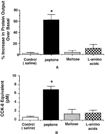
5% peptone caused a 62610% increase in protein secretion
(Fig. 2 A) and an elevation of plasma CCK levels to 6.961.8 pM (Fig. 2 B). Note that the peptone (mol wt , 1000) in the intestinal perfusate collected from the donor rat was removed by ultrafiltration which was confirmed by a negative ninhydrin staining.
In separate studies, we showed that intraduodenal infusion of concentrated perfusate collected from donor rats which were perfused with maltose (300 mM) or 5% l-amino acids did
not significantly increase pancreatic protein secretion or plasma CCK levels (Fig. 2, A and B). These observations indicate that
peptone but neither maltose nor l-amino acids stimulated
CCK-RP secretion.
Effects of vagotomy, atropine, guanethidine and hexametho-nium. To investigate the mechanisms by which peptone stimu-lates CCK-RP secretion, acute bilateral subdiaphragmatic vag-otomy was performed in the donor rats 30 min before
intraduodenal perfusion of 5% peptone. The administration of concentrated perfusate collected from the acute vagotomized donor rats caused a 64%612 increase in pancreatic protein output over basal and elevated plasma CCK levels to 7.361.1 pM (Fig. 3, A and B). These values were similar to those ob-served in control experiments indicating that the vagus nerves do not play a role in the mediation of CCK-RP secretion. Sim-ilarly, administration of concentrated perfusate collected from donor rats pretreated with guanethidine increased plasma CCK levels to 6.460.7 pM and caused a 60%612 increase in protein output over basal in the recipient rats (Fig. 3, A and B). In contrast, administration of concentrated perfusates col-lected from donor rats pretreated with atropine or hexametho-nium failed to significantly increase pancreatic amylase secre-tion or plasma CCK levels in the recipient rats (Fig. 3, A and B). These observations indicate that peptone acts at a
[image:4.612.58.417.60.205.2]presyn-Figure 1. Pancreatic protein output and plasma CCK levels in rats perfused with casein or peptone solutions between the fifth and eighth hour post diversion of bile pancreatic juice. Values are mean6SE for six rats in each group. *P, 0.01.
[image:4.612.315.496.437.668.2]aptic site of the extravagal cholinergic pathway (enteric ner-vous system) to stimulate CCK-RP secretion.
Effects of mucosal anesthetic and serosal application of ben-zalkonium chloride (BAC).Administration of concentrated peptone perfusate collected from donor rats pretreated with local anesthetic (lidocaine) failed to increase the plasma CCK levels and pancreatic protein secretion in recipient rats (Fig. 4, A and B). Thus topical application of lidocaine prevented CCK-RP secretion stimulated by 5% peptone. This indicates that a local enteric neural circuitry is involved in mediating the secretion of CCK-RP. In contrast to the observation made with mucosal application of lidocaine, intraduodenal adminis-tration of concentrated perfusate collected from donor rats pretreated with serosal benzalkonium chloride which ablates myenteric neurons resulted in a 64612% increase in protein secretion and elevated plasma CCK level to 6.060.5 pM (Fig. 4, A and B). These values were similar to levels observed in control experiments. Therefore it appears that the submucous neural plexus but not the myenteric plexus is involved in the mediation of peptone stimulated CCK-RP secretion.
Effects of 5HT and substance P antagonists.To delineate the local neural reflex in the submucous plexus which mediates CCK-RP secretion, we examined the effects of 5HT and sub-stance P antagonists since both 5HT and subsub-stance P are found in abundance in the enterochromaffin cells and the enteric neural plexuses. Intraduodenal administration of concentrated perfusates collected from donor rats pretreated with ket-anserin (5HT2 antagonist) or ICS 205-930 (5HT3 antagonist)
resulted in partial or no increase in pancreatic protein output and plasma CCK levels in the recipient rats (Fig. 5, A and B).
On the other hand, infusion of intestinal perfusates obtained from donor rats pretreated with 5HTP-DP-acetyl (5HT1p
an-tagonist) resulted in normal increases in pancreatic protein se-cretion and plasma CCK levels in the recipient rats (Fig. 5, A and B). This suggests that 5HT3 and possibly 5HT2, but not
5HT1p receptors are involved in the mediation of CCK-RP
se-cretion.
In separate studies, we showed that intestinal perfusates collected from donor rats pretreated with the substance P an-tagonist 96,345) but not its inactive enantiomer (CP-96,344), when infused into the recipient rats resulted in no in-crease in pancreatic protein secretion (Fig. 6). This suggests that substance P containing neurons are involved in the local neural reflex mediating the secretion of CCK-RP.
Discussion
It has been previously demonstrated that protein is the major dietary factor stimulating CCK release in the rats (1), although the responsible mechanism(s) remains to be determined. Re-cent studies suggest that protein does not act directly on CCK containing cells (19) but is likely to involve some intermediary luminal factor(s). In species such as rats where feedback inhi-bition of pancreatic secretion occurs, CCK release appears to be controlled by active intraluminal proteases (4). Since pro-teins may bind and thus inhibit intraluminal endopeptidases, it has been proposed that dietary protein in the intestine by
[image:5.612.317.494.57.329.2]com-Figure 3. Pancreatic protein secretion (A) and plasma CCK levels (B) in the recipient rats in response to intraduodenal administration of concentrated intestinal perfusates obtained from donor rats per-fused with 5% peptone solution. Donor rats were pretreated with acute vagotomy, atropine (100 mg/kg per hour), guanthidine (5 mg/ kg) or hexamethonium (15 mg/kg bolus plus 7.5 mg/kg per hour iv in-fusion) before peptone infusion. Values are mean6SE for eight rats in each group. *P, 0.01.
[image:5.612.58.238.59.295.2]peting for trypsin which would otherwise inactivate the CCK-RP, causes an increase in luminal CCK-RP which, in turn, re-leases CCK and stimulates pancreatic secretion (4). This rep-resents the major mechanism by which intact proteins such as casein stimulate CCK release. Our current study clearly showed that in contrast to intact protein which binds pro-teolytic enzymes but does not directly stimulate CCK-RP se-cretion, peptone stimulates CCK-RP release into the lumen. In contrast, maltose and L-amino acids failed to evoke secre-tion of CCK-RP into the lumen. These findings are consistent with the observations that protein and peptides but not carbo-hydrates (1) or amino acids (1) elevate plasma CCK levels in the rats.
Our studies showed that in rats with diversion of bile pan-creatic juice, casein was ineffective to stimulate CCK release and pancreatic secretion. This supports the proposal that casein stimulates CCK release by protecting the intraluminal CCK-releasing peptides from inactivation (2, 20). It is interest-ing to note that Hara et al. (21) who reported that casein stim-ulated pancreatic secretion in rats with chronic (7 d) division of bile pancreatic juice. This unusual observation requires confir-mation since it differs from the earlier studies by Schneeman and Lyman (22) who showed that the pancreas failed to re-spond to trypsin inhibitors under conditions of chronic bile pancreatic juice diversion. Hara et al. proposed that the longer period of bile pancreatic juice diversion in their model may re-store the responsiveness of acinar cells to CCK (21). In their studies however neither CCK nor CCK-RP were measured making it difficult to explain their observation.
Similar to the observations of Spannagel and Green (23), and Levan et al. (24), we found that peptone could stimulate CCK secretion in rats with diversion of bile pancreatic juice. Peptides appear to mediate the effect of peptone because mixed amino acids were ineffective to evoke pancreatic secre-tion or plasma CCK release. It has been proposed that luminal nutrients may be necessary to maintain normal CCK synthetic activity and that the release of CCK occurs spontaneously in the absence of the suppressive effects of pancreatic proteases (24). Our studies clearly demonstrate that peptone in the duodenum acts by stimulating the luminal secretion of CCK-RP and this in turn stimulates the release of CCK. This may explain the observation that peptide digests of lactalbumin not intact lactalbumin were potent stimuli of pancreatic enzyme secretion (5). Therefore it appears that intact protein such as casein acts by competing for proteolytic enzymes which would otherwise inactivate the CCK-RP. The resulting increase of ac-tive CCK-RP in the intestine releases CCK and stimulates pancreatic secretion. On the other hand, peptone, a pancreatic protease digest of protein is capable of stimulating the secre-tion of CCK-RP. These two mechanisms acting in concert may be important in the postprandial increase of CCK secretion in rats.
Extrinsic innervation of the gut does not appear to play a role in controlling the release of CCK. Truncal vagotomy had no effect on basal or tryptophan stimulated CCK release in dogs (25). In anesthetized rats, vagotomy also did not affect endogenous CCK release in response to the diversion of bile pancreatic juice or casein feeding (26). Similarly, the basal CCK release was not controlled by the vagus and vagal
[image:6.612.58.235.60.318.2]stimu-Figure 5. Pancreatic protein outputs (A) and plasma CCK levels (B) in recipient rats in response to intraduodenal administration of con-centrated intestinal perfusates obtained from donor rats perfused with 5% peptone. Donor rats were pretreated with various 5HT re-ceptor antagonists, 5HTP-DP-acetyl (1 mg/kg iv), ketanserin (200 mg/ kg iv) and ICS 205–930 (200 mg/kg iv). Values are mean6SEfor six rats in each group. *P, .005.
[image:6.612.314.496.62.304.2]lation did not augment or diminish protein stimulated CCK re-lease in conscious rats (27). The demonstration that vagotomy did not affect the secretion of CCK-RP in the present study is consistent with the above observations. In addition we also demonstrated that adrenergic blockade also did not influence the secretion of CCK-RP in response to protein stimulation.
In contrast to vagotomy, the administration of atropine and hexamethonium completely abolished the secretion of CCK-RP. This suggests that the secretion of CCK-RP may involve a nicotinic synapse and a cholinergic secreto motor neuron. We have also shown previously that atropine inhibited the rise in plasma CCK and pancreatic secretion evoked by diversion of bile pancreatic juice in anesthetized rats (18). The effect of at-ropine on feedback regulation of pancreatic secretion however is controversial. Other investigators showed that atropine has no effect in conscious rats (28). Experimental design may ac-count for these differences. In the studies by Levan and Green (28), conscious rats were used and bile pancreatic juice was re-turned to the duodenum. Return of the bile pancreatic juice, in addition to the infusion of trypsin inhibitor, may contribute to the lack of effects of atropine in conscious rats observed by these investigators. Other factors generated by changes in the state of consciousness may also be responsible for these differ-ences.
We have previously demonstrated that intramural neural pathways are involved in mediating the secretion of CCK-RP, since infusion of tetrodotoxin into the superior mesenteric ar-tery inhibited the rise in plasma CCK and pancreatic secretion evoked by the diversion of bile pancreatic juice (18). The en-teric nervous system is composed of the myenen-teric and submu-cous plexuses. To identify the local neural plexus involved, we examined the effect of a topical anesthetic, lidocaine, applied directly to the duodenal mucosa. This technique has been shown to abolish the rise in plasma CCK and pancreatic secre-tion evoked by the diversion of bile pancreatic juice (18) but did not affect the functioning of the myenteric plexus since it did not inhibit duodenal contraction in response to vagal stim-ulation (unpublished data). In our study we showed that the intraduodenal application of lidocaine completely abolished the secretion of CCK-RP evoked by peptone. In contrast, se-rosal application of the surfactant, benzalkonium chloride, to the duodenum, which has been shown to ablate . 90% of the ganglia in the myenteric plexus without affecting the submu-cous plexus (12), did not influence the secretion of the CCK-RP. To demonstrate that we have successfully destroyed most of the myenteric plexus, we showed no contraction in the ben-zalkonium chloride treated intestinal segment in response to electrical field stimulation (unpublished data). These observa-tions indicate that the enteric circuitry controlling CCK-RP secretion is present in the submucous not myenteric plexus. Similar studies with intestinal perfusion of lidocaine and in-traarterial infusion of tetrodotoxin showed that the submucous nervous system is involved in mediating intestinal secretion in-duced by cholera toxin (29) or bile salts (30).
To delineate the local neural reflex in the submucous plexus which mediates CCK-RP secretion, we examined the effect of the 5HT3 receptor antagonist, ICS 205-930 which has
been shown to prevent 5HT-induced vagal afferent mediated inhibition of the nociceptive tailflick reflex and a complex se-ries of cardiovascular responses (13). Administration of ICS 205-930 markedly inhibited the secretion of CCK-RP in re-sponse to peptone stimulation, suggesting that 5HT is involved
in activating the neural reflex responsible for the secretion of CCK-RP. Others have shown that mucosal stroking or cholera toxin also released 5HT, which in turn activates a reflex cas-cade to induce secretion via an atropine-sensitive pathway (31–33). Myenteric neurons contain 5HT (34, 35), but none of the neurons in the submucous plexus are thought to be sero-tonergic (34–36). Since we have ruled out the involvement of the myenteric plexus, the most obvious source of 5HT is the mucosal enterochromaffin cells which are well situated for sensing intraluminal chemical or mechanical events. The re-ceptors responsible for the 5HT mediated reflex appears to be the 5HT3 receptor since administration of 5HT1p receptor
an-tagonists failed to affect the secretion of CCK-RP. The dose of 5HTP-DP-acetyl (5HT1p antagonist) used has been previously
shown to significantly increase the rate of gastric emptying (37). It is interesting to note that ketanserin, a 5HT2 receptor
antagonist partially inhibited the secretion of CCK-RP. This suggests that 5HT may concurrently activate both 5HT2 and
5HT3 receptor subtypes located in the submucous plexus to
stimulate the secretion of CCK-RP. Although activation of two receptor subtypes to elicit a single response may at first seem unusual, there has been a number of reports using recep-tor-selective agonists and antagonists at a-adrenoreceptor (38), dopamine (39), cholinergic (40), adenosine (41), and his-tamine (42) receptors that have clearly shown that dual activa-tion of two receptor subtypes may be involved to elicit a single response. In fact, recently it has been shown that the periph-eral co-nociceptive actions of 5HT in the viscera require the activation of 5HT2 and 5HT3 receptor subtypes located in
car-diopulmonary capsaicin-sensitive vagal afferents (43). Further detailed dose response studies are needed to examine if dual activation of 5HT2 and 5HT3 receptor subtypes is necessary to
evoke the secretion of CCK-RP.
Recently studies demonstrated that primary afferent neu-rons exist in the submucous plexus. These neuneu-rons contain ac-tylcholine as well as neuropeptides, including substance P and calbinin (32). Their morphological and functional characteris-tics are consistent with their role as sensory neurons (44, 45). They are multipolar and receive few synaptic inputs indicating they are neither relayed or final neurons in a reflex pathway. Previous studies have demonstrated that administration of the substance P antagonist CP-96,345, markedly reduced the vas-cular inflammatory response to an allograft (15) and reduced toxin-A-induced secretion and mucosal permeability mediated by substance P (16). These findings indicate that sub P contain-ing afferent neurons in the submucous plexus may mediate various physiological processes of the intestinal mucosa. We showed that administration of the substance P antagonist CP-96,345 not its enantiomer SCP-96,344 inhibited the secretion of CCK-RP. This suggests that substance P containing neurons in the submucous plexus are involved in the local neural reflex mediating the secretion of CCK-RP.
Acknowledgments
The investigation was supported in part by U.S. Public Health Service grants DK-32838 and P30 DK-349333 from the National Institute of Dibetes, Digestive and Kidney Disease.
References
1. Liddle, R.A., G.M. Green, C.K. Conrad, and J.A. Williams. 1986. Pro-teins but not amino acids, carbohydrates, or fats stimulate cholecystokinin secre-tion in the rat. Am. J. Physiol. 251 (Gastrointest. Liver Physiol. 14):G243–G248. 2. Lu, L., D. Louie, and C. Owyang. 1989. A cholecystokinin releasing pep-tide mediates feedback regulation of pancreatic secretion. Am. J. Physiol. 256: G430–G435.
3. Miyasaka, K., D. Guan, R.A. Liddle, and G.M. Green. 1989. Feedback regulation by trypsin: evidence for intraluminal CCK releasing peptide. Am. J. Physiol. 256:G175–G181.
4. Owyang, C., K. Herzig, L. Lu, and D. Louie. 1990. Pancreatic enzymes in feedback regulation of cholecystokinin release. In Gastrointestinal Endocrinol-ogy: Receptors and Post-Receptor Mechanisms. J.C. Thompson, editor. Aca-demic Press, San Diego, CA. 297–306.
5. Green, G., S. Taguchi, and D. Guan. Effect of pachydrolyes of dietary protein on pancreatic secretion in the rat. In The Gastrointestinal Response to Injury, Starvation, and Eternal Nutrition. Report of the Eighth Ross Confer-ence on Medical Research. A. Roche, editor. Ross Laboratories, Columbus, OH.
6. Bradford, M.A. 1976. Rapid and sensitive method for the quantitation of microgram quantities of protein utilizing the principle of protein dye binding.
Anal. Biochem. 72:248–254.
7. Louie, D.S., J.A. Williams, and C. Owyang. 1985. Action of pancreatic polypeptide on rat pancreatic secretion: in vivo and in vitro. Am. J. Physiol. 249: G489–G495.
8. Li, Y., Y. Hao, and C. Owyang. 1995. Evidence for autoregulation of cholecystokinin secretion during diversion of bile pancreatic juice in rats. Gas-troenterology. 109:231–238.
9. Westfall, T.C., and H. Osada. 1968. Influence of adrenalectomy on the re-pletion of norepinephrine following treatment with metaraminol or guanethi-dine. Fed. Proc. 27:601.
10. Leung, F. 1993. Primary sensory nerves mediate in part the protective mesenteric hyperemia after intraduodenal acidification in rats. Gastroenterol-ogy. 105:1737–1745.
11. Luck, M.S., J. White, and P. Bass. 1993. Gastrointestinal transit is not impaired by regional loss of myenteric neurons in rat jejunum. Am. J. Physiol.
256 (Gastrointest. Liver Physiol. 28):G654–G659.
12. See, N.A., B. Greenwood, and P. Bass. 1990. Submucosal plexus alone integrates motor activity and epithelial transport in rat jejunum. Am. J. Physiol.
259 (Gastrointest. Liver Physiol. 22):G593–G598.
13. Meller, S.T., S. Lewis, M. Brody, and G.F. Gebhart. 1992. Vagal affer-ent-mediated inhibition of a nociceptive reflex by i.v. serotonin in rat. II. Role
of 5-HT receptor subtypes. Brain Research. 585:71–86.
14. McLean, S., A.H. Ganong, T.F. Seeger, D.K. Bryce, K.G. Pratt, L.S. Reynolds, C.J. Siok, J.A. Lowe, III, and J. Heym. 1991. Activity and distribu-tion of binding sites of a non-peptide substance P (NK1) receptor antagonist. Science (Wash. DC). 251:4.7–439.
15. Cuenoud, H.F., J.W. Budrow, III, I. Joris, G. Majno, R.M. Snider, J.A. Lowe III, and S.E. Leem. 1992. Inflammatory venular leakage is reduced by a substance P antagonist (CP-96,345) (Abstract). FASEB J. 6:2338.
16. Pothoulakis, C., I. Castagliuolo, J.T. LaMont, A. Jaffer, J.C. O’Keane, R.M. Snider, and S.E. Leeman. 1994. CP-96,345, a substance P antagonist, in-hibits rat intestinal responses to Clostridium difficile toxin A but not cholera toxin. Proc. Natl. Acad. Sci. USA. 91(3):947–951.
17. Liddle, R.A., J.D. Goldfine, and J.A. Williams. 1984. Bioassay of plasma cholecystokinin in rats: effects of food, trypsin inhibitor, and alcohol. Gastroen-terology. 27:542–549.
18. Louie, D.S., D. May, P. Miller, and C. Owyang. 1986. Cholecystokinin mediates feedback regulation of pancreatic enzyme secretion in rats. Am. J. Physiol. 250 (Gastrointest. Liver Physiol. 13):G252–G259.
19. Sharara, A.I., E. Bouras, M.A. Misukonis, and R.A. Liddle. 1993. Evi-dence for indirect dietary regulation of cholecystokinin release in rats. Am. J. Physiol. 265 (Gastrointest. Liver Physiol. 28):G107–G112.
20. Iwai, K., T. Fushiki, and S. Fukuoka. 1988. Pancreatic enzyme secretion mediated by novel peptide: Monitor peptide hypotheses. Pancreas. 6:720–728.
21. Hara, H., H. Narakina, and S. Kiriyama. 1994. Enhancement of pancre-atic secretion by dietary protein in rats with chronic diversion of bile-pancrepancre-atic juice from the proximal small intestine. Pancreas. 9:275–279.
22. Schneeman, B.O., and R.L. Lyman. 1975. Factors involved in the intesti-nal feedback regulation of pancreatic enzyme secretion in the rat. Proc. Soc. Exp. Biol. Med. 148:897–903.
23. Spannagel, A.W., and G.M. Green. 1994. Role of intraluminal nutrients in feedback regulation of pancreatic enzyme secretion. Ann. NY Acad. Sci. 713: 424–426.
24. Levan, V.H., R.A. Liddle, and G.M. Green. 1987. Jejunal bypass stimu-lation of pancreatic growth and cholecystokinin secretion in rats: Importance of luminal nutrients. Gut. 28:25–29.
25. Singer, M.V., W. Nielbel, J.B.M.J. Jansen, D. Hoffmeister, S. Gotthold, H. Goebell, and C.B.H.W. Lamers. 1989. Pancreatic secretory response to in-travenous caerulein and intraduodenal tryptophan: studies before and after stepwise removal of the extrinsic nerves of the pancreas in dogs. Gastroenterol-ogy. 96:925–934.
26. Li, Y., and C. Owyang. 1994. Endogenous cholecystokinin stimulated pancreatic enzyme secretion via a vagal afferent pathway. Gastroenterology.
107:525–531.
27. Lewis, L., and J. Williams. 1990. Regulation of cholecystokinin secretion by food, hormones, and neural pathways in the rat. Am. J. Physiol. 258 (Gas-trointest. Liver Physiol. 21):G512–G518.
28. Levan, V., and G. Green. 1986. Effect of atropine on rat pancreatic secretory response to trypsin inhibitors and protein. Am. J. Physiol. 261 (Gas-trointest. Liver Physiol. 14):G64–G69.
29. Cassuto, J., M. Jodd, R. Tuttle, and O. Lundgren. 1961. On the role of intramural nerves in the pathogenesis of cholera toxin-induced intestinal secre-tion. Scand. J. Gastroenterol. 16:377–384.
30. Karlstrom, L., J. Cassuto, M. Jodd, and O. Lundgren. 1983. The impor-tance of the enteric nervous system for the bile-salt-induced secretion in the small intestine of the rat. Scand. J. Gastroenterol. 18:117–123.
31. Sidhu, M., Y.Z. Wang, and H.J. Cooke. 1994. Submucous reflexes: sen-sory transduction mechanisms and neural pathways influencing mucosal strok-ing-induced secretion in the guinea pig colon (Abstract). Gastroenterology. 106: A1050.
32. Kirschgessner, A.L., H. Tamir, and M.D. Gershon. 1992. Identification and stimulation by serotonin of intrinsic sensory neurons of the submucosal plexus of the guinea pig gut: activity-induced expression of Fos immunoreactiv-ity. J. Neurosci. 12:235–248.
33. Lundgren, O., J. Svanvik, and L. Jivegard. 1989. Enteric nervous system I. Physiology and pathophysiology of the intestinal tract. Dig. Dis. Sci. 34:264– 288.
34. Cooke, H.J. 1994. Neuroimmune signaling in regulation of intestinal ion transport. Am. J. Physiol. 266:G167–G178.
35. Costa, M., J.B. Furness, A.C. Cuello, A.A.J. Vershofstad, H.W.J. Stein-busch, and R.P. Elde. 1982. Neurons with 5-hydroxytryptamine-like immunore-activity in the enteric nervous system: their visualization and reactions to drug treatment. Neuroscience. 7:351–363.
36. Read, N.W., and K.A. Gwee. 1994. The importance of 5HT receptors in the gut. Pharmacol. Ther. 62:159–173.
37. Mawe, G.M., T.A. Brancheck, and M.D. Gershon. 1989. Blockade of 5-HT-mediated enteric slow EPSPs by BRL 24924: Gastrokinetic effects. Am. J. Physiol. 257 (Gastrointest. Liver Physiol.):G386–G396.
38. Gesek, F.A., E.J. Cragoe, Jr., and J.W. Strandhov. 1989. Synergistic al-pha-1 and alpha-2 adrenergic stimulation of rat proximal nephron Na1/H1 ex-change. J. Pharmacol. Exp. Ther. 249:694–700.
39. Walters, J.R., D.A. Bergström, J.H. Carlson, T.N. Chase, and A.R. Braun. 1987. D1 dopamine receptor activation required for postsynaptic
[image:8.612.58.269.54.225.2]sion of D2 agonist effects. Science (Wash. DC). 236:719–722.
40. Gilberg, P.G., T. Gordh, Jr., P. Hartvig, I. Jansson, J. Petterson, and C. Post. 1989. Characterization of the antinociception induced by intrathecally ad-ministered carbachol. Pharmacol. Toxicol. 64:340–343.
41. Fujiwara, Y., M. Sato, and S. Otsuki. 1986. Interaction of carbam-azepine and other drugs and adenosine (A1 and A2) receptors. Psychopharma-cology. 90:332–335.
42. Alvarez, E.O., and A.M. Banzan. 1986. Histamine in dorsal and ventral hippocampus. II. Effects of H1 and H2 histamine antagonist on exploratory be-havior in male rats. Physiol. Behav. 37:39–45.
43. Meller, S.T., S. Lewis, M. Brody, and G.F. Gebhart. 1991. The periph-eral nociceptive actions of intravenously administered 5-HT in the rat requires dual activation of both 5-HT2 and 5-HT3 receptor subtypes. Brain Research.
561:61–68.
44. Bornstein, J.C., J.B. Furness, and M. Costa. 1989. An electrophysiologi-cal comparison of substance P-immunoreactive neurons in the guinea-pig sub-mucous plexus. J. Auton. Nerv. Syst. 26(2):113–120.
45. Furness, J.B., J.C. Bornstein, R. Murphy, and S. Pompolo. 1992. Roles of peptides in transmission in the enteric nervous system. Trends Neurosci.