JOURNAL OFCLINICALMICROBIOLOGY, May 2007, p. 1556–1560 Vol. 45, No. 5 0095-1137/07/$08.00⫹0 doi:10.1128/JCM.02116-06
Copyright © 2007, American Society for Microbiology. All Rights Reserved.
Evaluation of a Novel Chromogenic Agar Medium for Isolation and
Differentiation of Vancomycin-Resistant
Enterococcus faecium
and
Enterococcus faecalis
Isolates
䌤
Nathan A. Ledeboer,
1Kingshuk Das,
1Michael Eveland,
2Ce
´line Roger-Dalbert,
3Sandrine Mailler,
3Sonia Chatellier,
3and William Michael Dunne
1*
Department of Pathology and Immunology, Washington University School of Medicine,1and Barnes-Jewish Hospital,2
St. Louis, Missouri, and Microbiology Research and Development, bioMe´rieux, La-Balme-les-Grottes, France3
Received 15 October 2006/Returned for modification 20 December 2006/Accepted 15 February 2007
The development of reliable and rapid methods for the identification of patients colonized with vanco-mycin-resistant enterococci (VRE) is central to the containment of this agent within a hospital environ-ment. To this end, we evaluated a prototype chromogenic agar medium (VRE-BMX; bioMe´rieux, Marcy l’Etoile, France) used to recover VRE from clinical specimens. This medium can also identify isolated colonies as either vancomycin-resistant Enterococcus faecium orEnterococcus faecalis, based on distinct colony colors. We compared the performance of VRE-BMX with bile esculin azide agar supplemented with vancomycin (BEAV). For this study, 147 stool samples were plated on each test medium and examined after 24 and 48 h of incubation. At 24 h, the sensitivity and specificity of each medium were as follows: BEAV, 90.9% and 89.9%, respectively; VRE-BMX, 96.4% and 96.6%, respectively. The positive predictive values (PPV) of VRE-BMX and BEAV at 24 h were 89.8% and 80.7%, respectively. VRE-BMX provided the identification of 10 isolates of vancomycin-resistantE. faecalisand 4 isolates of vancomycin-resistantE. faeciumthat were not recovered by BEAV. Further, VRE-BMX was capable of identifying patients colo-nized with bothE. faecium andE. faecalis, a feature useful for infection control purposes that is not a function of BEAV. In terms of the recovery of vancomycin-resistant E. faecium and E. faecalis, the sensitivity and PPV were as follows: BEAV, 75.7% and 74.6%, respectively; VRE-BMX, 95.5% and 91.3%, respectively. In this initial evaluation, we found that VRE-BMX provided improved recovery of VRE from stool specimens, with the added advantage of being able to differentiate between vancomycin-resistantE. faecalisandE. faecium. Extending the incubation period beyond 24 h did not significantly improve the recovery of VRE and resulted in decreased specificity.
Enterococcusspecies are members of the normal intestinal flora and are the most common aerobic gram-positive cocci found in the large bowel of humans. The organisms have gained more notoriety, however, as nosocomial pathogens, now grouped as the third most common cause of bacteremia in the United States (5). Of the clinically important
entero-cocci, E. faecium is more likely to acquire resistance to
glycopeptides, whileE. faecalisis linked more frequently to
serious disease (12). The most common form of
glycopep-tide resistance is mediated byvanA, a transposon-mediated,
inducible gene that leads to high-level resistance to
vanco-mycin and teicoplanin. vanA can be plasmid mediated or
chromosomal (10, 18). Glycopeptide resistance stemming
fromvanBis less common and confers resistance to
vanco-mycin at moderate to high levels but not to teicoplanin (6, 12). Less commonly acquired vancomycin resistance genes
includevanD,vanE,vanF, andvanG, which confer moderate
to low-level resistance to glycopeptides.
The clinical impact of infection by vancomycin-resistant en-terococci (VRE) has been examined in several studies (2, 3, 8,
9, 14–16, 19), with the most notable consequences being in-creases in mortality, length of hospital stay, and cost of hospi-talization. While a clear link between colonization with
glyco-peptide-resistantEnterococcusand increased mortality has not
been clearly established (12), antimicrobial therapy does pro-mote selection and proliferation of VRE in the hospital envi-ronment (11, 12). Patients who become colonized with VRE can become asymptomatic carriers for months to years (12).
Until recently, detection of VRE colonization relied on culture techniques using selective/differential media (17). To date, the most effective medium identified for screening for VRE has been bile esculin azide agar supplemented with
6g of vancomycin/ml (BEAV) (11). While being
reason-ably sensitive, the use of selective culture techniques can be time consuming (the average recovery and identification time is 24 to 72 h) and labor intensive. Recent advances in the use of highly specific chromogenic substrates with suf-ficient sensitivity to identify VRE in a selective agar medium led to the development of VRE-BMX. The chromogenic substrates incorporated in VRE-BMX are targeted by
en-zymes specific forE. faeciumorE. faecalis.Enzymatic
deg-radation generates either purple (E. faecium) or blue-green
(E. faecalis) by-products that distinguish the colony pigmen-tation of each species.
In this study, we compared the performance characteristics of VRE-BMX to BEAV for the detection and differentiation
* Corresponding author. Mailing address: Department of Pathology and Immunology, Division of Laboratory and Genomic Medicine, Washington University School of Medicine, Campus Box 8118, 660 South Euclid, St. Louis, MO 63110. Phone: (314) 362-1547. Fax: (314) 362-1461. E-mail: [email protected].
䌤Published ahead of print on 28 February 2007.
1556
on May 16, 2020 by guest
http://jcm.asm.org/
of VRE from stool samples obtained from hospitalized pa-tients.
MATERIALS AND METHODS
Study enrollment and collection of clinical specimens.Study subjects were selected from inpatients at Barnes-Jewish Hospital, a 1,442-bed tertiary care hospital in St. Louis, MO. Patients were considered for inclusion in the study if fecal specimens were submitted to the Barnes-Jewish microbiology laboratory for routine VRE screening. Collected specimens were transported and stored at 4°C for up to 24 h before being cultured. This study was approved by the Washington University Human Studies Committee (IRB no. 05-1091).
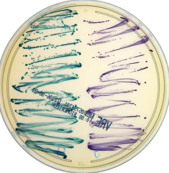
Media and culture conditions.We compared the performance of an experi-mental chromogenic medium, VRE-BMX (bioMe´rieux, Marcy l’Etoile, France), with that of BEAV (Remel, Lenexa, KS). VRE-BMX and BEAV plates were directly inoculated by submerging a sterilized 10-l inoculating loop in the specimen and streaking for isolation by the quadrant technique. This procedure was repeated for each medium type. Plates were incubated at 35°C in ambient air and examined for growth at 24 and 48 h. Colonies on VRE-BMX plates with purple or green pigmentation were presumptively identified as vancomycin-resistantE. faeciumorE. faecalis, respectively (Fig. 1). The appearance of black colonies on BEAV plates was presumed to indicate positivity for VRE. Following observation of the colony morphologies, individual unique colonies were subcul-tured onto CPS ID 3 medium (bioMe´rieux), a chromogenic medium for pre-sumptive identification of enterococci. Individual colonies were also subcultured
to tryptic soy agar plates supplemented with 5% sheep blood (Remel) for con-firmatory identification and antimicrobial susceptibility testing with the VITEK 2 automated identification and susceptibility system (bioMe´rieux). Additionally, the identification of isolated colonies and glycopeptide resistance were con-firmed genotypically by PCR or PCR-restriction fragment length polymorphism (RFLP) analysis.
DNA isolation for PCR analysis.Bacteria were grown overnight at 35°C on Columbia 5% sheep blood agar plates (bioMe´rieux). A bacterial suspension equivalent to a 0.5 McFarland standard was prepared, and cells were disrupted by mechanical lysis with glass beads of different sizes. After vortexing of the bacterial suspensions with beads for 2 min, samples were placed on ice and the supernatant was saved for amplification.
Multiplex PCR for detection of glycopeptide resistance genes and identifica-tion ofE. faeciumandE. faecalis.Multiplex PCR was adapted from the method previously described by Depardieu et al. (7). Briefly, the multiplex PCR consisted of 25l of 2⫻reaction mix (QIAGEN Multiplex PCR master mix; QIAGEN, Courtaboeuf, France), 0.2M concentrations of each primer, 5l of 5⫻ Q-solution (QIAGEN), and 5l of template DNA in a total volume of 50l. Samples were amplified as follows: an initial denaturation step at 95°C for 15 min; 35 cycles of denaturation at 94°C for 30 s, annealing at 54°C for 90 s, and elongation at 72°C for 1 min; and a final elongation at 72°C for 10 min. Amplified products were detected and quantified with a 2100 BioAnalyzer system (Agilent, Massy, France), according to the manufacturer’s instructions.
[image:2.585.120.468.72.428.2]Identification of enterococci by PCR-RFLP of thesodAgene.A 429-bp frag-ment internal to the sodAgene was amplified with degenerate primers, as
FIG. 1. VRE-BMX medium plated withE. faecalisandE. faecium.This figure demonstrates the ability of VRE-BMX to identify E. faeciumandE. faecalisbased on the color of the colony; on VRE-BMX, colonies ofE. faecalisappear blue-green and colonies ofE. faecium appear purple. For the purposes of this study, fecal specimens were plated directly to VRE-BMX and BEAV and observed for growth at 24 and 48 h.
on May 16, 2020 by guest
http://jcm.asm.org/
previously described by Poyart et al. (13). For RFLP analysis, 8l of PCR product was digested with 1l (10 units/l) of MseI endonuclease (New England BioLabs, Ipswich, MA) in a total volume of 20l, according to the manufactur-er’s instructions. Digested products were then visualized by electrophoresis with a 2100 BioAnalyzer system (Agilent). RFLP patterns were analyzed by compar-ing the number and size of fragments between enterococcal species.
Statistical analysis.To determine whether the results from each medium were significantly different, the significance of results was determined by MacNemar’s test or by binomial distribution with the significance level fixed at 5% (ifPwas ⬍5%, then the differences in performance were considered statistically signifi-cant).
RESULTS
Isolation of VRE from fecal samples.Stool samples from 147 patients were cultured for VRE according to the protocol. Colonies on VRE-BMX were screened for a purple
(vanco-mycin-resistant E. faecium) or blue-green
(vancomycin-resis-tant E. faecalis) hue (Fig. 1). Purple or blue-green colonies were easily differentiated from other flora. BEAV plates were screened for colonies causing a blackening of the medium around the colony and were easily distinguished from sur-rounding contaminants. Following observation of the colony morphologies, suspect colonies of VRE were identified and tested for glycopeptide resistance with VITEK 2 and con-firmed by molecular techniques, as described in Materials and Methods.
VRE-BMX performance characteristics. Upon completion of the medium evaluation, an analysis was performed to de-termine the sensitivity, specificity, positive predictive value (PPV), and negative predictive value (NPV) at 24 and 48 h. The sensitivity of each medium was calculated for detection of
VRE as well as for the detection of vancomycin-resistantE.
faeciumandE. faecalis. After molecular confirmation, the sen-sitivity of VRE-BMX for the detection of VRE at 24 and 48 h was 96.4% and 94.8%, respectively (Table 1). The sensitivity of BEAV for the detection of VRE was 90.9% at 24 h and 91.4% at 48 h (Table 1). The differences in sensitivity between 24 and 48 h were not statistically significant.
Further, the sensitivity of each medium was analyzed for the
detection ofEnterococcus species that carry clinically
signifi-cant glycopeptide resistance determinants (vanA,vanB), most
notably in vancomycin-resistantE. faeciumandE. faecalis. The
sensitivity of VRE-BMX for the detection ofE. faeciumwas
94.4% at 24 h, and that of BEAV was 85.7% (Table 2). There was no significant change in sensitivity when the incubation period of either medium was extended to 48 h (Table 2). The
sensitivity of VRE-BMX for the detection ofE. faecalis
colo-nization was 100% at both 24 and 48 h, while BEAV had a calculated sensitivity of 16.7 and 13.3% after 24 and 48 h of incubation, respectively (Table 2). The marked difference in
the sensitivity of BEAV between vancomycin-resistantE.
fae-cium and E. faecalis can be explained because VRE-BMX
identified 11 patients that were colonized with bothE. faecium
andE. faecalis. While VRE-BMX can easily distinguish dual colonization because of the differential properties of the chro-mogenic substrates, BEAV has no such differential capabili-ties, making it impossible to differentiate vancomycin-resistant
E. faeciumfromE. faecaliswithout further testing.
As with the sensitivity analysis, the specificity of VRE-BMX compared to BEAV was calculated at 24 and 48 h. The spec-ificity analysis showed that VRE-BMX was 96.6% and 73.9% specific at 24 and 48 h, respectively (Table 1). The specificity of BEAV at 24 h was 89.9%, but it dropped to 77.2% after extended incubation (Table 1). The drop in specificity of each medium was due to the proliferation of contaminant flora that may cause false-positive results. These organisms included gram-negative bacilli and yeast on VRE-BMX and
gram-pos-itive bacilli, gram-posgram-pos-itive cocci, and Enterococcus species
other thanE. faecalisorE. faeciumon BEAV.
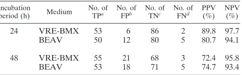
[image:3.585.300.542.89.200.2]The PPV and NPV of both media are shown in Table 3.
TABLE 1. Sensitivity and specificity analysis of VRE-BMX and BEAV for VRE at 24 and 48 h
Incubation
period (h) Medium
No. of TPa No. of FNb Sensitivity (%) Specificity (%)
24 VRE-BMX 53 2 96.4 96.6
BEAV 50 5 90.9 89.9
48 VRE-BMX 55 3 94.8 73.9
BEAV 53 5 91.4 77.2
aA true positive (TP) is defined as a blue-green or purple colony on
VRE-BMX or a gray to black colony on BEAV that was identified as VRE by VITEK 2 and confirmed by PCR.
bA false negative (FN) is defined as an isolate that was confirmed as VRE on
[image:3.585.44.283.91.164.2]one medium but did not grow on another medium.
TABLE 2. Sensitivity analysis of VRE-BMX and BEAV for E. faeciumorE. faecalisat 24 and 48 h
Incubation
period (h) Medium Isolate
No. of TPa No. of FNb Sensitivity (%)
24 VRE-BMX E. faecium 51 3 94.4
E. faecalis 12 0 100.0 BEAV E. faecium 48 8 85.7 E. faecalis 2 10 16.7
48 VRE-BMX E. faecium 54 3 94.7
E. faecalis 14 0 100.0 BEAV E. faecium 51 6 89.5 E. faecalis 2 13 13.3
aA true positive (TP) is defined as a blue-green or purple colony on
VRE-BMX or a gray to black colony on BEAV that was identified as VRE by VITEK 2 and confirmed by PCR.
bA false negative (FN) is defined as an isolate that was confirmed as VRE on
one medium but did not grow on another medium.
TABLE 3. PPV and NPV of VRE-BMX and BEAV for VRE at 24 and 48 h
Incubation
period (h) Medium
No. of TPa No. of
FPb No. of
TNc No. of
FNd PPV
(%) NPV
(%)
24 VRE-BMX 53 6 86 2 89.8 97.7
BEAV 50 12 80 5 80.7 94.1
48 VRE-BMX 55 21 68 3 72.4 95.8
BEAV 53 18 71 5 74.7 93.4
aA true positive (TP) is defined as a blue-green or purple colony on
VRE-BMX or a gray to black colony on BEAV that was identified as VRE by VITEK 2 and confirmed by PCR.
bA false positive (FP) is defined as an isolate that exhibited typical coloration
on the respective medium but was not identified as VRE by VITEK 2 or confirmed by PCR.
cA true negative (TN) is defined as the lack of a typically colored colony. dA false negative (FN) is defined as an isolate that was confirmed as VRE on
one medium but did not grow on another medium.
1558 LEDEBOER ET AL. J. CLIN. MICROBIOL.
on May 16, 2020 by guest
http://jcm.asm.org/
[image:3.585.304.543.576.651.2]VRE-BMX produced a PPV of 92.7% for vancomycin-resis-tantE. faecium (vanA or vanB) and 75.0% for
vancomycin-resistantE. faecalis(vanAorvanB) at 24 h. By comparison, the
PPV of BEAV for all VRE was 76.9% at 24 h and did not change significantly when the incubation period was extended to 48 h.
Growth of contaminants.Breakthrough growth of contami-nants on each medium was assessed at 24 and 48 h to deter-mine the efficacy of the antimicrobial agents in VRE-BMX to suppress proliferation of normal stool flora (non-VRE). Con-taminants were classified into two groups: (i) those that were considered potential false positives because they produced col-onies with any hue of blue-green or purple on VRE-BMX that
could be misinterpreted as vancomycin-resistantE. faeciumor
E. faecalis, or resulted in black colonies on BEAV, and (ii) colorless isolates. While BEAV grew 50% more contaminants at 24 h than did VRE-BMX, extending the incubation period to 48 h resulted in 24 and 26 cultures with breakthrough growth, respectively, on BEAV and VRE-BMX (Fig. 2). Iso-lates most likely to cause false-positive results on VRE-BMX
includedCandidaspp. and gram-negative rods. Contaminants
causing potential false-positive results on BEAV included
En-terococcus spp. other thanE. faecium and E. faecalis,
gram-positive rods, and Streptococcus spp. Most breakthrough
growth on VRE-BMX consisted of lightly green-colored colo-nies in the first quadrant of growth. However, contaminants on BEAV were found on all quadrants and were more likely to be
Enterococcusspp. other thanE. faeciumandE. faecalis.
DISCUSSION
Recognizing the clinical impact of the VRE epidemic, the CDC drafted recommendations in 1995 to assist infection control per-sonnel and hospital epidemiologists with the rapid spread of this organism within the hospital environment (4). Fundamental to these recommendations were strategies to contain cases or out-breaks of VRE and decrease the rates of transmission, including the isolation of VRE-infected or -colonized patients. The devel-opment of a sensitive method for detection of VRE colonization/ infection, and preferably one that is rapid and simple to perform so as to facilitate screening of large numbers of patient samples with prompt isolation, is central to this goal.
The identification of VRE from colonized patients can be accomplished by screening cultures of stool or rectal swabs with differential and/or selective media. One such medium, BEAV, has the advantage of suppressing the normal fecal flora and allowing for growth of organisms carrying clinically
signif-icant glycopeptide resistance genes, such as vanA or vanB.
[image:4.585.87.501.64.347.2]However, the disadvantage of BEAV is that additional confir-matory tests are required to identify isolates and confirm glyco-peptide resistance. Performing such tests on all of the colony morphotypes consistent with VRE can be time consuming and labor intense. In response to this problem, a number of manu-facturers have developed novel methods of detecting VRE colo-nization that do not require confirmatory testing. These include molecular techniques, such as real-time PCR, and novel culture media.
FIG. 2. Growth of contaminants on each medium at 24 and 48 h. Contaminants are classified as potential false-positive isolates and colorless isolates. Isolates with any hue of blue-green or purple were classified as potential false positives on VRE-BMX; isolates with a black hue on BEAV were classified as potential false positives.
on May 16, 2020 by guest
http://jcm.asm.org/
While real-time gene amplification methods are currently being developed for the detection of agents such as VRE, they often add a level of cost and complexity that are not in line with the clinical relevance of the targeted organism. Though these methods offer the benefit of decreased turn-around time and have been shown to be significantly more sensitive than culture for many viruses, their utility for bacteria, especially organisms that comprise the normal gut flora, is more nebulous. For example, nucleic acid amplification techniques can identify antimicrobial resistance genes in the absence of a viable or-ganism or when the resistance determinant is carried by an organism other than the targeted bacterium. In the latter
ex-ample, the detection ofvanAorvanBcarried by an organism
other than VRE in the presence of a pan-susceptible entero-coccus could provide misleading information.
To address these issues, the ideal candidate for a VRE screen would be selective and differential: able to differentiate
vancomycin-resistantEnterococcus faecalisfromEnterococcus
faeciumwithout the additional step of identification and resis-tance confirmation. Further, the screen would have reasonable sensitivity for screening purposes and specificity to limit the need for additional testing.
The data presented in this article show that a chromogenic
medium, VRE-BMX, from bioMe´rieux provides a viable
alter-native for screening of patients for gut colonization with VRE. This medium is able to identify and differentiate
vancomycin-resistantE. faeciumfromE. faecaliswhile inhibiting growth of
vancomycin-susceptible Enterococcus spp. Because this
me-dium reliably identifies enterococci to species level and con-firms glycopeptide resistance, it is not necessary to pursue additional biochemical analysis or determine antimicrobial susceptibilities in the clinical laboratory.
In this evaluation, BEAV culture combined with PCR was used as the “gold standard” for detection and confirmation of VRE from fecal specimens. After incubation of the plates for 24 and 48 h, the sensitivity, specificity, PPV, and NPV were calculated. In each of these analyses, we determined that VRE-BMX was at least a comparable, if not a superior, alter-native to BEAV for the detection of VRE. Since BEAV does
not identify Enterococcus to the species level and requires
additional steps to obtain these data for infection control pur-poses, we conclude that VRE-BMX provides value-added qualities to a VRE screening agar. Our comparison showed that incubation of VRE-BMX beyond 24 h did not improve the sensitivity of the medium and actually reduced the specificity due to breakthrough growth of normal glycopeptide-resistant
flora, such asCandidaspecies. We noted that this was
primar-ily observed in the first quadrant of the medium. When spec-imens were inoculated onto the medium, residual stool gener-ally remained in the first quadrant. The excess specimen likely overwhelmed the inhibitory level of the vancomycin within the
medium. To circumvent this problem, we would recommend first placing samples in a sterile diluent such as saline before plating and limiting the evaluation to 24 h of incubation.
ACKNOWLEDGMENT
W.M.D. is a consultant for bioMe´rieux, Inc., which provided support for the performance of this study.
REFERENCES
1. Reference deleted.
2.Bhavnani, S. M., J. A. Drake, A. Forrest, J. A. Deinhart, R. N. Jones, D. J. Biedenbach, and C. H. Ballow.2000. A nationwide, multicenter, case-control study comparing risk factors, treatment, and outcome for vancomycin-resis-tant and -susceptible enterococcal bacteremia. Diagn. Microbiol. Infect. Dis. 36:145–158.
3.Carmeli, Y., G. Eliopoulos, E. Mozaffari, and M. Samore.2002. Health and economic outcomes of vancomycin-resistant enterococci. Arch. Intern. Med. 28:2223–2228.
4.Centers for Disease Control and Prevention.1995. Recommendations for preventing the spread of vancomycin resistance: recommendations of the Hospital Infection Control Practices Advisory Committee (HICPAC). Am. J. Infect. Control.23:87–94.
5.Centers for Disease Control and Prevention.1999. National nosocomial infections surveillance (NNIS) system report, data summary from January 1990 to May 1999. Am. J. Infect. Control.27:520–532.
6.Courvalin, P. 2005. Genetics of glycopeptide resistance in gram-positive pathogens. Int. J. Med. Microbiol.294:479–486.
7.Depardieu, F., B. Perichon, and P. Courvalin.2004. Detection of thevan alphabet and identification of enterococci and staphylococci at the species level by multiplex PCR. J. Clin. Microbiol.42:5857–5860.
8.Garbutt, J. M., M. Ventrapragada, B. Littenberg, and L. M. Mundy.2000. Association between resistance to vancomycin and death in cases of Entero-coccus faeciumbacteremia. Clin. Infect. Dis.30:466–472.
9.Lautenbach, E., W. B. Bilker, and P. J. Brennan.1999. Enterococcal bacte-remia: risk factors for vancomycin resistance and predictors of mortality. Infect. Control Hosp. Epidemiol.20:318–323.
10.Leclercq, R., E. Derlot, J. Duval, and P. Courvalin.1988. Plasmid-mediated resistance to vancomycin and teicoplanin inEnterococcus faecium. N. Engl. J. Med.319:157–161.
11.Moellering, R. C.1992. Emergence ofEnterococcusas a significant pathogen. Clin. Infect. Dis.14:1173–1178.
12.Moellering, R. C.2005.Enterococcusspecies,Streptococcus bovis, and Leu-conostocspecies, p. 2411–2421.InG. L Mandell, J. R. Bennett, and R. Dolin, Principles and practices of infectious diseases, 6th ed., vol 2. Elsevier Churchill Livingstone, Philadelphia, PA.
13.Poyart, C., G. Quesne, C. Boumaila, and P. Trieu-Cuot.2001. Rapid and accurate species-level identification of coagulase-negative staphylococci by using thesodAgene as a target. J. Clin. Microbiol.39:4296–4301. 14.Sloan, L. M., J. R. Uhl, E. A. Vetter, C. D. Schleck, W. S. Harmsen, J.
Manahan, R. L. Thompson, J. E. Rosenblatt, and F. R. Cockerill III.2004. Comparison of the Roche LightCyclervanA/vanBdetection assay and cul-ture for detection of vancomycin-resistant enterococci from perianal swabs. J. Clin. Microbiol.42:2636–2643.
15.Song, X., A. Srinivasan, D. Plaut, and T. M. Perl.2003. Effect of nosocomial vancomycin-resistant enterococcal bacteremia on mortality, length of stay, and costs. Infect. Control Hosp. Epidemiol.24:251–256.
16.Stosor, V., L. R. Peterson, M. Postelnick, and G. A. Noskin.1998. Entero-coccus faeciumbacteremia. Does vancomycin resistance make a difference? Arch. Intern. Med.158:522–527.
17.Teixeira, L. M., and R. M. Facklam.2003.Enterococcus, p. 422–433.InP. R. Murray, E. J. Baron, J. H. Jorgensen, M. A. Pfaller, and R. H. Yolken (ed.), Manual of clinical microbiology, 8th ed., vol 1. ASM Press, Washington, DC. 18.Uttley, A. H. C., C. H. Collins, J. Naidoo, and R. C. George.1988.
Vanco-mycin-resistant enterococci. Lanceti:57–58.
19.Webb, M., L. W. Riley, and R. B. Roberts.2001. Cost of hospitalisation for and risk factors associated with vancomycin-resistantEnterococcus faecium infection and colonization. Clin. Infect. Dis.33:445–452.
1560 LEDEBOER ET AL. J. CLIN. MICROBIOL.