BRAIN OF CHICK EMBRYOS
J.Sravani1, K.Padmaja2 and Ch. Srilatha3
1,2
Department of Veterinary Biochemistry, C.V.Sc., SVVU, Tirupati 3
Department of Veterinary Pathology, C.V.Sc., SVVU, Tirupati
*Corresponding Author: J. Sravani1 Abstract
An experiment was conducted to study the effect of Bisphenol-A (BPA) on oxidative stress during embryonic development in chicks. Fertile eggs were procured and eighty (80) eggs in two sets were assigned randomly into 4 groups of 20 each. Group I served as control and received 10 µl of distilled water, groups II to IV were treated with BPA dissolved in distilled water in three concentrations of 100µM, 250µM and 500µM respectively. A single dose of 10 µl volume injected into the aircell using microsyringe separately to 11th day old and 14th day old chick embryos. The developed embryos were sacrificed after 24 and 48 hours of BPA exposure and liver and brain tissues were collected and preserved in 10% formalin for histopathological studies. In histopathological sections, degenerative, necrotic changes with mononuclear cell infiltration, congestion and proliferation of fibroblasts are prominent in liver of both 11th and 14th day BPA treated chick embryos. Neuronophagia, satellitosis, extensive demyelinating changes, proliferation of the capillaries, perineuronal vacuolation was observed in brain of BPA treated chick embryos. The study concludes that the toxic effects of BPA are dose dependent and are inversely effective with age during the embryonic development in chicks.
Key words: Bisphenol-A, Brain, Chick embryos, Histopathology, Liver
I. INTRODUCTION
Environmentally released manmade chemicals like Polychlorinated biphenyls (PCBS) and Bisphenol-A (BPA) has serious effects on endocrine and reproductive systems in domestic animals. Among endocrine disrupting chemicals, BPA is one of the most studied because of its extensive use (Gioiosa et al., 2013). Since last two decades, there has been increasing scientific concern regarding the adverse effects of chemical pollutants in the environment which interferes with normal functioning of different organs in animals and humans.
Chemically bisphenol-A is 2,2-bis-(4-hydroxyphenyl)-propane. It is one of the most common chemicals exposed in everyday life. It is a component of polycarbonate plastics and epoxy resin coatings and is produced at approximately 3.2 million metric tons/year. Maximum tolerable dose of BPA is found to be 0.2% in diet in male and female mice (Furukawa et al., 1994). The current reference dose for humans is 50µg/kg b.wt./day. It is used in a wide variety of consumer products like food storage containers, hard plastic water bottles, medical equipment, canned foods, PVC pipes, dental sealants, nail polish, electronic compact discs and digital versatile discs etc (Calafat et al., 2009). The chemical bonds between BPA molecules are unstable and the chemical leaches from bottles into materials with time of storage and use. It comes into contact with, heating plastic bottles, presence of acidic or basic foods and beverages in plastic and repeated washing increases the rate of BPA leaching from bottles (Gibson, 2007).
early puberty in females, low sperm counts, increased susceptibility to reproductive tract cancers and altered brain development in males and females (Newbold et al., 2009).
Bisphenol-A induced mullerian duct malformation in female quail embryos and feminization of the left testis in male chicken embryos (Berg et al., 2001). Highest concentrations of BPA was found in adipose tissue and liver (100%) followed by brain (70%) (Geens et al., 2011). It is a potential risk to early stages of mammalian life (Komada et al., 2012). There is evidence that several teratogens affect the developing embryo by increasing its oxidative stress, because of its relatively weak anti-oxidant defense especially at early stages of organogenesis, resulting in severe embryonic damage (Ornoy, 2007). Exposure to BPA during sensitive periods in organogenesis increases susceptibility to prostate and mammary gland cancers (Vom Saal et al., 2007). Early exposure to BPA leads to abnormalities in mammary tissue development (Vandenberg et al., 2008).
It was reported that BPA exposure during embryonic/fetal life and infancy induces tissue oxidative stress and peroxidation, ultimately leading to underdevelopment of the brain, kidney and testis (Kabuto et al., 2004). Exposure to high concentration of BPA results in the induction of oxidative damage and also causes damage to the biological membranes and cellular structures in brain tissues of chicken embryos. Histopathological examination of brain in BPA treated chicken embryos revealed hyperemia and demyelination in cerebellum and base cranium (Gharibi et al., 2013).
Chick embryo is an excellent in vivo model due to its low cost, easy accessibility and similarity to mammalian embryos and provides direct measured dosing of their absorption and monitoring of their effects on embryo development (Dennery, 2007).
II. MATERIAL AND METHODS
An experiment was planned to study the histopathological studies in BPA exposed chick embryos.
A. Chemicals:
All the chemicals used in the present study were of analytical grade (AR) and were obtained from Sisco research laboratories Pvt. Ltd., (Mumbai, India) and Himedia laboratories Pvt. Ltd., (Mumbai, India).
B. Source of Fertilized Eggs and Incubation Conditions:
The present study was conducted at the Department of Veterinary Biochemistry, College of Veterinary Science, Tirupati. Freshly laid wild Bobcock strain zero day old fertilized eggs were procured from Department of Poultry Science, College of Veterinary Science, Tirupati. They were incubated at 37.5±0.5˚C with a relative humidity of 65% in an egg incubator. The humidity of the incubator was maintained by keeping a tray full of water inside. The water was replaced every day and the water level was maintained to keep the same percentage of humidity throughout the incubation. Eggs were rotated manually three times a day and were examined through the candler every day for proper growth and viability of embryos. The eggs with dead embryos were removed immediately from the incubator.
C. Treatment to chick embryos:
Protocols of chicken embryo toxicity study has gained acceptance by several regulating agencies (Michelle et al., 2007). Preliminary experiments were carried out on chick embryos to arrive the toxic concentrations and standardized 100 µM, 250 µM and 500 µM as low, medium and high doses. Bisphenol-A dissolved in distilled water (water solubility is 0.04%) and prepared three
concentrations as 100µM, 250µM and 500µMsolutions.
Fertile eggs were purchased and eighty (80) eggs assigned randomly into four groups of 20 eggs each for BPA administration on 11th day. Similar set of 80 eggs simultaneously were kept for 14th day studies.
D. Experimental groups:
Group II : Bisphenol- A (100µM) Group III : Bisphenol -A (250µM) Group IV : Bisphenol -A (500µM)
E. Collection of Tissues:
From each group, six healthy and live chick embryos were randomly picked after 24 and 48
hours from 11th and 14th day BPA treated embryos. The embryos were sacrificed and liver and brain
tissues were collected for histopathological studies. The tissues were fixed in 10% neutral buffered formalin (pH 7.2) until further analysis. The samples were processed and sections of 4-7 µm were cut and stained with Haematoxylin and Eosin stain (H&E). The specimens were examined under light microscope (Singh and Sulochana, 1997).
III. RESULTS
Mild degenerative changes with micro vesiculation, focal areas of necrosis, karyorrhexis of the nucleus and focal infiltration of inflammatory cells were observed in liver tissue after 24 hours in Group-II (Plate 2) and III (Plate 3) compared to Group-I (Plate 1) in 11th day BPA injected chick embryos. In Group-III, the degenerative and necrotic changes with mononuclear cell infiltration are prominent and moderate in nature. Degenerative changes in hepatic cells and focal mononuclear cell infiltration are noticed in liver of Group-III and IV after 48 hours in 11th day BPA exposed chick embryos (Plate 4).
In 14th day BPA treated embryos congestion, extensive degenerative changes and
proliferation of the fibroblasts are noticed after 24 hours in liver tissue of Group-II (Plate 6), III (Plate 7) and IV (Plate 8) compared to Group-I (Plate 5).
In liver mild degenerative changes in hepatic cells in Group-II (Plate 9) and infiltration of mononuclear cells and karyorrhexis of hepatic cell nucleus are observed in Group-III (Plate 10) and
IV (Plate 11) after 48 hours in 14th day BPA treated chick embryos.
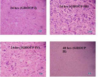
Neuronophagia, satellitosis and extensive demyelinating changes are noticed in brain after 24
hours in 11th day BPA treated embryo in Group III (Plate 13) and Group IV (Plate 14) compared to
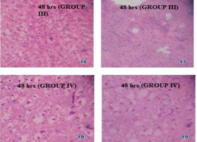
Group I (Plate 12) in a dose dependent manner. After 48 hours of BPA treatment proliferation of the capillaries, perineuronal vacuolation, extensive demyelinating changes giving spongyosis appearance
are observed in Group-II to IV (Plates 15, 16, 17, 18 and 19). In 14th day BPA exposed embryos
proliferation of the capillaries, atropy of the nerve cells, perinueronal vacuolation and extensive demyelinating changes observed after 24 hours in Group II (Plate 21) and Group III (Plate 22) respectively and after 48 hours in Group III (Plate 23) compared to control (Plate 20).
IV. DISCUSSION
A histopathological section of liver and brain tissues from the experimental groups has been evaluated to study the signs of development of oxidative damage caused in response to BPA toxicity. Mild degenerative changes with micro vesiculation, focal areas of necrosis, karyorrhexis of the nucleus and focal infiltration of inflammatory cells were observed in liver tissue after 24 hours in Group-II and III compared to Group-I in 11th day BPA injected chick embryos which are in accordance with Tyl et al. (2002) and Yamasaki et al. (2002) who observed chronic hepatic inflammation in rats. Demyelinating hyperemic and degenerative changes were noticed in the brain of BPA injected chick embryos. Similar results were also reported by Gharibi et al. (2013) with BPA treatment. Liver tissue impairment and vacuolation noticed in the present study are in agreement with Venkataswamy et al. (2013) where vacuolation of hepatocytes were more pronounced around the central vein in the developing chick embryos injected with acrylamide.
These results implied that exposure to BPA progressively increased the intensity of these degenerative changes such as increased dilatation and congestion of sinusoids and central vein and hydropic degeneration with focal array of hepatic cords. The results are in agreement with Abdel Hameed (2004) who described the vacuolation of hepatocytes as ballooning degeneration and
treated embryos congestion, extensive degenerative changes and proliferation of the fibroblasts are
noticed after 24 hours in liver tissue of Group-II to IV compared to Group-I. Degenerative changes
in hepatocytes might have appeared due to significant induction of oxidative stress after BPA exposure. Mild degenerative changes in hepatic cells in Group-II and infiltration of mononuclear
cells and karyorrhexis of hepatic cell nucleus are observed in Group-III and IV after 48 hours in 14th
day BPA treated chick embryos. It was reported that hepatic necrosis and congestion in liver of rats exposed to BPA is due to induction of ROS and disruption of balance between ROS and antioxidant
defense system (Korkmaz et al., 2010). These studies also revealed an increased damage in liver and
brain.
Fig. 1: Histopathology of liver tissue of different groups
Fig. 3: Histopathology of brain tissue of different groups
BIBLIOGRAPHY
[1] Abdel Hameed, T.F. 2004. “Light and electron microscopic studies on the effect of orally administered formalin on liver and kidney of guinea pig”. Journal of the Egyptian German Society of Zoology C. Histology and Histochemistry, 45: 203-224.
[2] Calafat, A M.; Weuve J.; Ye X.; Jia L T.; Hu H.; Ringer S.; Huttner K. and Hauser R. 2009. “Exposure to bisphenol A and other phenols in neonatal intensive care unit premature infants”. Environmental Health Perspectives, 117(4): 639–644.
[3] Chitra, K C.; Latchoumycandane C. and Mathur P P. 2003. “ Induction of oxidative stress by bisphenol-A in the epididymal sperm of rats”. Toxicology, 185: 119-127.
[4] Cipelli, R.; Harries L.; Okuda K.; Yoshihara S I.; Melzer D. and Galloway T. 2014. “Bisphenol A modulates the metabolic regulator oestrogen-related receptor-α in T- cells”. Reproduction, 147(4): 419-426.
[5] Dennery, P A. 2007. “Effects of oxidative stress on embryonic development”. Birth Defects Research Part C: Embryo Today: Reviews, 81(3): 155-162.
[6] Furukawa, F.; Nishikawa A.; Mitsui M., Sato M.; Suzuki J.; Imazawa T. and Takahashi M. 1994. “A 13-week subchronic toxicity study of bisphenol-A in B6C3F1 mice”. Eisei Shikensho Hokoku, 112: 89-96.
[7] Geens, T.; Goeyens L. and Covaci A. 2011. “Are potential sources for human exposure to bisphenol-A overlooked?” International journal of hygiene and environmental health, 214(5): 339-347.
[8] Gharibi S.; Dilmaghanian A.; Sadighara P. and Fard R M N. 2013. “The effect of bisphenol A on oxidative stress indices and pathological changes in the brain of chicken embryos”. World Applied Sciences Journal, 26(3): 345-351.
[9] Gibson, R L. 2007. “Toxic Baby Bottles: Scientific study finds leaching chemicals in clear plastic baby bottles.
Environment California Research and Policy Center”. http://WWW.environment California.org.
[10]Gioiosa, L.; Parmigiani S.; Vom Saal F S. and Palanza P. 2013. “The effects of bisphenol A on emotional behavior depend upon the timing of exposure, age and gender in mice”. Hormones and behavior, 63(4): 598-605.
[11]Kabuto, H.; Amakawa M. and Shishibori T. 2004. “Exposure to bisphenol A during embryonic/fetal life and infancy
increases oxidative injury and causes underdevelopment of the brain and testis in mice”. Life Sciences, 74(24): 2931-2940.
[12]Komada, M.; Asai Y.; Morii M.; Matsuki M.; Sato M. and Nagao T. 2012. “Maternal bisphenol A oral dosing relates
to the acceleration of neurogenesis in the developing neocortex of mouse fetuses”. Toxicology, 295(1): 31-38. [13]Korkmaz, A.; Ahbab M A.; Kolankaya D. and Barlas N. 2010. “Influence of vitamin C on bisphenol A,
nonylphenol and octylphenol induced oxidative damages in liver of male rats”. Food and Chemical Toxicology,
48(10): 2865-2871.
[14]Michelle, L.; Hui N Y. and Cheng S H. 2007. “Toxicity and cardiac effects of carbaryl in early developing zebrafish
(Danio rerio) embryos”. Toxicology and Applied Pharmacology, 222(2): 159-168.
[16]Tyl, R W.; Myer C B.; Marr M C.; Thomas B F.; Keimowitz A R.; Brine D R.; Veselica M M.; Fail P A.; Chang T Y.; Seely J C.; Joiner R L.; Butla J H.; Diamond S S.; Cagen S Z.; Shiotsuka R V.; Stropp G D. and Waechter J M. 2002. “Three-generation reproductive toxicity study of dietary bisphenol A in CD Sprague-Dawley rats”. Toxicological Sciences, 68(1): 121-146.
[17]Venkataswamy, M.; Meena bai M.; Divya K.; Pallavi C. and ThyagaRaju K. 2013. “Assessment of alterations in antioxidant enzymes and histology of liver and cerebral cortex of developing chick embryo in acrylamide toxicity”. International Journal of Advanced Research, 1(5): 256-264.
[18]Vom Saal, F S.; Akingbemi B T. and Belcher S M. 2007. “Chapel Hill bisphenol A expert panel consensus statement: Integration of mechanisms, effects in animals and potential to impact human health at current levels of exposure”. Reproductive Toxicology, 24(2): 131-138. \\