Open access:
www.balimedicaljournal.com
or
www.ojs.unud.ac.id
108
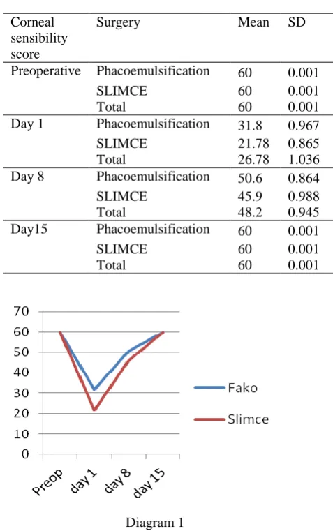
PHACOEMULSIFICATION AND SUTURELESS
LARGE-INCISION MANUAL CATARACT EXTRACTION
CHANGE CORNEAL SENSIBILITY
1
Anom-Supradnya, I G. N.,
1Jayanegara, W. G.,
1Sugiana, I G. N. M., and
2Raka-Widiana I G.
1Ophthalmology Department, Faculty of Medicine, Udayana University, Bali-Indonesia 2
Internal Medicine Department, Faculty of Medicine, Udayana University, Bali-Indonesia
Background:
Cataract is the leading cause of blindness worldwide, with surgery as a definitive
therapy. Incisions may damage the underlying tissue, including loss of corneal sensibility. The
purpose of this study was to determine differences in corneal sensibility decreased in patients after
phacoemulsification and SLIMCE.
Method:
This study was a randomized clinical trial assessing
changes of corneal sensibility in immature senile cataract patients after phacoemulsification and
SLIMCE at Sanglah and Indera Hospital Denpasar, Bali-Indonesia from March to August 2013.
Surgeons and examiners are experienced ophthalmologists. Examiner did not know the initial data
subjects. Preoperative assessment of corneal sensibility on the first, eighth, and fifteenth days were
assessed postoperative using Cochet-Bonnet aesthesiometer. Statistical analysis was performed by
applying mean difference of two independent groups test with repeated measures of multiple
comparisons (ANOVA). Samples from each group were 17 eyes.
Results
: There were significant
differences in corneal sensibility on the first day postoperative (33.2 mm), day 8 21.5 mm), day 15
(-11.8 mm) in both groups. Decrease in corneal sensibility in both groups was significant (
p
= 0.001).
There was decreasing in corneal sensibility with SLIMCE technique in the first (21.8 mm) and eighth
day (45.9 mm).
Conclusion:
In phacoemulsification technique, corneal sensibility was also decreased
in the first (31.8mm) and eighth day (50.6 mm) but returned to preoperative values on day 15 in both
groups. Decrease in corneal sensibility during follow-up period is influenced by cataract surgery
technique (
p
= 0.017) and was higher in SLIMCE technique compared with phacoemulsification.
Keywords
:
phacoemulsification, sutureless, large-incision, manual, cataract, extraction, corneal
INTRODUCTION
Cataract is the leading cause of blindness worldwide. World Health Organization (WHO) estimated there are 45 million people with blindness in the world. The most common cataract is senile cataract (90%) that occurs because of the degenerative process, from the age of 50 years.1,2,3,4 Definitive treatment of patients with senile cataract is cataract surgery. Currently developing cataract surgery with phacoemulsification technique that allows lens extraction with bimanual technique with smaller incision and does not require stitches so that healing process could be faster.5-7
Phacoemulsification requires expensive equipment investment and operating costs are also high. Lower economic capacity of developing countries and limited access to health facilities make the more often found cataract is large hard density cataract. Sutureless large-incision manual
Address for correspondence:
Anom-Supradnya, I G. N.
Ophthalmology Department, Faculty of Medicine, Udayana University, Bali-Indonesia E-mail: anom-supradnya@gmail.com
cataract extraction (SLIMCE) using scleral incision 8 mm width can remove the nucleus easily and safely, especially for large cataracts with hard density.1,2,8,9
Open access:
www.balimedicaljournal.com
or
www.ojs.unud.ac.id
109
and temporal incisions compared to inferiorincisions.15-17
Several studies have reported a decrease in corneal sensibility after phacoemulsification in the first week and first month. Corneal sensibility was back to near normal in the third month.18,19,20 Other study reported corneal sensibility was back to near normal in the first month.21 There is no research yet about corneal sensibility after SLIMCE.
The purpose of this study was to determine the differences in postoperative corneal sensibility reduction after cataract surgery between phacoemulsification techniques and SLIMCE.
PATIENTS AND METHOD
This study was a randomized clinical trial that assessed changes of corneal sensibility in immature senile cataract patients who underwent cataract surgery by phacoemulsification technique and SLIMCE in two places, Sanglah eye clinic General Hospital and Indera Hospital Denpasar, Bali-Indonesia from March to August 2013. Number of sample was 17 eyes for each group, calculated based on the formula of hypothesis test for two unpaired groups. All subjects in this research signed informed concern and ethical clearance was issued by local ethical committee Sanglah General Hospital. No mall clinical practice was observed in tjis study. Samples were selected consecutively from the affordable population after fulfilling the inclusion criteria until the required sample size was met. Inclusion criteria were all immature senile cataract patients. Exclusion criteria included patients with history of thermal trauma, chemical, or corneal perforation in the same eye, using contact lenses for more than three months, using non-ceasable topical β-blockers, suffered from diabetes mellitus, refused to participate in the study and was operated by operators other than those specified. Sample in accordance with the inclusion criteria not on any of the exclusion criteria and were willing to sign an informed consent were randomized with permutation block randomization into two groups.
Examinations were performed by two experienced ophthalmologists accompanied by the researchers. Examiner does not know the subjects’ initial data. Initial examination included visual acuity, corneal sensibility with Cochet-Bonnet aesthesiometer (Luneau Ophthalmologie, Chartres Cedex, Franc) and fluoresceine test to assess corneal erosion. Examination of corneal sensibility started from a scale of 60 mm. Cochet-Bonnet aesthesiometer nylon filament touched the central cornea gently. If there was no blinking reflex, the scale was reduced every five millimeters until blinking reflex were seen. The scale used is the longest one that resulted in a blinking reflex. Examinations were performed three times and mean values were calculated. Operators are two
experienced ophthalmologists. Procedures in phacoemulsification group, patients were anesthetized with topical anesthesia pantocain 2%. Corneal incision in the temporal side from central of the limbus with clear cornea incision technique using keratome 2.75 mm, followed by incision for second instrument. Injection of lidocaine 2% diluted with Ringer's lactate 1:1 into anterior chamber (AC) as much as 0.6 ml. Anterior capsule was stained with trypan blue. Irigation and aspiration was done with Ringer’s Lactate. Viscoelastic injection to anterior chamber (AC), followed by anterior capsulotomy with continous curvilinear capssulorhexis (CCC) technique. Then hydrodissection and hydrodealination were done. Nucleopraxis was done with quick chop technique or stop and chop, followed by emulsification, and cleaning of epinucleus with injection of dispersive type viscoelastic into anterior chamber. Foldable Intra Ocular Lens (IOL) was then injected into the capsular bag. Irrigation and aspiration was done to clean up the rest of viscoelastic followed by instillation of antibiotic and steroid eye drops, and then the eye was bandaged.
Group with SLIMCE technique underwent procedures according to the standard SLIMCE technique, described as follows. Patients were anesthetized with subconjunctival anesthesia using lidocaine 2% and peritomy was done in the superior conjunctiva and limbus. Half thickness scleral incision was done in the superior part about 8 mm length, frown-shaped with midpoint 2 mm from limbus using blade no.15, then sclerocorneal tunnel was made 4 mm wide from the scleral incision, extending 2 mm into the cornea. Side pockets on each edge of the incision were made to provide sufficient space to remove the nucleus. Then, side port were made with slit knife 15º at three or nine o'clock. Anterior capsule was stained with trypan blue, irrigation and aspiration with Ringer's lactate, viscoelastic injection to AC, followed by anterior capsulotomy 7-8 mm diameter by CCC technique. Dispersive type of viscoelastic was injected to maintain AC and facilitate nucleus extraction. Lens was extracted using vectis. The rest of the cortex was cleaned by aspiration and irrigation using simcoe cannula. IOL was put in the capsular bag. Irrigation and aspiration was done to clean up the rest of the viscoelastics. Corneoscleral tunnels were not stitched and continued by subconjunctival injection of aqueous solution containing dexamethasone disodium phosphate and gentamicin sulfate. Instillation of antibiotic and steroid eye drops and eye oinments were done, then the eye was bandaged.22
Open access:
www.balimedicaljournal.com
or
www.ojs.unud.ac.id
110
after procedure were recorded on theophthalmology status. Examinations after cataract surgery, were performed on the first, 8th and 15th day postoperatively.
Examinations after cataract surgery included visual acuity, slit-lamp examination, sensibility test of cornea and fluorescein examination. Slit lamp examination was done to identify complications and abnormalities caused by the surgery. Data before and after cataract surgery were recorded in the form of parent table. Statistical analysis was carried out using mean difference test of two independent groups with multiple comparisons of repeated measures (ANOVA). The amount of effects from treatment to outcome is stated with a mean difference with 95% confidence intervals as precise values. Level of significance (α) was determined at p<0.05.
RESULTS
The study included 34 eyes with each of the 17 eyes on phacoemulsification group and SLIMCE. Preoperative corneal sensibility were normal in both groups. Table 1 shows there were significant differences in corneal sensibility on follow up the first day, day 8, day 15 in both groups. There were significant decrease in corneal sensibility after cataract surgery by phacoemulsification technique and SLIMCE (p= 0,001).
Table .1
The mean difference of cornea sensibility score during follow-up in both groups (phacoemulsification and SLIMCE)
Follow up (1)
Follow up (2)
Mean Differ-ence
p
95% CI Lower
limit
Upper limit
Pre op Day 1 33.2 .001 28.8 37.7
Day 1 Day 8 -21.5 .001 -24.9 -18.0
Day 8 Day 15 -11.8 .001 -16.2 -7.3
Table 2 and Diagram 1 show that there was a higher decreasing in corneal sensibility by SLIMCE techniques and returned to normal after day 15 in both groups. Overall there is an effect between phacoemulsification techniques and SLIMCE to the decrease in corneal sensibility during follow-up period (p = 0.017).
In this study there was no corneal erosion after corneal sensibility examination in the preoperative and follow-up day 15 postoperative in both groups. Examination on the first day postoperative occurred five cases of corneal erosion in phacoemulsification technique and four cases in SLIMCE technique. Examination on the 8th day occurred one case of corneal erosion in SLIMCE technique.
Table 2
Changes in corneal sensibility in preoperative, postoperative day 1, day 8, day 15 between
phacoemulsification and SLIMCE groups
Corneal sensibility score
Surgery Mean SD
Preoperative Phacoemulsification 60 0.001
SLIMCE 60 0.001
Total 60 0.001
Day 1 Phacoemulsification 31.8 0.967 SLIMCE 21.78 0.865 Total 26.78 1.036 Day 8 Phacoemulsification 50.6 0.864 SLIMCE 45.9 0.988
Total 48.2 0.945
Day15 Phacoemulsification 60 0.001
SLIMCE 60 0.001
Total 60 0.001
Diagram 1
Chart for corneal sensibility changes preoperative, postoperative day 1, day 8, day 15 between groups
phacoemulsification and SLIMCE
DISCUSSION
Corneal sensibility before surgery in this study was 100% normal in both surgical techniques. This finding were likely due to all subjects having no other risk factors that could cause a decrease in corneal sensibility, except for age above 50 years.
Mean value of corneal sensibility obtained was significantly different in follow up on the first, 8th and 15th day in both groups. Postoperative corneal sensibility was decreased significantly in both groups.
Open access:
www.balimedicaljournal.com
or
www.ojs.unud.ac.id
111
of the eyeball can affect the trigeminal nerves, thusadding to decrease of cornea sensibility.11,15,23,24 The results of this study are consistent with several other studies reporting significant decrease of corneal sensibility on the first day after phacoemulsification surgery.16,20,21,25
SLIMCE techniques may also cause a decrease in corneal sensibility because conjunctival peritomy, cauterization of bleeding and scleral incision up to the corneal stroma were performed. This causes dissection of myelination nerve that goes to the cornea and lead to decrease in corneal sensibility.15, 17,26
Both surgical techniques affect the decrease in corneal sensibility postoperatively. Decrease in corneal sensibility is greater in the SLIMCE group compared to phacoemulsification with clear corneal incision on follow up the first and eighth day. This result is in contrast with the research hypothesis, probably due to the smaller width of the clear corneal incision in phacoemulsification technique, which is 2.75 mm. The decrease of corneal sensibility with corneal incision was also influenced by the width of the incisions. Smaller incisions means smaller decrease of corneal sensibility.13,19 In SLIMCE techniques, conjunctival peritomy was done with cauterization of bleeding and a wider scleral incision about 8 mm wide. These may damage the nerve and causes more extensive inflammation compared to phacoemulsification technique. Procedures performed on SLIMCE technique of making side port can also make dissect the corneal nerves. Nerve damage and more extensive inflammation as well as the side port in SLIMCE technique resulted in a greater decrease of corneal sensibility than phacoemulsification techniques.11,15,23,27 Different results were obtained in other studies, which reported larger decrease of corneal sensibility in corneal incision compared to scleral incision. This difference of result could be caused by larger clear cornea incision between 5-6 mm.17 Decrease of corneal sensibility begin to occur in the first day postoperative in both groups, and at day 15 postoperatively corneal sensibility returned back as preoperative levels in both groups. Corneal sensibility returned back to preoperative values may be because of wound healing and innervation recovery process. On the scleral incision, innervation will undergo recovery process after 25 hours post-operatively, and on day 8 to day 10 nerve recovery would be complete. Nerve recovery process on the cornea consists of two phases. The first phase of neurological recovery was in conjunction with surgical wound healing process, in which wound healing in corneal stroma occurs in the fifth day postoperatively. The second phase begins by formation of new neurites cells. These neurites cells begin to form on day 25 and play a role in the regeneration of corneal nerve axons. In
this phase, neural growth factors were also formed. After cataract surgery, lubricant application may be considered because the decrease in corneal sensibility and the quantity of tear film.15,17,24 Similar results were also reported that corneal sensibility in phacoemulsification technique achieved normal value on the seventh day postperatively.16 Another study in Jakarta reported corneal sensibility in scleral incision improved as preoperative level on the fifteenth day post operation, whereas in phacoemulsification technique it is still decreased in the fifteenth day.17 Different results of corneal sensibility in patients after phacoemulsification reported returning back to preoperative values after more than three months.18-21
Corneal sensibility examination with Cochet-Bonnet aesthesiometer can cause coneal erosion. At the preoperative examination there is no corneal erosions in both groups. Corneal erosions were obtained on the first day postoperative examination, in which corneal erosion occurred in five cases from phacoemulsification technique and four cases of SLIMCE techniques. On the eighth day examination there was one case from SLIMCE techniques. Corneal erosion was not found on follow-up day 15 postoperatively in both groups. Corneal erosion is likely to occur because of friction from Cochet-Bonnet aesthesiometer with the cornea. Corneal erosion will improve in the next six to 48 hours.28,29
CONCLUSION
Decrease in cornea sensibility after cataract surgery was higher in SLIMCE technique compared to the phacoemulsification technique with clear cornea incision.
ACKNOWLEDGEMENT
Acknowledgement is extended to the director of Indera Hospital Bali Province and staffs, who has given consent to conduct the study and to Harumi Purwa who has translated the manuscript.
REFERENCES
1. American Academy of Ophthalmology Staff. Lens and Cataract. United State of America: American Academy of Ophthalmology 2011-2012 ; p.71-125.
2. Khurana, A. K. Comprehensive Ophthalmologi. Fourth edition. New Delhi: New Age International 2007; p. 89-202
3. Pujiyanto. “Faktor-faktor Risiko yang Berpengaruh terhadap Kejadian Katarak Senilis” (tesis). Semarang: Universitas Diponogoro; 2004.
Open access:
www.balimedicaljournal.com
or
www.ojs.unud.ac.id
112
5. Sukardi, I., Hutauruk,J.A. Transisi MenujuFakoemulsifikasi, Langkah-langkah menguasai teknik dan menghindari komplikasi. Edisi 1. Jakarta. Kelompok Yayasan Obor Indonesia; 2004.
6. Combes, A., Gartry D. Cataract Surgery in: Susan Lightman, series editor. Fundamental of Clinical Ophthalmology. MBJ. London; 2003;P 13-182.
7. Ilyas, S. Ilmu penyakit mata. Edisi ke-3.Jakarta. Balai Penerbit FKUI; 2004. p.205-8
8. Dennis, S., Rao,S.K., Fan,H., Congdon, N.G., Wong,V., Liu,Y. et al. Endotelial Cell Loss and Surgically Induced Astigmatsm After Suturless Large-Incision Manual Cataract Extraction (SLIMCE). Arch Ophthalmol 2009; 127(10): 1284-1289
9. Haripriya, A., Chang,D., Reena,M., Shekhar,M. Complication Rates of Phacoemulsification and Manual Small Incision Cataract Surgery at Aravind Eye Hospital. J Cataract Refract Surg
2012, 38:1360-1369.
10.Tuisku, I.S. “Corneal Nerves in Refractive Surgery and Dry Eye” (dissertation). Finland. University of Helsinki; 2008.
11.Kim, H.H., Paiva,C.S., Yen,M.T. Effect of Upper Eyelid blepharoplasty on Ocular Surface Sensation and tear production. Can J Ophthalmol 2007, 42:739-42
12.Gharaee, H., Mousavi,M., Daneshvar,R., Hosseini,M., Sazande,S, Effect of Clear Corneal Incision location on Tear Film following Phacoemulsification Surgery.Iranian Journal of Ophthalmology 2009, 21(3):29-34 13.Rosita,Y. “Perubahan Sensibilitas Kornea dan
Konjungtiva Pasca Operasi Strabismus dengan Tehnik Insisi Konjungtiva Daerah Limbus” (tesis) Surabaya. Universtas Airlangga. 2008. 14.Lawrenson, J.G.,Ruskell,G.L. Investigation of
Limbal Touch sensitivity using a Cochet-Bonnet aesthesiometer. Br J of Ophthalmol
1993, 77:339-43
15.Zhang, S., Li, Y.Z. Incision Cataract Surgery Effect of Ocular Surface. Int J Ophthalmol
2010, 10(9): 1719-21.
16.Setyowibowo. “Perubahan Sensitivitas Kornea Pra dan Paska Bedah Ekstraksi Katarak Fakoemulsifikasi” (tesis). Surabaya. Universtas Airlangga; 2010.
17.Grace. “Perubahan Sensibilitas Kornea dan Lapisan Air Mata akibat Perbedaan Insisi pada Bedah Katarak Insisi Kecil Manual & Fakoemulsifikasi” (tesis). Jakarta: Universitas Indonesia ; 2006.
18.Kim,Y.M.,Kim,S.W.,Kim,T.I., Kim,E.K., Seo,K.R. The Change of Corneal Sensitivity and Recovery of Corneal Nerve after Cataract Surgery. J Korean Ophthalmol Soc. Korean
2007, 48(1):13-18
19.Khanal, S., Tomlison,A., Esakowitz,L., Bhatt,P., Jones,D., Nabili,S., et al. Changes in Corneal Sensitivity and Tear Physiology after Phacoemulsification. Ophthalmic Physiol Opt
2008, 28(2):127-34.
20.Kim, J.H., Chung,J.L., Kang,S.Y Change in Corneal Sensitivity and Corneal Nerve after Cataract Surgery.Cornea 2009, 28:20-5. 21.Oh, T., Jung,Y., Chang,D., Kim,J., Kim,H.
Changes in the tear film and ocular surface after cataract surgery. Jpn J Ophthalmol 2012, 56:113–8
22.Dennis,S., Congdon,N.G., Rao,S.K., Fan, H., Liu,Y., Zhang,L. et al. Visual Outcomes and Astigmatism After Sutureless. Manual Cataract Extraction in Rural China. Study of Cataract Outcomes and Up-Take of Services (SCOUTS) in the Caring Is Hip Project. Report 1. Arch Ophthalmol 2007, 125(11):1539-1544
23.Belmonte,C.Neural Basis Of Sensation in intact and Injured Corneas. Experimental Eye Research 2004,78:513-25.
24.Muller,L.J., Marfurt C.F., Kruse F., Tervo T.M. Corneal nerves: Structure, Contents and Function. Experimental Eye Research 2003; 76: 521-42
25.Paramastri, A., Norma, D.H., Siti, S.S. Pengaruh Operasi Katarak Insisi Lebar terhadap Sensibilitas Kornea dan Kejadian Dry Eye. Med Hosp 2012, 1(2); 103-7.
26.Kohlhaas, M. Corneal Sensation after cataract and refractive surgery. J Cataract Refractive Surg 1998, 24:1399-409
27.Acosta, M.C., Peral A., Luna C., Pintor J., Belmonte C., Gallar J. Tear secretion Induced by Selective Stimulation of Corneal and Conjunctival Sensory Nerve Fibers. Invest Ophthalmol Vis Sci 2004; 45: 2333-6.
28.Daniel, G.D., Mitchell A.W., Dayle H.G., Henry F.F. Cornea and Sclera. In: William,T., Edward,A.J., editors. Duane’s Ophthalmologi. Philadelphia. 2007; Chapter 4.
29.Triharpini, N.Y. “Perbandingan antara Bandage Contact Lens dan Bebat Tekan terhadap Luas Erosi dan Skala Nyeri Erosi Kornea” (Tesis). Denpasar: Universitas Udayana; 2013.