European Journal of Biological Research 2017; 7 (4): 291-298
Comparison of biofilm-producing
Enterococcus faecalis
,
Enterococcus faecium
, and unusual
Enterococcus
strains
Anna Sieńko*, Dominika Ojdana, Piotr Majewski, Paweł Sacha, Piotr Wieczorek,
Elżbieta Tryniszewska
Department of Microbiological Diagnostics and Infectious Immunology, Faculty of Pharmacy with the Division of Laboratory Medicine, Medical University of Bialystok, 15a Waszyngtona Street, 15-269 Bialystok, Poland
*Corresponding author: Anna Sieńko; Tel.: + 48 85 746 85 71; E-mail: [email protected]
ABSTRACT
The present study focused on determining the prevalence of biofilm-forming ability in
Enterococ-cus faecalis, E. faecium, and unusual EnterococEnterococ-cus
clinical isolates, and comparison of resistance and the prevalence of selected virulence factors among biofilm-positive strains. The ability to form biofilm
was detected in 13.3% of E. faecalis, 90% of
E. faecium, and 57.1% of unusual Enterococcus
strains (p=0.026). All E. faecalis strains were susceptible to β-lactams, while 37.5% of unusual and all E. faecium isolates were resistant to these antibiotics. Resistance to gentamicin was detected in 75% of E. faecalis, 55.5% of E. faecium, and 25% of other strains; resistance to streptomycin in 25%, 83.3%, and 50%, respectively. Analysis of the virulence revealed that the enterococcal surface protein (esp) gene was found in all E. faecium, 75.0% of E. faecalis, and 37.5% of other strains; collagen adhesin gene (ace) in 100%, 25.0%, and 37.5%; and hyaluronidase gene (hyl) in 83.3%, 0%, and 37.5%, respectively. Analysis of the resistance and virulence patterns showed that E. faecium isolates had the greatest variety of virulence and resistance determinants, while the lowest variety was exhibited by unusual strains. These findings
indicate that unusual biofilm-producing
Entero-coccus strains have lower resistance and virulence
potency than E. faecalis and E. faecium.
Keywords: Enterococcus faecalis; Enterococcus
faecium; Biofilm; Resistance; Virulence.
1. INTRODUCTION
Today, Enterococcus spp. are the fourth most common etiological factor in nosocomial infections in Europe [1]. Although these cocci are members of the microbiota of the human gastrointestinal tract, they often infect the bloodstream, surgical sites, and urinary tract, due to their multiresistance to many antimicrobials [2, 3]. Enterococcus spp. have an intrinsic resistance to cephalosporins, lincosamides, and low levels of aminoglycosides, and they can easily acquire resistance, most prominently to glycopeptides and aminoglycosides (high-level resistance), by means of mutations or as a result of transfer and incorporation of genes located on mobile genetic elements, such as plasmids and transposons [1, 4]. Moreover, these bacteria have the ability to form strong biofilm structures, and to produce several virulence factors, such as entero-coccal surface protein (Esp), aggregation substance Received: 14 July 2017; Revised submission: 25 September 2017; Accepted: 02 October 2017
Copyright: © The Author(s) 2017. European Journal of Biological Research © T.M.Karpiński 2017. This is an open access article licensed under the terms of the Creative Commons Attribution Non-Commercial 4.0 International License, which permits
unrestricted, non-commercial use, distribution and reproduction in any medium, provided the work is properly cited.
European Journal of Biological Research 2017; 7 (4): 291-298 (As), collagen adhesion (Ace), hyaluronidase (Hyl),
and gelatinase (GelE) [5-7]. Esp is the factor that mediates the colonization, and, together with GelE, has been suggested to be involved in biofilm formation [5-7]. Ace and EfaA are principal viru-lence traits associated with infective endocarditis, whereas Hyl causes tissues damages [5-7]. The majority of nosocomial enterococcal infections are caused by E. faecalis and E. faecium. However, today there is an increasing prevalence of infections caused by other rarely isolated species, for example:
E. avium, E. gallinarum, E. durans, and E. casseli-flavus [8-10].
Biofilm is an assemblage of microbial cells enclosed in a self-produced polysaccharide matrix and attached to a biotic or abiotic surfaces,
provi-ding an optimal microenvironment for growth, and facilitates transmission of mobile determinants between microorganisms [11, 12]. Evidence sug-gests that bacteria in biofilms are more resistant to antimicrobials and hosts factors than other microorganisms and are extremely difficult to eradicate [13]. Likewise, among Enterococcus, it is suggested that an ability to produce biofilm is a very important virulence factor which has a major impact on the course of nosocomial infections [5, 7]. Unfortunately, our knowledge about the
mecha-nisms and determinants involved in the process of biofilm formation among enterococci is still
insufficient [14]. The ability to create biofilm has
been suggested to occur less frequently among
E. faecium strains compared to E. faecalis strains,
but, astonishingly, data about biofilm-forming ability among unusual enterococcal species are very limited and unclear [14, 15]. Furthermore, there are only a few reports about the differences in resistance and virulence of various biofilm-producing
Entero-coccus species [13, 16, 17]. This prompted us to
determine the prevalence of biofilm-forming ability among E. faecalis, E. faecium, and unusual
Entero-coccus spp. clinical isolates. Then, we focused on
the comparison of the antibiotic resistance, the ability to hemolyze, and the presence of selected virulence genes among these three groups of biofilm-producing Enterococcus spp. strains. More-over, the next goals of this study were to determine their exact resistance profiles, and to indicate the antibiotic with the highest activity against these strains.
2. MATERIAL AND METHODS
2.1. Strains
Tests were performed on sixty-four entero-coccal isolates: thirty E. faecalis, twenty E. faecium, and fourteen others (five E. avium, three E.
casse-liflavus, three E. gallinarum, three E. durans),
isolated from clinical specimens from patients hospitalized at the University Hospital in Bialystok (Poland) from December 2013 to January 2015. Isolates were recovered from various clinical materials, mostly blood, peritoneal fluid, broncho-alveolar lavage (BAL), feces, urine, and pus. Most of the collected isolates were gathered from the intensive care unit and a hematology clinic.
2.2. Identification and susceptibility testing
The identification and susceptibility testing were conducted on the automated VITEK 2 system (bioMérieux, France) according to the manufactu-rer’s instruction using VITEK 2 GP and AST-P516 cards, respectively. Susceptibility to ampicillin, imipenem, gentamicin, streptomycin, vancomycin, teicoplanin, linezolid, and tigecycline was inter-preted according to the European Committee on Antimicrobial Susceptibility Testing (EUCAST) recommendations (breakpoint tables for interpreta-tion of minimum inhibitory concentrainterpreta-tions, MIC, and zone diameters; version 5.0, 2015; http://www.eucast.org).
2.3. Biofilm and hemolysin production
The tube method [18, 19] and Congo red agar (CRA) method [20, 21] were used to assess the ability of tested isolates to biofilm formation. Each experiment was repeated three times for each strain. Strains that demonstrated the ability to produce biofilm by both methods were considered as biofilm positive (BIO+) isolates. Hemolysin production was determined on Columbia blood agar supplemented with 5% sheep blood (OXOID, United Kingdom) [22].
2.4. DNA extraction
European Journal of Biological Research 2017; 7 (4): 291-298 from overnight E. faecium cultures using a Genomic
Mini Kit (A&A Biotechnology, Poland) according to the manufacturer’s guidelines.
2.5. PCR detection of virulence genes
Then, PCR assays were performed to detect the following virulence genes: gelE, ace, hyl, esp,
as, and cyl. The primers sequences are listed in
Table 1. PCR amplification was performed in 25 µl mixtures using 2 µl of DNA solution, 1 µl of each primer, 8.5 µl of nuclease-free water, and 12.5 µl of PCR master mix (DNA Gdańsk, Poland). Samples were subjected to an initial denaturation at 94ᴼC for 5 min, followed by 30 cycles of denaturation at 94ᴼC for 1 min, annealing at an appropriate temperature for 1 min, and elongation at 72ᴼC for 1 min using a DNA thermocycler (SensoQuest GmbH, Germany).
PCR products were separated electrophore-tically on the Sub-Cell GT apparatus (Bio-Rad, USA) at 5 V/cm for 100 min on a 1.5% agarose gel (Sigma-Aldrich, USA) containing 0.5% ethidium bromide (MP Biomedicals, USA) in Tris-borate-EDTA (ethylenediaminetetraacetic acid) buffer. Then, amplicons were visualized and photographed using the ChemiDoc XRS imaging system and Quantity One 1-D analysis software (Bio-Rad). The positions of obtained products were estimated with the molecular weight marker PerfectTM 100-1000 bp DNA ladder (EURx, Poland). To confirm the presence of the above-mentioned virulence genes, DNA sequencing was carried out on selected PCR products by the GENOMED S.A. company in Poland. The sequences were aligned and compared with reference sequences achieved using GenBank with the Basic Local Alignment Search Tool (BLAST) algorithm.
Table 1. PCR primers, annealing temperatures, and product sizes for detection of virulence genes. Virulence
gene Primers
Product size (bp)
Annealing
temperature (̊C) Reference
gelE 5’ AAT TGC TTT ACA CGG AAC GG 3’
5’ GAG CCA TGG TTT CTG GTT GT 3’ 548
52 [23]
ace 5’ GGC CAG AAA CGT AAC CGA TA 3’
5’ CGC TGG GGA AAT CTT GTA AA 3’ 353
hyl 5’ ACA GAA GAG CTG CAG GAA ATG 3’
5’ GAC TGA CGT CCA AGT TTC CAA 3’ 276
55 [24]
esp 5’ AGA TTT CAT CTT TGA TTC TTG G 3’
5’ AAT TGA TTC TTT AGC ATC TGG 3’ 510
as 5’ CACGCTATTACGAACTATGA 3’
5’ TAAGAAAGAACATCACCACGA 3’ 375
cyl 5’ TGG ATG ATA GTG ATA GGA AGT 3’
5’ TCT TTC ATC ATC TGA TAG TA 3’ 517
2.6. Statistical analysis
STATA 13.1 (StataCorp LP, USA) was used for statistical analysis. Differences among
E. faecalis, E. faecium, and unusual enterococcal
strains were assessed by the Chi-square test and Fisher’s exact test. Results with p<0.05 were considered significant.
3. RESULTS AND DISCUSSION
The present study focused on determining the
prevalence of biofilm-forming ability among three enterococcal groups: E. faecalis, E. faecium, and other clinical isolates, and on comparison of the antibiotic resistance and the prevalence of selected virulence traits between BIO+ strains from these groups. Interestingly, we found that the ability to form biofilm occurred in 4/30 (13.3%) E. faecalis, 18/20 (90%) E. faecium, and 8/14 (57.1%) rarely isolated strains: 5 E. avium, 1 E. durans, 1 E.
casse-liflavus, and 1 E. gallinarum (statistically significant
European Journal of Biological Research 2017; 7 (4): 291-298 produce biofilm was found in 60.9% of E. faecalis
isolates [25], in Italy - in 80% of E. faecalis strains [26]. In the case of E. faecium isolates, in India, Italy, Turkey, and Spain, this ability occurred much less frequently (0%, 28.8%, 48%, and 75%, respectively) [5, 8, 27, 28]. Lleo et al. [17] described the biofilm-forming ability among four out of twelve unusual enterococcal strains (33.3%), which our study supports. However, in contrast to our findings, Dworniczek et al. [29] indicated the lack of these features in rare species. These varied results indicate that the level of the ability to produce biofilm among different Enterococcus species varies with geographic location.
In the next step of our research, only BIO+ strains (30/64) were chosen for further investigation.
A comparison of antibiotic resistance among
E. faecalis, E. faecium and other isolates showed
that all E. faecalis strains were susceptible to tested ß-lactams, while 37.5% of other strains and all
E. faecium isolates were resistant to these
antibio-tics. These results strongly overlap with results recently published by us [30] and other researchers [9, 16, 31]. Resistance to gentamicin was detected in 75% of E. faecalis, 55.5% of E. faecium, and 25% of other strains; resistance to streptomycin in 25%, 83.3%, and 50%, respectively. Findings from our previous work showed that more E. faecium isolates were resistant to aminoglycosides: 76% to gentamicin, and 91.4% to streptomycin [30]. Interestingly, a study by Tan et al. [9] demonstrated that all unusual enterococcal isolates from blood were susceptible to gentamicin and around 80% of them were susceptible to ß-lactams. Therefore, the authors concluded that combination therapy (penicillin with aminoglycosides) could be easily used for the treatment of serious infections caused by rare species of Enterococcus, such as bacteremia and sepsis. This finding is not confirmed in our survey. We revealed that resistance to glycopeptides occurred only in the case of four (22.2%) E. faecium
isolates; two strains from the rare group,
E. gallinarum and E. casseliflavus, showed intrinsic
resistance to vancomycin. Similar results were obtained by other authors [9, 32]. We concluded that tigecycline and linezolid had the highest activity against all studied isolates (100% susceptibility), including those resistant to glycopeptides and aminoglycosides. Many studies confirmed that these
antibiotics are a valuable therapeutic option in serious enterococcal infections [33-35]. Unfortu-nately, resistance to these drugs has been recently reported [34, 36, 37], indicating that resistance to newer antibiotics is also increasing, and develop-ment of new targeted enterococcal drugs is needed.
Our comparative analysis of the prevalence of virulence genes among E. faecalis, E. faecium, and other strains revealed that the esp gene was found in all E. faecium, 75% of E. faecalis, and 37.5% of other strains. Similar proportions were seen by other researchers [6, 11, 25, 38, 39, 40]. These findings indicate that this gene has a connection with biofilm-forming ability, especially in E. faecium strains. However, many authors found that there is no association between the presence of the esp gene and biofilm production [5, 14, 29, 41]. These conflicting results suggest that esp requires inte-ractions with other virulence traits to result in biofilm enhancement.
Considering the presence of other virulence factors in our studied BIO+ groups, we found that the ace gene occurred in all E. faecium, 25% of
E. faecalis, and 37.5% of unusual isolates; hyl in
83.3%, 0%, and 37.5%, respectively. The gelE gene was detected only in E. faecalis strains. According to the literature, the presence of gelE and as genes among E. faecalis is very common, whereas they are extremely rarely present in E. faecium and rare enterococcal isolates; consequently, they are not necessary in the process of biofilm formation among these species [5, 25, 28, 42, 43]. These assumptions are confirmed by our survey. However, some researchers imply that there is a strong relationship between gelE and the ability to form biofilm [12, 15]. Other virulence genes, cyl and as, were also found only in E. faecalis isolates, which is in accordance with other studies [22, 40, 44].
European Journal of Biological Research 2017; 7 (4): 291-298 three or more antibiotics and had the ability to
hemolyse. Different results were seen in recent research by Tsikrikonis et al. [25], who detected only 1.9% of hemolysin-producing E. faecium
clinical isolates. We also found that one E. faecalis isolate and three E. avium isolates were susceptible to all tested antibiotics.
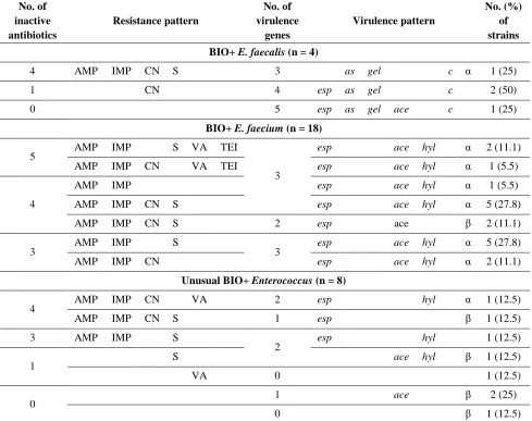
Table 2. Characteristics of resistance and virulence patterns among BIO+ E. faecalis, BIO+ E. faecium, and other
BIO+ Enterococcus strains. AMP, ampicillin; IMP, imipenem; CN, gentamicin; S, streptomycin; VA, vancomycin; TEI, teicoplanin; esp, enterococcal surface protein; as, aggregation substance; gel, gelatinase; hyl, hyaluronidase,
ace, collagen adhesin; c, cytolysin; α, β, types of hemolysis.
No. of inactive antibiotics
Resistance pattern
No. of virulence
genes
Virulence pattern
No. (%) of strains
BIO+ E. faecalis (n = 4)
4 AMP IMP CN S 3 as gel c α 1 (25)
1 CN 4 esp as gel c 2 (50)
0 5 esp as gel ace c 1 (25)
BIO+ E. faecium (n = 18)
5 AMP IMP S VA TEI
3
esp ace hyl α 2 (11.1)
AMP IMP CN VA TEI esp ace hyl α 1 (5.5)
4
AMP IMP esp ace hyl α 1 (5.5)
AMP IMP CN S esp ace hyl α 5 (27.8)
AMP IMP CN S 2 esp ace β 2 (11.1)
3 AMP IMP S 3 esp ace hyl α 5 (27.8)
AMP IMP CN esp ace hyl α 2 (11.1)
Unusual BIO+ Enterococcus (n = 8)
4 AMP IMP CN VA 2 esp hyl α 1 (12.5)
AMP IMP CN S 1 esp β 1 (12.5)
3 AMP IMP S
2 esp hyl 1 (12.5)
1 S ace hyl β 1 (12.5)
VA 0 1 (12.5)
0
1 ace β 2 (25)
0 β 1 (12.5)
In conclusion, we observed that the proportion of isolates producing biofilm was the highest among E. faecium isolates, at the middle level among the unusual Enterococcus spp. group, and the lowest in E. faecalis isolates. Interestingly, our data demonstrated that unusual biofilm-forming
Enterococcus strains have lower resistance to
antibiotics and are characterized by possession of lower virulence capabilities than BIO+ E. faecalis and BIO+ E. faecium clinical isolates. Moreover,
E. faecium strains showed the highest resistance and
biofilm-European Journal of Biological Research 2017; 7 (4): 291-298 forming ability among unusual Enterococcus species,
observed in this study, indicates that these isolates could also stay in the medical environment and, consequently, slowly acquire resistance and viru-lence traits. Therefore, the infections caused by these strains should not be underestimated, and determination of their susceptibility should always be performed. The changing epidemiology and increasing resistance to antibiotics among
Entero-coccus species stress the need to search in new
directions for the treatment and new methods for preventing the spread of enterococcal nosocomial infections.
ACKNOWLEDGEMENTS
We thank Steven J. Snodgrass for editorial assistance. The results of this work were presented in part at the Biofilms 7 Conference 2016, Porto, Portugal (26-28.06.2016).
AUTHOR'S CONTRIBUTION
AS: Conception and design, Development of methodology, Acquisition of data, Analysis and interpretation of data, Writing, review and/or revision of the manuscript; DO: Acquisition of data, Analysis and interpretation of data, Administrative, technical, or material support; PM: Analysis and interpretation of data, Administrative, technical, or material support; PS and PW: Writing, review and/or revision of the manuscript; ET: Writing, review and/or revision of the manuscript, Study supervision. The final manuscript has been read and approved by all authors.
SOURCE OF FUNDING
This work was supported by funds from Medical University of Bialystok (Poland), and from the Leading National Research Centre (137/KNOW/ 2015) in Bialystok.
TRANSPARENCY DECLARATION
The authors have no conflict of interest to declare.
REFERENCES
1. Orsi GB, Ciorba V. Vancomycin resistant
enterococci healthcare associated infections. Ann Ig. 2013; 25: 485-492.
2. Amyes SG. Enterococci and streptococci. Int J Antimicrob Agents. 2007; 29: 43-52.
3. Marschall J, Piccirillo ML, Fraser VJ, Doherty JA, Warren KW. Catheter removal versus retention in the management of catheter-associated enterococcal bloodstream infections. Can J Infect Dis Med. 2013; 24(3): e83-e87.
4. Fernandes SC, Dhanashree B. Drug resistance and virulence determinants in clinical isolates of
Enterococcus species. Indian J Med Res. 2013; 137:
981-985.
5. Di Rosa R, Creti R, Venditti M, D'Amelio R, Arciola CR, Montanaro L, et al. Relationship between biofilm formation, the enterococcal surface protein (Esp) and gelatinase in clinical isolates of
Enterococcus faecalis and Enterococcus faecium.
FEMS Microbiol Lett. 2006; 256: 145-150.
6. Fisher K, Phillips C. The ecology, epidemiology and virulence of Enterococcus. Microbiology. 2009; 155: 1749-1757.
7. Sava IG, Heikens E, Huebner J. Pathogenesis and immunity in enterococcal infections. Clin Microbiol Infect. 2010; 16: 533-540.
8. Prakash VP, Rao SR, Parija SC. Emergence of unusual species of enterococci causing infections, South India. BMC Infect Dis. 2005; 5: 14.
9. Tan CK, Lai CC, Wang JY, Lin SH, Liao SH, Huang Y, et al. Bacteriemia caused by non-faecalis and non-faecium Enterococcus species at a medical center in Taiwan, 2000 to 2008. J Infect. 2010; 61: 34-43.
10. Kenzaka T, Takamura N, Kumabe A, Takeda K. A case of subacute infective endocarditis and blood access infection caused by Enterococcus durans. BMC Infect Dis. 2013; 13: 594.
11. Heikens E, Bonten MJ, Willems RJ. Enterococcal surface protein Esp is important for biofilm formation of Enterococcus faecium E1162. J Bacteriol. 2007; 189: 8233-8240.
European Journal of Biological Research 2017; 7 (4): 291-298
14. Almohamad S, Somarajan SR, Singh KV,
NallapareddySR, Murray BE. Influence of isolate origin and presence of various genes on biofilm
formation by Enterococcus faecium. FEMS
Microbiol Lett. 2014; 353: 151-156.
15. Kafil HS, Mobarez AM. Assessment of biofilm formation by enterococci isolates from urinary tract infections with different virulence profiles. J King Saud Univ Sci. 2015; 27: 312-317.
16. Iwen PC, Kelly DM, Linder J, Hinrichs SH, Dominguez EA, Rupp ME, et al. Change in prevalence and antibiotic resistance of Enterococcus species isolated from blood cultures over an 8-year period. Antimicrob Agents Chemother. 1997; 41: 494-495.
17. Lleo M, Bonato B, Tafi MC, Caburlotto G, Benedetti D, Canepari P. Adhesion to medical device materials and biofilm formation capability of
some species of enterococci in different
physiological states. FEMS Microbiol Lett. 2007; 274: 232-237.
18. Christensen GD, Bisno AL, Simpsom WA, Beachey
EH. Adherence of slime producing strains of
Staphylococcus epidermidis to smooth surfaces.
Infect Immun. 1982; 37: 318-326.
19. Oliveira A, Cunha MD. Comparison of methods for the detection of biofilm production in coagulase-negative staphylococci. BMC Res Notes. 2010; 3: 260.
20. Freeman DJ, Falkiner FR, Keane CT. New method for detecting slime production by coagulase negative staphylococci. J Clin Pathol. 1989; 48: 872-874.
21. Cabrera-Contreras R, Morelos-Ramirez R, Galicia-Camacho AN, Melendez-Herrada E. Antibiotic resistance biofilm production in Staphylococcus
epidermidis strains, isolated from a Tertiary Care
Hospital in Mexico City. ISRN Microbiol. 2013; 13: 1-5.
22. Vergis EN, Shankar N, Chow JW, Hayden MK, Snydman DR, Zervos MJ, et al. Association between the presence of enterococcal virulence factors gelatinase, hemolysin, and enterococcal surface protein and mortality among patients with bacteremia due to Enterococcus faecalis. Clin Infect Dis. 2003; 35: 570-575.
23. Camargo ILBC, Gilmore MS, Darini ALC.
Multilocus sequence typing and analysis of putative virulence factors in vancomycin-resistant and vancomycin-sensitive Enterococcus faecium isolates from Brazil. Clin Microbiol Infect. 2006; 12: 1123-1130.
24. Zou LK, Wang HN, Zeng B, Li JN, Li XT, Zhang AY, et al. Erythromycin resistance and virulence genes in Enterococcus faecalis from swine in China. New Microbiol. 2011; 34: 73-80.
25. Tsikrikonis G, Maniatis AN, Labrou M, Ntokou E, Michail G, Daponte A, et al. Differences in biofilm formation and virulence factors between clinical and fecal enterococcal isolates of human and animal origin. Microb Pathog. 2012; 52: 336–343.
26. Baldassarri L, Cecchini R, Bertuccini L,
Ammendolia MG, Iosi F, Arciola CR, et al.
Enterococcus spp. produces slime and survives in
rat peritoneal macrophages. Med Microbiol
Immunol. 2001; 190: 113-120.
27. Latasa C, Solano C, Penadés JR, Lasa I. Biofilm-associated proteins. CR Biol. 2006; 329(11): 849-857.
28. Diani M, Esiyok OG, Ariafar MN, Yuksel FN, Altuntas EG, Akcelik N. The interactions between
esp, fsr, gelE genes and biofilm formation and pfge
analysis of clinical Enterococcus faecium strains. Afr J Microbiol Res. 2014; 8: 129-137.
29. Dworniczek E, Wojciech Ł, Sobieszczańska B, Seniuk A. Virulence of Enterococcus isolates collected in Lower Silesia (Poland). Scand J Infect Dis. 2005; 37: 630-636.
30. Sieńko A, Wieczorek P, Majewski P, Ojdana D, Wieczorek A, Olszańska D, et al. Comparison of antibiotic resistance and virulence between biofilm-producing and non-biofilm-producing clinical isolates of E.
faecium. Acta Biochim Pol. 2015; 62: 859-866.
31. BillströmH, SullivanA, LundB. Cross-transmission of clinical Enterococcus faecium in relation to esp and antibiotic resistance. J Appl Microbiol. 2008; 105: 2115-2122.
32. Contreras GA, Diaz Granados CA, Cortes L, Reyes J, Vanegas S, et al. Nosocomial outbreak of
Enterococcus gallinarum: untaming of rare species
of enterococci. J Hosp Infect. 2008; 70: 346-352. 33. Franiczek R, Dolna I, Dworniczek E, Krzyżanowska
B, Seniuk A, Piątkowska E. In vitro activity of tigecycline against clinical isolates of Gram-positive and Gram-negative bacteria displaying different resistance phenol-types. Adv Clin Exp Me. 2008; 17: 545-551.
34. Freitas AR, Coque TM, Novais C, Hammerum AM,
Lester CH, Zervos MJ, et al. Human and swine hosts share vancomycin-resistant Enterococcus
faecium CC17 and CC5 and Enterococcus faecalis
CC2 clonal clusters harboring Tn1546 on
European Journal of Biological Research 2017; 7 (4): 291-298
35. Sieńko A, Wieczorek P, Wieczorek A, Sacha P, Majewski P, Ojdana D, et al. Occurrence of high-level aminoglycoside resistance (HLAR) among
Enterococcus species strains. Prog Health Sci. 2014;
4: 179-187.
36. Werner G, Grforer S, Fleige C, Witte W, Klare I. Tigecycline-resistant Enterococcus faecalis strain isolated from a German Intensive Care Unit Patient. J Antimicrob Chemother. 2008; 61: 1182-1183. 37. Baldir G, Engin DO, Kucukercan M, Inan A, Akcay
S, Ozyuker S, et al. High-level resistance to aminoglycoside, vancomycin and linezolid in enterococci strains. J Microbiol Infect Dis. 2013; 3: 100-103.
38. Dupre I, Zanetti S, Schito AM, Fadda G, Sechi LA. Incidence of virulence determinants in clinical
Enterococcus faecium and Enterococcus faecalis
isolates collected in Sardinia (Italy). J Med Microbiol. 2003; 52: 491-498.
39. Tendolkar PM, Baghdayan AS, Gilmore MS,
Shankar N. Enterococcal surface protein, Esp, enhances biofilm formation by Enterococcus
faecalis. Infect Imm. 2004; 72: 6032-6039.
40. Hallgren A, Claesson C, Saeedi B, Monstein HJ, Hanberger H, Nilsson LE. Molecular detection of aggregation substance, enterococcal surface protein,
and cytolysin genes and in vitro adhesion to urinary catheters of E. faecalis and E. faecium. Int J Med Microbiol. 2009; 299: 323-332.
41. Raad II, Hanna HA, Boktour M, Chaiban G, Hachem RY, Dvorak T, et al. Vancomycin-resistant
Enterococcus faecium: catheter colonization, esp
gene, and decreased susceptibility to antibiotics in biofilm. Antimicrob Agents Chemother. 2005; 49: 5046-5050.
42. Vankerckhoven V, Van Autgaerden T, Vael C, Lammens C, Chapelle S, Rossi R, et al. Development of a multiplex PCR for the detection of asa1, gelE, cylA, esp, and hyl genes in enterococci and survey for virulence determinants among european hospital isolates of Enterococcus
faecium. J Clin Microbiol. 2004; 42: 4473-4479.
43. Comerlato CB, Resende MC, Caierão J, d'Azevedo PA. Presence of virulence factors in Enterococcus
faecalis and Enterococcus faecium susceptible and
resistant to vancomycin. Mem Inst Oswaldo Cruz. 2013; 108: 590-595.
44. Archimbaud C, Shankar N, Forestier C, Baghdayan A, Gilmore MS, Charbonne F, et al. In vitro adhesive properties and virulence factors of
Enterococcus faecalis strains. Res Microbiol. 2002;