Downloaded from ht
tp://www.jr
i.ir
Original Article
J Reprod Infertil. 2017;18(2):218-224
Antimicrobial Properties of Amniotic and Chorionic Membranes: A Comparative
Study of Two Human Fetal Sacs
Majid Zare-Bidaki 1, Sajad Sadrinia 2, Soheila Erfani 3, Ehsan Afkar 4, Nahid Ghanbarzade 3
1- Infectious Diseases Research Center, Faculty of Paramedical Sciences, Birjand University of Medical Sciences, Birjand, Iran 2- Faculty of Dentistry, Birjand University of Medical Sciences, Birjand, Iran
3- Faculty of Medicine, Birjand University of Medical Sciences, Birjand, Iran
4- Deputy of Research and Technology, Birjand University of Medical Sciences, Birjand, Iran
Abstract
Background: There is evidence of antibacterial properties of human chorioamniotic
layer. However, the distinctive contribution of its individual parts, amniotic and cho-rionic membranes, to these effects is still unknown. The aim of present study was comparison of the antibacterial effects between amniotic and chorionic membranes.
Methods: Chorioamniotic layer was removed from placenta belonging to 43 healthy
mothers whose infants were delivered by caesarean section. Their amniotic and cho-rionic fetal tissues were manually peeled in sterile conditions. The antibacterial ef-fects of all membrane samples were evaluated on 8 standard strains of bacterial col-lection using disk diffusion method on bacteriologic media. Results of bacterial growth inhibition in the presence of amniotic or chorionic membranes were meas-ured and recorded as median±IQR. For data analysis and statistical comparison of samples, Kruskal-Wallis and Mann-Whitney U-test were applied using SPSS (v. 18).
Results: Amniotic and chorionic membranes significantly showed different level of
growth inhibitory effects on 8 bacterial strains including seven pathogens: E. coli,
Bacillus cereus, Klebsiella pneumonia, Streptococcus pyogenes, Pseudomonas
aeru-ginosa, Staphylococcus aureus, Shigella flexneri and one probiotic: Lactobacillus
plantarum (p=0.018 and p<0.001, respectively). The number of bacterial growth
in-hibition zones around chorionic membranes was more than of what found around amniotic membranes.
Conclusion: The superiority of antibacterial effects of the chorionic membrane
compared with the amniotic membrane can represent the key role of maternal part in placenta in protecting the fetus against possible infections. The antimicrobial effect of amniotic and chorionic membranes is significantly different on various bacteria.
Keywords: Amniotic membrane, Antibacterial effect,Chorionic membrane.
To cite this article: Zare-Bidaki M, Sadrinia S, Erfani S, Afkar E, Ghanbarzade N. Antimi-crobial Properties of Amniotic and Chorionic Membranes: A Comparative Study of Two Human Fetal Sacs. J Reprod Infertil. 2016;18(2):218-224.
Introduction
lthough preterm birth can have many differ-ent causes, there is growing evidence that in-fection is a leading cause of preterm labors. Bacteria get into the amniotic cavity during preg-nancy in various ways, such as crossing the pla- centaandhematogenousspread,aswellasthrough vaginal colonization and they cause infection (1-3). Subclinical amniotic fluid infections may lead to premature deliveries and preterm labors (4).
The fact that such infections are rare during preg-nancy and fetus remains in a sterile environment until birth even when the potentially normal flora and dangerous bacteria have been colonized in vaginal area, indicates the factors preventing in-fectious organisms from growth in uterus and ges-tational sac.
The role of local defense mechanisms and fac-tors, such as immunoglobulins and cytokines in
* Corresponding Author:
Nahid Ghanbarzade, Faculty of Medicine, Birjand University of Medical Sciences, Birjand, Iran
E-mail:
Nghanbarzadeh@gmail. com
Received: Oct. 20, 2016
Downloaded from ht
tp://www.jr
i.ir
J Reprod Infertil, Vol 18, No 2, Apr-Jun 2017 219 Zare-Bidaki M, et al.
JRI
preventing infant infection has been demonstrated (5). One of the factors playing an important natu-ral role in disinfection of fetus or prevention of infection is an amniotic fluid, in which the fetus is floating. The other factor is chorionic and amniot-ic membranes surrounding this fluid.
The chorioamniotic mesodermal separation is indeed a two-layer, thin, semi-transparent, yet sturdy and high tensile strength membrane. The inner layer is known as amniotic membrane and is adjacent to the amniotic fluid. The outer layer which is called chorionic membrane is next to the uterus and is considered as a maternal part of the placenta. Amniotic and chorionic membranes are strongly attached to each other (6).
Although several studies including our previous study have reported the presence of antimicrobial properties in the chorioamniotic membrane (7-10), the search in international scientific data banks showed no cases of quantitative compara-tive study between the antibacterial properties of amniotic and chorionic membranes. Therefore, the present study aimed to evaluate and compare the antibacterial properties of the two amniotic and chorionic fetal membranes in vitro.
Methods
Study design and sampling: The present study was an experimental study conducted in 2015. Chorio-amniotic membranes of placenta belonging to 43 healthy mothers, whose infants had been born by Caesarean section, were sampled. C-section was selected due to the high risk of contamination of fetal membranes with maternal vaginal and intes-tinal flora during vaginal delivery. The mothers were selected randomly. Detailed examination of mothers was performed by a physician and rele-vant questionnaires were completed by the re-searcher. Although the placenta is a discardable tissue, the written consent of the mothers was the first condition for their entry into the study. Other inclusion criteria were ages between 19 (included) and 39 (included) years, completion of normal pregnancy, lack of tobacco and opioid addiction, lack of any infectious blood-transmitted diseases, no history of risky sexual behaviors and non-use of antibiotics within one month before sampling. The various steps of the research were conducted after approval of the Ethics Committee of Birjand University of Medical Sciences.
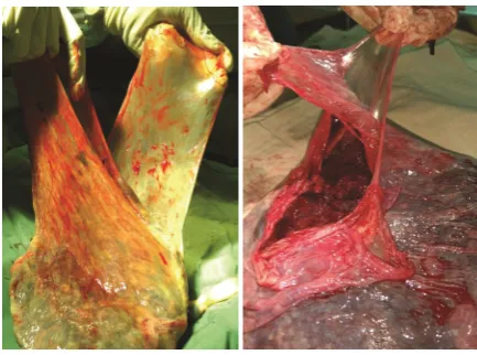
Fetal chorioamniotic tissues of each placenta re-moved during c-section were manually peeled in sterile conditions (Figure 1). The peeled
mem-branes were washed immediately and separately using sterile phosphate buffered saline solution (PBS) so that debris, blood and possibly remnants of amniotic fluid were washed away (8). The tis-sue samples were transferred to the laboratory at an interval of half an hour and at 4°C. Then, at least four 1x1 cm sized pieces were rapidly re-moved from each peeled amniotic and chorionic membranes using scalpel in a Class II biological safety cabinet. The resulting tissue pieces were kept in sterile PBS solution for a maximum of two hours at 4°C until the time for transition to bacte-riologic media.
Studiedbacteria:Thetissuesampleswerechanged to suspension at first. Antimicrobial effects of all fetal tissue samples were tested on eight standard bacterial strains including seven pathogens: E. coli (ATCC25922), Bacillus cereus (ATCC11778),
Klebsiella pneumoniae (ATCC700603),
Streptoco-ccus pyogenes (ATCC19615), Pseudomonas
aeru-ginosa (ATCC27883), Staphylococcus aureus (AT
CC29213), Shigella flexneri (ATCC12022) and one probiotic: Lactobacillus plantarum (PTCC1745). All the strains were obtained in lyophilized stocks from Microbial Collection Department of Pasteur Institute of Iran.
Cultivation of bacteria: Bacterial recovery was carried out by inoculating lyophilized bacteria in nutrient broth medium (Merck, Germany) and incubating them for 24 hr at 37°C. The disk diffu-sion method was used to test the antibacterial ef-fects of amniotic and chorionic membranes indi-vidually. Initially, 100 μl of each bacterial suspen-sion equal to the McFarland 0.5 standard (1.5X10⁸
CFU/ml) was prepared in normal saline. Then, the
bacterial suspensions were cultured in plastic petri dishes (80 mm DIC) containing Mueller Hinton Agar medium (Merck, Germany) using spread
Figure 1. Peeling chorionic and amniotic membranes from
Downloaded from ht
tp://www.jr
i.ir
220 J Reprod Infertil, Vol 18, No 2, Apr-Jun 2017
Antimicrobial Properties of Fetal Sacs
JRI
plate technique (8, 11). Exceptionally, the blood agar medium (Merck, Germany) was used to cul-ture Streptococcus pyogenes. In the next step, the frag-mentedamnioticandchorionicmembranesof each tissue sample were placed on the cultured petri dishes, so that their possible antibacterial components spread on the medium. For each bac-terial strain, minimum of 4 pieces of amniotic membrane and 4 pieces of chorionic membrane of each placenta were used. Each of these quaternary sets was called a tissue group. Umbilical cord and a collection of antibiotic discs including erythro-mycin,ciprofloxacin,cefixime,ceftriaxone,cepha- lexin, amikacin, imipenem were used as negative and positive controls, respectively. All procedures were performed in accordance with Clinical and Laboratory Standards Institute (CLSI) guidelines (12).
In all culture cases, the plates were incubated at 37°C for 24 hr and finally the zone of inhibition (ZOI) of each bacterial strain around individual fragments of amniotic and chorionic membranes was measured using a caliper and recorded (13). In examining the culture results, only cases were recorded as growth inhibition, in which growth inhibition zone was a minimum of 1 mm around at least two out of four fragmented membranes of each tissue group.
In each plate, on which growth inhibition zone was observed, a swab was taken from the agar surface where a fetal membrane was laid as well as the marginal inhibition zone. The swabs were re-cultured and incubated for 24 hr at 37°C in the absence of any fetal membrane. The results were recorded in order to evaluate the bacteriostatic or bactericidal activity of the growth inhibition.
Data analysis: Initially, the normality of the data was controlled by the One-Sample K-S (Kolmo-gorov-Smirnov) Test. Results of bacterial growth inhibition in the presence of amniotic or chorionic membranes were reported as median±IQR. Data analysis was performed by SPSS (v.18) using Kruskal-Wallis and Mann-Whitney U-test.
Results
Growth inhibition: The size of bacterial growth inhibition zones differed, depending on the type of bacteria, type of membrane (amnion or cho-rion) and the placenta sample. The inhibition zone size ranged between 0-12 mm (Figures 2 and 3).
Growth inhibition zones around the amniotic membranes were observed in more than 70% of
samples for all the bacteria other than
Staphylo-coccus aureus. Growth inhibition zones around
chorionic membranes were observed in more than 80% of samples for all the bacteria other than
Staphylococcus aureus and Streptococcus
pyo-genes (Figure 4).
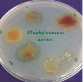
Figure 2. Formation of inhibitory zone of Bacillus cereus
(ATCC11778) around amniotic tissue
Figure 3. Formation of inhibitory zone of Staphylococcus
aureus (ATCC29213) around amniotic tissue
Figure 4. Comparing number of tissue groups having zone of
inhibition with the total number of groups (%) in chorionic and amniotic membranes based on bacterial strain. The min-imum and the maxmin-imum sizes of inhibitory zone were 1 mm
Downloaded from ht
tp://www.jr
i.ir
J Reprod Infertil, Vol 18, No 2, Apr-Jun 2017 221 Zare-Bidaki M, et al.
JRI
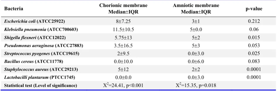
Sizes of the inhibitory zones: Each of the amniotic and chorionic membranes showed different sizes of inhibitory zone on the eight bacterial strains (Table1).Thenumberofgrowthinhibitionsaround chorionic membranes was significantly more than that of the amniotic membranes. These effects were mostly found in the chorionic membranes (Figure 1).
Using Kruskal-Wallis test, the inhibition zones around the amniotic membranes were significant-ly different among the bacteria (p=0.018). Post-hoc analysis following a Mann-Whitney U-test showed that the inhibition zone for Bacillus cere-us and Klebsiella pneumoniae was significantly greater than the one found for Staphylococcus aureus (p<0.01).
Performing similar analysis, the inhibition zone around chorionic membranes was significantly different among the bacteria (p=0.001). The inhi-bition zone for Staphylococcus aureus was signif-icantly more than the one found for E. coli (p= 0.012) and Streptococcus pyogenes (p<0.001). Also the inhibition zone for Pseudomonas
aeru-ginosa was more than the one found for
Strepto-coccus pyogenes (p<0.05).
Lactobacilli plantarum revealed the minimum
size of inhibitory zone around both amniotic and chorionic membranes. This strain significantly did not show zone of inhibition around any of the chorionic membrane samples (p<0.001).
No positive culture was found on plates inocu-lated by the swabs taken from agar surfaces where amniotic or chorionic membranes were laid.
Discussion
The findings of the present study revealed that amniotic and chorionic membranes independently provide growth inhibitory effects on a varied and
different range of bacteria. It also disclosed that the antimicrobial effects of fetal membranes are significantly stronger on some certain bacteria. Growth inhibition of E. coli and Bacillus cereus
was found nearly at a similar level around both types of fetal membranes, which in some part was in agreement with what Parthasarathy et al. ob-served (11). However, Pseudomonas aeruginosa,
Staphylococcus aureus and Shigella flexneri were
more intensively under inhibitory effects of chori-onic membrane which was not observed by Par-thasarathy et al. This disagreement could rise due to some different reasons. They did not report the number of their tissue samples and also did not conduct any statistical analysis which might be due the low number of their tissue samples. More-over, they worked only on clinical pathogens which might have different effects in comparison with the standard bacterial strains. In concordance with our study, in Kjaegaard et al.’s work, the growth inhibition was more pronounced for some of bacteria than for others (8). Similar findings of ours were previously reported for amniotic mem-brane by Tehrani et al., whereas they did not study antibacterial effects of chorionic membrane (14).
The most important finding of the current study was the superiority observed in antibacterial ef-fects of the chorionic membrane compared with the amniotic membrane. The only previous study in this field is a descriptive and non-quantitative study conducted by Kjaergaard et al. in 2001 (8). Although they demonstrated antibacterial effects in both amniotic and chorionic membranes, more severe yet limited antibacterial effects was report-ed for chorionic membrane which had been ob-served only in the liquid medium. Perhaps the small sample size in their study made it impossi-ble to analyze their data quantitatively for
com-Table 1. The median values of growth inhibition zones around the amniotic and chorionic membranes
Bacteria Chorionic membrane MedianIQR Amniotic membrane MedianIQR p-value
Escherichia coli (ATCC25922) 87.25 31 0.212
Klebsiella pneumonia (ATCC700603) 11.510.5 50.0 0.06
Shigella flexneri (ATCC12022) 5.7513 52 0.015
Pseudomonas aeruginosa (ATCC27883) 3.516.5 53 0.053
Streptococcus pyogenes (ATCC19615) 29.5 0.03.0 0.025
Bacillus cereus (ATCC11778) 0.010.0 0.06.0 0.083
Staphylococcus aureus (ATCC29213) 512 22 0.0001
Lactobacilli plantarum (PTCC1745) 0.00.0 0.03.0 0.0001
Downloaded from ht
tp://www.jr
i.ir
222 J Reprod Infertil, Vol 18, No 2, Apr-Jun 2017
Antimicrobial Properties of Fetal Sacs
JRI
paring the antibacterial effects of the two fetal membranes on solid medium.
According to our findings, when the swabs taken from the agar surface below the fetal membrane as well as the marginal inhibition zone, were cul-tured on a new agar plate, no bacterial growth was observed. One might speculate that the antibacte-rial effects in both amniotic and chorionic mem-branes are bactericidal. In the present study, which is an example of an in vitro model to test the antibacterial effects of fetal membranes, the above theory was presumably proved.
The nature of antibacterial factors in amniotic and chorionic membranes and the quantitative or substantive differences that are likely to exist be-tween these fetal membranes are disputed. Kjaer-gaard et al. demonstrated that for applying antimi-crobial effects of fetal membranes, no direct con-tact between the bacteria and tissue is needed. They managed to apply this property even through a filter on bacteria (8). Also, Tehrani et al. demon-strated that there is no difference between mesen-chymal and optimal levels of amniotic membrane in terms of antibacterial effects (14). Generally, these findings suggest that the antibacterial effects in the fetal membranes are not tissue-dependent.
The presence of some specific productions of immune system, such as IgA in the fetal mem-branes in the edge detached from the placenta has beenproved(15).EvenBerezoswskietal.have de-monstrated the presenceof considerable amounts of this immunoglobulin in chorioamniotic mem-branes of women with premature rupture of fetal membrane (16). However, it is rather implausible that IgA could be a major factor of bacterial growth inhibition around the fetal membranes of healthy and full-term pregnancies in non-inflam-matory conditions. Some other antimicrobial com-pounds have been reported in the fetal mem-branes. Human beta-defensin is a large group of natural antibacterial proteins produced by a num-ber of epithelial cells including the chorioamniotic membranes (17, 18). HBD3 compound is a domi-nant defensin in the amniotic epithelial. Defen-sin’s inhibitory effects, especially β3-defensin, have been shown against a number of pathogens (18, 19).
Elafin and SLPI (Secretory Leukocyte Protease Inhibitor) are other anti-bacterial agents, the pres-ence of which in amnion tissue has been reported (5, 20). In vitro studies have shown that SLPI is excreted by both Decidua levels, including Pari-talis and Basalis (20). Both Elafin and SLPI are
among the peptides that have anti-protease and elastase inhibition activity and as components of the innate immune system protect surfaces in con-tact with contamination by controlling the inflam-matory responses in the mucosal surface. It has been shown that Elafin is secreted by the endome-trial epithelial cells and amniotic membrane (5, 21). Elafin's inhibitory effects against
Staphylo-coccus aureus and Pseudomonas aeruginosa have
been previously reported (22).
In addition to anti-bacterial protein compounds, the presence of compounds such as lactoferrin and hyaluronic acid, both of which are known as anti-inflammatory and antimicrobial properties, has been reported in the amniotic membrane (23, 24). In addition to antimicrobial compounds known in fetal membranes, several antibacterial factors have been also reported in amniotic fluid. The role of antimicrobial agents, including transferrin, lyso-zyme, 7S immunoglobulin, globulin β1c/β1a, IgA and also some peptides, such as α-defensins (HNP1-3) and calprotectin has been demonstrated in amniotic fluid (25-27). However, it does not seem that after the withdrawal of amniotic fluid, these agents still remain at the fetal membranes surfaces and play a role in the appearance of its experimental antibacterial effects on the culture media. More importantly, in the present study, the fetal membranes were carefully washed in PBS solution before use, in order to ensure that the amniotic fluid does not stay on their surface.
One might argue that the superiority of antibac-terial effects of the chorionic membrane compared with the amniotic membrane can represent more effective and key role in maternal part of placenta in protecting the fetus against possible infections.
Despite conclusive experimental evidence indi-cating the existence of antimicrobial factors in the fetal membranes, no investigation report of quan-titative comparisons of the type and frequency of antimicrobial factors between chorionic and am-niotic membranes was found in international da-tabases. Further researches are needed to clarify the exact contribution of each antimicrobial com-pound found in amniotic and chorionic mem-branes in the emergence of antibacterial properties of these membranes in vitro as well as to obtain a quantitative understanding of antimicrobial activi-ties in each fetal membrane.
Downloaded from ht
tp://www.jr
i.ir
J Reprod Infertil, Vol 18, No 2, Apr-Jun 2017 223 Zare-Bidaki M, et al.
JRI
other limitation was the elastic nature of fetal membranes which made it difficult to cut off the membrane samples into fragments exactly at the same size.
Conclusion
This study aimed to compare the antibacterial properties of amniotic and chorionic membranes using placental tissue of 43 Iranian women in Bir-jand, South Khorasan Province. This study re-vealed that not only the antimicrobial effect of amniotic and chorionic membranes is significantly different on various bacteria, but also the antibac-terial effect level of chorionic membrane is great-er than amniotic one.
Acknowledgement
This article is results of a research project ap-proved and granted (Code: 29/93) by the vice chancellor of Birjand University of Medical Sci-ences. Hereby, authors sincerely appreciate Ms. Nahid Askari, the senior technologist in medical microbiology research laboratory, the staff in gen-ecology ward of Vali-asr Hospital and Mr. Alireza Ehteshampour, BSc of health sciences, all at Birjand University of Medical Sciences for their sincere cooperation.
Conflict of Interest Authors declare no conflict of interest.
References
1. Stirling KM, Hussain N, Sanders MM, Campbell W. Association between maternal genital mycoplasma colonization and histologic chorioamnionitis in pre-term births. J Neonatal Perinatal Med. 2016;9(2): 201-9.
2. Nadeau HC, Subramaniam A, Andrews WW. Infec-tion and preterm birth. Semin Fetal Neonatal Med. 2016;21(2):100-5.
3. Vinturache AE, Gyamfi-Bannerman C, Hwang J, Mysorekar IU, Jacobsson B; Preterm Birth Interna-tional Collaborative (PREBIC). Maternal micro-biome - A pathway to preterm birth. Semin Fetal Neonatal Med. 2016;21(2):94-9.
4. Yoneda N, Yoneda S, Niimi H, Ueno T, Hayashi S, Ito M, et al. Polymicrobial amniotic fluid infection with Mycoplasma/Ureaplasma and other bacteria in-duces severe intra-amniotic inflammation associated with poor perinatal prognosis in preterm labor. Am J Reprod Immunol. 2016;75(2):112-25.
5. Stock SJ, Kelly RW, Riley SC, Calder AA. Natural antimicrobial production by the amnion. Am J Ob-stet Gynecol. 2007;196(3):255.e1-6.
6. Sadler TW. Langman's medical embryology. 12th ed. Philadelphia: Lippincott Williams & Wilkins; 2015. 400 p.
7. Talmi YP, Sigler L, Inge E, Finkelstein Y, Zohar Y. Antibacterial properties of human amniotic mem-branes. Placenta. 1991;12(3):285-8.
8. Kjaergaard N, Hein M, Hyttel L, Helmig RB, Sch-onheyder HC, Uldbjerg N, et al. Antibacterial prop-erties of human amnion and chorion in vitro. Eur J Obstet Gynecol Reprod Biol. 2001;94(2):224-9. 9. Sangwan VS, Basu S. Antimicrobial properties of
amniotic membrane. Br J Ophthalmol. 2011;95(1): 1-2.
10. Zare Bidaki M, Lessani T, Khazaie Z. [Evaluation of anti-bacterial effects of chorionic membranes in vitro]. Birjand Univ Med Sci J. 2012;19(2):140-6. Persian.
11. Parthasarathy M, Sasikala R, Gunasekaran P, Raja J. Antimicrobial activity of human amniotic and chorionic membranes. J Acad Ind Res. 2014;2(10): 545-7.
12. Clinical and Laboratory Standards Institute (CLSI). Performance standard forantimicrobial susceptibil-ity testing standard M02-A12. Clinical and Labora-tory Standards Institute; 2015. 16 p.
13. Soltan-Dallal MM, Kalafi Z, Rastegar-Lari A, Hos-seini S, Rahimi-Foroushani A, Deilami-Khiabani Z, et al . The effect of reduced bacterial dilution on human amniotic membrane antibacterial activity, in vitro. Zahedan J Res Med Sci. 2013;15(5):6-8. 14. Asi Tehrani F, Peirovi H, Niknejad H.
[Determina-tionofantibacterialeffectoftheepithelialand mes-enchymal surfaces of amniotic membrane on Esch-erichia coli, Staphylococcus aureus]. Qom Univ Med Sci J. 2013;7(4):12-22. Persian.
15. Cunha SP, Berezowski AT, Costa MW, Costa JC, Ribeiro dos Santos R, Duarte G. Demonstration of the presence of IgA in the human chorioamniotic membrane. Int J Gynaecol Obstet. 1984;22(2):107-10.
16. Berezoswski AT, Cunha SP, da Costa JC, Bacchi CE.QuantificationofimmunoglobulinAin chorio-amnioticmembraneofpatientswithpremature rup-ture of membranes. Int J Gynaecol Obstet. 1994;47 (1):23-6.
17. Ganz T. Defensins: antimicrobial peptides of in-nate immunity. Nat Rev Immunol. 2003;3(9):710-20.
Downloaded from ht
tp://www.jr
i.ir
224 J Reprod Infertil, Vol 18, No 2, Apr-Jun 2017
Antimicrobial Properties of Fetal Sacs
JRI
19. Buhimschi IA, Jabr M, Buhimschi CS, Petkova AP, Weiner CP, Saed GM. The novel antimicrobial peptide beta3-defensin is produced by the amnion: a possible role of the fetal membranes in innate im-munityoftheamnioticcavity.AmJObstet Gynecol. 2004;191(5):1678-87.
20. DenisonFC,KellyRW,CalderAA,RileySC. Se-cretory leukocyte protease inhibitor concentration increases in amniotic fluid with the onset of labour in women: characterization of sites of release with-in the uterus. J Endocrwith-inol. 1999;161(2):299-306. 21. Majchrzak-Gorecka M, Majewski P, Grygier B,
Murzyn K, Cichy J. Secretory leukocyte protease inhibitor (SLPI), a multifunctional protein in the host defense response. Cytokine Growth Factor Rev. 2016;28:79-93.
22. Williams SE, Brown TI, Roghanian A, Sallenave JM. SLPI and elafin: one glove, many fingers. Clin Sci (Lond). 2006;110(1):21-35.
23. Kanyshkova TG, Buneva VN, Nevinsky GA. Lac-toferrin and its biological functions. Biochemistry (Mosc). 2001;66(1):1-7.
24. Higa K, Shimmura S, Shimazaki J, Tsubota K. Hy-
aluronic acid-CD44 interaction mediates the adhe-sionoflymphocytesbyamnioticmembranestroma. Cornea. 2005;24(2):206-12.
25. Yoshio H, Tollin M, Gudmundsson GH, Lagercra-ntz H, Jornvall H, Marchini G, et al. Antimicrobial polypeptides of human vernix caseosa and amniot-ic fluid: implamniot-ications for newborn innate defense. Pediatr Res. 2003;53(2):211-6.
26. Espinoza J, Chaiworapongsa T, Romero R, Edwin S, Rathnasabapathy C, Gomez R, et al. Antimicro-bial peptides in amniotic fluid: defensins, calpro-tectin and bacterial/permeability-increasing protein in patients with microbial invasion of the amniotic cavity, intra-amniotic inflammation, preterm labor and premature rupture of membranes. J Matern Fe-tal NeonaFe-tal Med. 2003;13(1):2-21.