E X T E N D E D G E N O M E R E P O R T
Open Access
High-quality genome sequence of the
radioresistant bacterium
Deinococcus ficus
KS 0460
Vera Y. Matrosova
1,2†, Elena K. Gaidamakova
1,2†, Kira S. Makarova
3, Olga Grichenko
1,2, Polina Klimenkova
1,2,
Robert P. Volpe
1,2, Rok Tkavc
1,2, Gözen Ertem
1, Isabel H. Conze
1,4, Evelyne Brambilla
5, Marcel Huntemann
6,
Alicia Clum
6, Manoj Pillay
6, Krishnaveni Palaniappan
6, Neha Varghese
6, Natalia Mikhailova
6, Dimitrios Stamatis
6,
TBK Reddy
6, Chris Daum
6, Nicole Shapiro
6, Natalia Ivanova
6, Nikos Kyrpides
6, Tanja Woyke
6, Hajnalka Daligault
7,
Karen Davenport
7, Tracy Erkkila
7, Lynne A. Goodwin
7, Wei Gu
7, Christine Munk
7, Hazuki Teshima
7, Yan Xu
7,
Patrick Chain
7, Michael Woolbert
1,2, Nina Gunde-Cimerman
8, Yuri I. Wolf
3, Tine Grebenc
9, Cene Gostin
č
ar
8and Michael J. Daly
1*Abstract
The genetic platforms ofDeinococcusspecies remain the only systems in which massive ionizing radiation (IR)-induced genome damage can be investigated in vivo at exposures commensurate with cellular survival. We report the whole genome sequence of the extremely IR-resistant rod-shaped bacteriumDeinococcus ficusKS 0460 and its phenotypic characterization.Deinococcus ficusKS 0460 has been studied since 1987, first under the nameDeinobacter grandis, thenDeinococcus grandis. TheD. ficusKS 0460 genome consists of a 4.019 Mbp sequence (69.7% GC content and 3894 predicted genes) divided into six genome partitions, five of which are confirmed to be circular. Circularity was determined manually by mate pair linkage. Approximately 76% of the predicted proteins contained identifiable Pfam domains and 72% were assigned to COGs. Of allD. ficusKS 0460 proteins, 79% and 70% had homologues in
Deinococcus radioduransATCC BAA-816 andDeinococcus geothermalisDSM 11300, respectively. The most striking differences betweenD. ficusKS 0460 andD. radioduransBAA-816 identified by the comparison of the KEGG pathways were as follows: (i)D. ficuslacks nine enzymes of purine degradation present inD. radiodurans, and (ii)D. ficuscontains eight enzymes involved in nitrogen metabolism, including nitrate and nitrite reductases, thatD. radioduranslacks. Moreover, genes previously considered to be important to IR resistance are missing inD. ficusKS 0460, namely, for the Mn-transporternramp, and proteins DdrF, DdrJ and DdrK, all of which are also missing inDeinococcus deserti.
Otherwise,D. ficusKS 0460 exemplifies theDeinococcuslineage.
Keywords:Deinococcus-Thermus,Deinococcaceae,Deinococcus ficus, Radiation-resistant, Rod-shaped, Phenotype characterization, Genome analysis, Phylogenetic analysis
* Correspondence:[email protected] †Equal contributors
1Uniformed Services University of the Health Sciences, School of Medicine, Bethesda, MD, USA
Full list of author information is available at the end of the article
Introduction
Species of the genusDeinococcushave been studied for their extreme IR resistance since the isolation of Deinococcus
radioduransin 1956 [1]. Since then, many other species of
the same genus have been isolated. The current number of recognized Deinococcus species is greater than 50 while there are more than 300 non-redundant 16S rRNA sequences of the familyDeinococcaceaein the ARB project database [2]. Apart from Deinococcus ficus KS 0460, only a few other representatives have been studied in detail for their oxidative-stress resistance mechanisms:
D. radiodurans,Deinococcus geothermalisandDeinococcus
deserti[3].The picture that has emerged for the life cycle
of most Deinococcus species is one comprised of a cell-replication phase that requires nutrient-rich conditions, such as in the gut of an animal, followed by release, drying and dispersal [1]. Desiccated deinococci can endure for years, and, if blown by winds through the atmosphere, are
expected to survive and land worldwide. As reported, some deinococci become encased in ice, and some entombed in dry desert soils. High temperatures also are not an obstacle to the survival of some deinococcal species.D. geothermalis
andDeinococcus murrayiwere originally isolated from hot
springs in Italy and Portugal, respectively [1]. The pros-pects of harnessing the protective systems ofD. radiodur-ansfor practical purposes are now being realized.
The complete genome sequence presented here is forD.
ficus KS 0460, originally named Deinobacter grandis KS
0460, isolated in 1987 from feces of an Asian elephant
(Elephas maximus) raised in the Ueno Zoological Garden,
Tokyo, Japan (Table 1) [4]. Later,Deinobacter grandiswas renamed Deinococcus grandis [5]. Strain KS 0460 was acquired by USUHS from the originating laboratory in 1988 by Kenneth W. Minton and has been the subject of study here ever since. As a candidate for bioremediation of radioactive DOE waste sites [6] and a target of study for
Table 1Classification and general features ofDeinococcus ficusKS 0460 according to MIGS recommendations [49]
MIGS ID Property Term Evidence codea
Classification DomainBacteria TAS [50]
PhylumDeinococcus-Thermus TAS [51,52]
ClassDeinococci TAS [53,54]
OrderDeinococcales TAS [5]
FamilyDeinococcaceae TAS [5,55]
GenusDeinococcus TAS [5,55]
SpeciesDeinococcus ficus TAS [4,9]
Strain: KS 0460
Gram stain Variable TAS [4,9]
Cell shape Rod TAS [4,9]
Motility Non-motile TAS [4,9]
Sporulation None TAS [4,9]
Temperature range Mesophile TAS [4,9]
Optimum temperature 30-37 °C TAS [4,9]
pH range; Optimum e.g. 5.5–10.0; 7.0 TAS [4,9]
Carbon source Glucose, fructose TAS [9]
MIGS-6 Habitat Elephas maximusfeces TAS [4]
MIGS-6.3 Salinity 1% NaCl (w/v) TAS [4]
MIGS-22 Oxygen requirement Aerobic TAS [4]
MIGS-15 Biotic relationship Free-living NAS
MIGS-14 Pathogenicity Non-pathogen NAS
MIGS-4 Geographic location Tokyo/Japan TAS [4]
MIGS-5 Sample collection 1987 TAS [4]
MIGS-4.1 Latitude Non reported
MIGS-4.2 Longitude Non reported
MIGS-4.4 Altitude Non reported
a
DNA repair [7], D. ficusKS 0460 was chosen for whole genome sequencing. The D. ficus KS 0460 genome now adds to the growing number of sequenced Deinococcus species needed to decipher the complex extreme IR resist-ance phenotype. To date, a genetic explanation for the complex survival tactics of deinococci has not been provided by comparative genomics or transcriptomics [8].
Organism information Classification and features
In a chemotaxonomic study published in 1987, an isolate (strain KS 0460) from γ-irradiated feces of an Asian elephant yielded an IR-resistant bacterium with a wall structure, cellular fatty acid composition, and GC content typical of members of the genusDeinococcus[4]. However, strain KS 0460 was rod-shaped and grew as pink-pigmented colonies, whereas most other deinococci grow as diplococci/tetracocci and yield red colonies. The ori-ginal isolate was namedDeinobacter grandis, but was later renamedDeinococcus grandisbased on its close phylogen-etic relationship (16S rRNA sequences) with deinococci [5]. Strain KS 0460 was subsequently included in experi-mental IR survival studies together with otherDeinococcus species, where it was referred to as grandis[7]. Our 16S rRNA phylogenetic analysis confirms that strain KS 0460
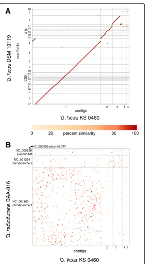
belongs to the genusDeinococcus, most closely related to the type strain of Deinococcus ficus DSM 19119 (also referred to as CC-FR2-10) (Fig. 1).
Consistent with the original description of D. ficus KS 0460, the rod-shaped cells are 0.5 to 1.2 μm by 1.5 to 4.0μm (Fig. 2a) and grow as pink colonies [4, 9].D. ficus KS 0460 was shown to have a D10 of approximately 7 kGy (Co-60) (Fig. 2b) and is capable of growth under chronic γ-irradiation at 62 Gy/h (Cs-137) (Fig. 2c). The cells are aerobic, incapable of growth under anaerobic conditions on rich medium, irrespective of the presence or absence of chronic IR (Fig. 2c). The general structure of the D. ficus KS 0460 genome was analyzed by PFGE of genomic DNA prepared from embedded cells. The plugs containing digested cells were exposed to 200 Gy prior to electrophoresis, a dose gauged in vitro to induce approximately 1 DNA double strand break per chromo-some in the range 0.5 - 2 Mbp [10]. Fig. 2d shows the presence of the five largest genomic partitions: main chromosome (~2.8 Mbp), 3 megaplasmids (~500 kb, ~400 kb and ~200 kbp) and one plasmid (~98 kbp), predicting a genome size ~4.0 Mbp. We did not observe the smallest genome partition (0.007 Mbp) by PFGE. The growth characteristics ofD. ficus KS 0460 in liquid culture at 32 and 37 °C (Fig. 2e) are very similar to D.
-8 -6 -4 -2 0
0 5 10 15 20 25
lo
g
su
rv
iv
a
l
kGy
0 10 20 30 40 50 60
DR DF
concentration (µ
g/L)
Mn Fe
A
B
C
D
E
F
M DF
1 2 3
4 5 6
7 8 9
G
0.5 m
net AUC
0.098
Mbp
2.2 1.6 1.125 0.945 0.825 0.75
0.45 0.365 0.285 0.225 No IR 62 Gy/h
O-2
O
+
2
DR DF
Mn/Fe ratio
0.4
0 0.1 0.2 0.3
EC
DF DR
EC
DF DR
EC
DF DR
EC
DF DR
H
Mbp
2.84
0.49 0.39
0.2
0 10 20 30 40 50 60 70
0 0.1 0.2 0.3
ultrafiltrate concentration 0
1 2 3 4 5
0 20 40 60 80 100 120
OD
600
hours
Fig. 2Deinococcus ficusKS 0460 (EXB L-1957) phenotype.aTransmission electron micrograph.D. ficusgrown in TGY, early-stationary phase.b Survival ofD. radioduransBAA-816 (red),D. ficus(blue), andE. coli(strain K-12, MG1655) (black) exposed to acute IR. The indicated strains were inoculated in liquid TGY and grown to OD600~ 0.9. Cells were then irradiated on ice with Co-60.cD. ficusis an aerobe capable of growth under
62 Gy/h. DR,D. radiodurans; DF,D. ficus; EC,E. coli.dPFGE of genome partitions in a 0.9% agarose gel. PFGE conditions: 0.5 × TBE, 6 V/cm with a 10 to 100 s switch time ramp at an included angle of 120°, 14 °C, 18 h. M, markerS. cerevisiaeYNN (BioRad).eGrowth curves at 37 °C (blue) and 32 °C (black) in TGY medium.fICP-MS on Mn and Fe content ofD. radioduransBAA-816 andD. ficus. Inset: Mn/Fe ratios.gProtease secretion assay. Halos indicate activity of proteases [60]. Strains: 1.D. radioduransBAA-816, 2.D. geothermalisDSM 11300, 3.D. ficusKS 0460, 4.D. murrayi (MD591), 5.D. radiopugnans(MD567), 6.D. radiodurans(MD878, SX-108-7B-1, [61]), 7.D. proteolyticus(MD568), 8.D. proteolyticus(MD628, [62]), and 9.D. proteolyticus(MD869).hAntioxidant capacities ofD. radioduransBAA-816 (red),D. ficus(blue), andE. coli(strain K-12, MG1655)
(black) ultrafiltrates assessed by antioxidant assay as described previously [63, 64]. Net AUC is an integrative value of a total fluorescence during
radiodurans [11]. It is unknown if strain D. ficus KS 0460 is genetically tractable because the cells are natur-ally resistant to the antibiotics tetracycline, chloram-phenicol and kanamycin at concentrations needed to select for plasmids and integration vectors designed for
D. radiodurans[12] (data not shown). D. ficusKS 0460,
like other deinococci, accumulate high concentrations of Mn2+(Fig. 2f ) [7, 13]. Bacterial Mn2+accumulation was previously shown to be important to extreme IR resist-ance, mediated by the Mn transport gene nramp and ABC-type Mn-transporter gene [14]. We also showed
thatD. ficusKS 0460 produces proteases, as detected in a
protease secretion assay on an indicator plate containing skimmed milk (Fig. 2g). For example, in D. radiodurans, the products of proteases–peptides–form Mn2+-binding ligands of Deinococcus Mn antioxidants, which protect proteins from IR-induced ROS, superoxide in particular [8, 13, 15]. Finally, we show thatD. ficusKS 0460 cells have a high intracellular antioxidant capacity (Fig. 2h), which is a strong molecular correlate for IR resistance [1, 11].
Extended feature descriptions
16S rDNA gene phylogenetic analysis was based on sequences from 22 type strains of genus Deinococcus including ten from completely sequenced genomes, and two from Deinococcus ficus strains KS 0460 and DSM 19119; andTruepera radiovictrixDSM 17093, the distinct species shown to be an outgroup to theDeinococcusgenus [16]. The maximum-likelihood phylogenetic trees were reconstructed using two approaches: (i) the FastTree pro-gram [17], with GTR substitution matrix and gamma-distributed evolutionary rates and maximum-likelihood algorithm; and (ii) PHYML program with the same parameters (Fig. 1 and Additional file 1: Figure S1) [18].
BothD. ficusstrains, as expected, group together, but the
position of this pair in both trees is poorly resolved (37 support value for FastTree method and 44 for PHYML method) potentially because of the long branch of this clade. In both trees, however, theD. ficusclade confidently groups deep in the Deinococcus tree within the branch
withD. gobiensisas a sister clade.
Genome sequencing information Genome project history
Deinococcus ficusKS 0460 was obtained from the Oyaizu
laboratory and was entered into the Daly strain collec-tion at USUHS on November 18, 1997. The strain was submitted to the EX Culture Collection, Mycosmo, Slovenia, on December 29, 2016 and was issued an accession number EXB L-1957. The genome of D. ficus KS 0460 was sequenced at the JGI. The project was initi-ated in 2009, the genome was released on August 26, 2012 as “Deinococcus sp. 2009”. The genome ofD. ficus KS 0460 has the status of an improved high-quality
draft. The genome assembly and annotation can be accessed through the JGI genome portal [19] and also GenBank [20]. The genome is considered to be near-complete. The search for bacterial Benchmarking Universal Single-Copy Orthologs [21] found a compar-able number of orthologs inD. ficusKS 0460 and in ten complete Deinococcusspecies genomes. Furthermore, of the 875 genes representing the core genome of the same ten complete Deinococcus species as determined by the GET_HOMOLOGUES pipeline [22], only five genes were missing fromD. ficusKS 0460.
Growth conditions and genomic DNA preparation
D. ficus KS 0460 was recovered from a glycerol frozen
stock on TGY solid rich medium (1% bactotryptone, 0.1% glucose, and 0.5% yeast extract, 1.5% w/v bacto agar) (3 days, 32 °C) with following inoculation of 25 ml TGY medium. The culture was grown up to OD600~ 0.9. Sub-sequently, 19 ml were used to inoculate 2 L of TGY medium and the culture was grown at 32 °C, overnight in aerated conditions in a shaker incubator (200 rpm). The cells were harvested at OD600 ~ 1.6. The DNA was iso-lated from a cell pellet (5.6 g) using Jetflex Genomic DNA Purification Kit (GENOMED, Germany). The final DNA concentration was 80μg ml−1, in a volume of 800μl. The DNA was RNA free and passed quality control.
Genome sequencing and assembly
were corrected with manual editing in Consed [25–27]. Gap closure was accomplished using repeat resolution software [Wei Gu, unpublished], and sequencing of bridging PCR fragments with Sanger and/or PacBio technologies [Cliff Han, unpublished]. A total of 21 PCR PacBio consensus sequences were completed to close gaps and to raise the quality of the final sequence.
Genome annotation
The genome sequence was annotated using the JGI Prokaryotic Automatic Annotation Pipeline [28] and further reviewed using the Integrated Microbial Genomes -Expert Review platform [29]. Genes were predicted using Prodigal [30], followed by a round of manual curation using the JGI GenePRIMP pipeline [31]. The genome sequence was analyzed and released publicly through the Integrated Microbial Genomes platform [32]. BLASTClust was used to identify internal clusters with thresholds of 70% covered length and 30% sequence identity [33]. SignalP [34] and TMHMM [35] were used to predict signal peptides and transmembrane helices, respectively.
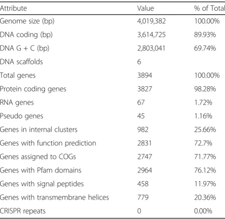
Genome properties
TheD. ficus KS 0460 genome consists of a 4,019,382 bp
sequence which represents six genome partitions: 2.84, 0.49, 0.39, 0.20, 0.098 and 0.007 Mbp (Table 3), consistent with PFGE (Fig. 2d); note, the smallest partition (0.007 Mbp) was too small to resolve by PFGE. The final assembly was based on 4946 Mbp of Illumina draft data, which provided
Table 3Summary of genome: one chromosome and five
plasmids
Label Size (Mbp) Topology INSDC identifier RefSeq ID
Chromosome 2.84 circular ATTJ01000001 ATTJ01000001
Megaplasmid 1 0.49 circular ATTJ01000002 ATTJ01000002
Megaplasmid 2 0.39 circular ATTJ01000003 ATTJ01000003
Megaplasmid 3 0.20 unknown ATTJ01000004 ATTJ01000004
Plasmid 1 0.098 circular ATTJ01000005 ATTJ01000005
Plasmid 2 0.007 circular ATTJ01000006 ATTJ01000006
Table 2Project information
MIGS ID Property Term
MIGS 31 Finishing quality High-Quality Draft
MIGS-28 Libraries used Illumina Standard (short insert paired-end) and Illumina CLIP (long insert paired-end)
MIGS 29 Sequencing platforms Illumina HiSeq 2000 (CLIP library); Illumina HiSeq 2000 (Standard library); PacBio
MIGS 31.2 Fold coverage 1237×
MIGS 30 Assemblers Allpaths r38445 and Velvet 1.1.05
MIGS 32 Gene calling method Prodigal within JGI Prokaryotic Automatic Annotation Pipeline
Locus Tag DEINO
Genbank ID ATTJ00000000.1
GenBank Date of Release 07/09/2013
GOLD ID Gp0007971
BIOPROJECT PRJNA157079
MIGS 13 Source Material Identifier EXB L-1957
Project relevance DNA repair mechanisms, bioremediation
D. ficus KS 0460
D. ficus
DSM 19119
percent similarity
0 20 80 100
1 2 3 4 5
27 2 9 16 11 308 2325 2421 2014 10 7 6 1 4 12 19 2215
2613
17 3 5 18
contigs
scaffolds
D. ficus KS 0460
D. radiodur
ans
BAA-816 NC_001263 chromosome 1
NC_001264 chromosome 2
NC_000958 plasmid M1
NC_000959 plasmid CP1
1 2 3 4 5
contigs
A
B
an average of 1237× coverage of the genome. The total genomic GC content was 69.7% and was similar across all but the smallest contig, which contained 62.5% GC. The genome contains 3827 predicted protein-coding genes and 67 RNA-coding genes (total 3894).
Insights from the genome sequence
Comparative genomic analysis of strain KS 0460 confirmed the observations made on the basis of the 16S rDNA sequence (Fig. 1)–that the sequenced strain belongs toD.
ficusand not toD. grandis, as originally reported. This is
exemplified by the existence of long syntenic regions between the genomes of D. ficusstrain KS 0460 and the type strain of D. ficus DSM 19119 (Fig. 3a), supporting near-identity between the strains; 16S rDNA sequences of these two strains are 99% identical. A close relationship between the strains is also supported by the high (97.8%) genome-wide average nucleotide identity between the two genomes as well as the high (0.84) fraction of orthologous genes (alignment fraction) between them. The suggested cutoff values for average nucleotide identity and alignment fraction between genomes belonging to the same species are 96.5% and 0.60, respectively [36]. The comparison betweenD. ficusKS 0460 andD. radioduransBAA-816 re-vealed almost no synteny between these genomes (Fig. 3b). Approximately 76% of the predicted proteins contained identifiable Pfam domains, and 72% were assigned to COGs (Tables 4 and 5). Of allD. ficusKS 0460 proteins, 3059 and 2717 had homologues inD. radiodurans BAA-816 andD.
geothermalis DSM 11300, respectively. Two regions with
coordinates 150,375-159,184 and 2,690,525-2,700,151 on the 2.84 Mbp chromosome [20] were identified as likely prophages of Myoviridae family using PHAST program
[37]. The largest number of transposable elements belongs to IS3 family (COG2801). There are 13 copies of this element in the genome. This transposon is absent in the genomes of D. radiodurans BAA-816 and D. geothermalis DSM 11300.
Extended insights
The mapping of D. ficus KS 0460 genes to KEGG path-ways by KOALA [38] showed that the strain contains the same DNA replication and repair genes asD. radiodurans, which were previously shown to be unremarkable [39] (Additional file 2: Table S1). The most striking differences between D. ficus KS 0460 andD. radiodurans BAA-816 identified by the comparison of the KEGG pathways were in purine degradation and nitrogen metabolism. Specific-ally, compared to D. radiodurans, D. ficuslacks guanine deaminase, xanthine dehydrogenase/oxidase, urate oxidase 5-hydroxyisourate hydrolase,
2-oxo-4-hydroxy-4-carboxy-Table 4Genome statistics
Attribute Value % of Total
Genome size (bp) 4,019,382 100.00%
DNA coding (bp) 3,614,725 89.93%
DNA G + C (bp) 2,803,041 69.74%
DNA scaffolds 6
Total genes 3894 100.00%
Protein coding genes 3827 98.28%
RNA genes 67 1.72%
Pseudo genes 45 1.16%
Genes in internal clusters 982 25.66%
Genes with function prediction 2831 72.7%
Genes assigned to COGs 2747 71.77%
Genes with Pfam domains 2964 76.12%
Genes with signal peptides 458 11.97%
Genes with transmembrane helices 779 20.36%
CRISPR repeats 0 0.00%
Table 5Number of genes associated with general COG functional categories
Code Value %age Description
J 226 6% Translation, ribosomal structure and biogenesis
A 0 0% RNA processing and modification
K 166 4% Transcription
L 97 3% Replication, recombination and repair
B 0 0% Chromatin structure and dynamics
D 43 1% Cell cycle control, Cell division, chromosome partitioning
V 71 2% Defense mechanisms
T 228 6% Signal transduction mechanisms
M 146 4% Cell wall/membrane biogenesis
N 25 1% Cell motility
U 23 1% Intracellular trafficking and secretion
O 125 3% Posttranslational modification, protein turnover, chaperones
C 152 4% Energy production and conversion
G 179 5% Carbohydrate transport and metabolism
E 280 7% Amino acid transport and metabolism
F 90 2% Nucleotide transport and metabolism
H 149 4% Coenzyme transport and metabolism
I 116 3% Lipid transport and metabolism
P 138 4% Inorganic ion transport and metabolism
Q 58 2% Secondary metabolites biosynthesis, transport and catabolism
R 217 6% General function prediction only
S 145 4% Function unknown
- 1080 28% Not in COGs
5-ureidoimidazoline decarboxylase, allantoinase, allantoate deiminase, and the entire urease operon (DRA0311-DRA0319 in D. radiodurans). In D. ficus KS 0460, these metabolic disruptions might contribute to the accumula-tion of Mn2+ antioxidants involved in the protection of proteins from radiation/desiccation-induced ROS [8]. In contrast,D. ficusKS 0460 contains eight genes involved in nitrogen metabolism, namely MFS transporter of NNP family, nitrate/nitrite transporter NarK, nitrate reductase/ nitrite oxidoreductase alpha subunit, nitrous oxide-forming nitrite reductase, nitrous oxide reductase, nitrite reductase (cytochrome c-5 52), nitronate monooxygenase, hydroxylamine reductase Hcp, and assimilatory nitrate reductase catalytic subunit NapA, that D. radiodurans BAA-816 lacks. Other genes present in D. ficus KS 0460 but absent in D. radiodurans BAA-816 are listed in Additional file 3: Table S2.
Despite the high intracellular Mn concentrations of
Deinococcusspecies (Fig. 2f), one of the proteins missing
in D. ficus KS 0460 is the homologue of the D.
radiodurans nrampMn-transporter (DR1709), previously
identified as critical to extreme IR resistance [40, 41]. On the other hand, D. ficusKS 0460 encodes a manganese/ zinc/iron ABC transport system (KEGG Module M00319) that is also encoded in theD. radiodurans genome. This points to the existence of diverse genetic routes to the complex phenotype of extreme IR resistance even if the physico-chemical defense mechanisms (accumulation of Mn and small metabolites) may be the same [42].
The largest protein families expanded in D. ficus KS 0460 include several signal transduction proteins (e.g. CheY-like receiver domains, diguanylate cyclase, bacteriophytochrome-like histidine kinase), several families of acetyltransferases and a stress response protein DinB/ YfiT family (Fig. 4a). Many of these families are known to be specifically expanded in previously characterized
Deinococcus species (Fig. 4b). Thus, D. ficus displays
the same trend.
In addition to the nramp transporter, other genes previously considered to be important to IR resistance
A
B
Fig. 4Expanded protein families inD. ficusKS 0460.aProtein families with 15 or more paralogs inD. ficusgenome. COG number and family name are indicated on theleft.bComparison of protein families found to be specifically expanded inDeinococcusspecies. Numbers of proteins correspond to a sum of all COG members indicated inparenthesis on the left. Abbreviations: DF,D. ficusKS 0460; DR,D. radioduransBAA-816; DG,
D. geothermalisDSM 11300; DD,D. desertiVCD115; TT,Thermus thermophilesHB27. Results for DinB/YfiT family were identified using COG2318
are missing in the genome ofD. ficusKS 0460, namely, the proteins DdrF, DdrJ and DdrK, all of which are also missing in D. deserti [3, 40]. DdrO and IrrE proteins found to be key players in regulation of irradiation responses in D. radiodurans and D. deserti [43, 44] are present in D. ficus KS 0460 (DeinoDRAFT_1503 and DeinoDRAFT_1002, respectively). This suggests that the same regulatory pathways are likely active in D. ficus KS 0460.
Conclusions
Twenty years have passed since the extremely IR-resistant bacterium D. radiodurans became one of the first free-living organisms to be subjected to whole genome sequen-cing [45]. Since then, comparative analyses between D.
radiodurans and other high-quality draft and complete
Deinococcusgenomes have continued, but with few novel
findings [10]. Deinococcus ficusKS 0460 hereby becomes the eleventh Deinococcusreference genome. We confirm by transmission electron microscopy that the very IR-resistant strain KS 0460 grows as single bacillus-shaped cells, whereas deinococci typically grow as diplococci and tetracocci. Our 16S rRNA phylogenetic analysis confirms that strain KS 0460 belongs to the genusDeinococcus, its ribosomal RNA being almost identical to the type strain
of D. ficus DSM 19119. The D. ficus KS 0460 genome
(4.019 Mbp) is 28% larger than D. radiodurans BAA-816 and is divided into six genome partitions compared to four partitions in D. radiodurans. Of the 875 genes representing the core genome of tenDeinococcusspecies, only five genes are missing fromD. ficusKS 0460. In other words, D. ficus KS 0460 exemplifies the Deinococcus lineage. In particular,D. ficusKS 0460 contains the same DNA replication and repair genes, and antioxidant genes (e.g. Mn-dependent superoxide dismutase and catalase) as
D. radiodurans, which were previously shown to be
unre-markable [10]. The most striking genomic differences between D. ficus KS 0460 andD. radiodurans BAA-816 are metabolic: (i) D. ficus lacks nine genes involved in purine degradation present in D. radiodurans, possibly contributing to the accumulation of small metabolites known to be involved in the production of Mn2+ antioxi-dants, which specifically protect proteins from IR-induced ROS; and (ii) D. ficus contains eight genes in nitrogen metabolism that are absent fromD. radiodurans, includ-ing nitrate and nitrite reductases, suggestinclud-ing thatD. ficus has the ability to reduce nitrate, which could facilitate sur-vival in anaerobic/microaerophilic environments. We also show thatD. ficusKS 0460 accumulates high Mn concen-trations and has a significantly higher antioxidant capacity than IR-sensitive bacteria. However,D. ficusKS 0460 lacks
the homologue of the D. radiodurans nramp
Mn-transporter, previously identified as critical to extreme IR resistance [40, 41], butD. ficusKS 0460 encodes at least
one alternative manganese transport system. Thus, like previous Deinococcusgenome comparisons, our D. ficus analysis demonstrates the limited ability of genomics to predict complex phenotypes, with the pool of genes consistently present in radioresistant, but absent from radiosensitive species of the phylum shrinking further [3, 10]. With D. ficus KS 0460, the number of com-pletedDeinococcus genomes is now sufficiently large to determine the core genome and pangenome of these remarkable bacteria. We anticipate that these fresh gen-omic insights will facilitate approaches applying
Deino-coccus Mn antioxidants in the production of irradiated
vaccines [46, 47] and as in vivo radioprotectors [48].
Additional files
Additional file 1: Figure S1.16S rRNA phylogenetic tree of the
Deinococcusgenus. The multiple alignment of 16S rRNA sequences
was constructed using MUSCLE program [58] with default parameters. The maximum-likelihood phylogenetic tree was reconstructed using the PHYML program [18], with GTR substitution matrix, empirical base frequencies, and gamma-distributed site rates; support values were computed using the aBayes method.Truepera radiovictrixwas chosen as an outgroup.D. ficusKS 0460 is marked in red,D. ficusDSM 19119 in green, completely sequenced genomes (according to GenBank) in purple. (PDF 416 kb)
Additional file 2: Table S1.DNA repair genes that are present inD.
ficusKS 0460 and inD. radioduransBAA-816. (XLSX 13 kb)
Additional file 3: Table S2.Genes that are present inD. ficusKS 0460 but absent inD. radioduransBAA-816. (XLSX 44 kb)
Abbreviations
COGs:Clusters of Orthologous Groups; D10: Dose yielding 10% survival;
IR: Ionizing radiation; KOALA: KEGG Orthology And Links Annotation; Mn2
+: Manganous ions; Net AUC: Net area under the fluorescence decay curve;
PFGE: Pulsed-field gel electrophoresis; ROS: Reactive oxygen species; USUHS: Uniformed Services University of the Health Sciences
Acknowledgements
We thank Dr. Alexander Vasilenko for transmission electron microscopy ofD.
ficusKS 0460.
Funding
The work performed at the Uniformed Services University of the Health Sciences was supported by a Defense Threat Reduction Agency grant HDTRA-18774-M. CG, NGC and TG acknowledge the support of the Slovenian Research Agency (BI-US/12-13-003, BI-US/14-15-009, Infrastructural Centre Mycosmo, MRIC UL) and the Research Program in Forest Biology, Ecology and Technology (P4-0107). The work conducted by the U.S. Department of Energy Joint Genome Institute, a DOE Office of Science User Facility, is supported by the Office of Science of the U.S. Department of Energy under Contract No. DE-AC02-05CH11231.
Disclaimer
The opinions expressed herein are those of the authors, and are not necessarily representative of those of the USUHS, the Department of Defense; or, the United States Army, Navy, or Air Force.
Authors’contributions
Competing interests
The authors declare they have no competing interests.
Publisher’s Note
Springer Nature remains neutral with regard to jurisdictional claims in published maps and institutional affiliations.
Author details
1Uniformed Services University of the Health Sciences, School of Medicine, Bethesda, MD, USA.2Henry M. Jackson Foundation for the Advancement of Military Medicine, Bethesda, MD, USA.3National Center for Biotechnology Information, National Library of Medicine, National Institutes of Health, Bethesda, MD, USA.4University of Bielefeld, Bielefeld, Germany.5Leibniz Institute DSMZ - German Collection of Microorganisms and Cell Cultures, Braunschweig, Germany.6DOE Joint Genome Institute, Walnut Creek, CA, USA.7Los Alamos National Laboratory, Los Alamos, NM, USA.8Department of Biology, Biotechnical Faculty, University of Ljubljana, Ljubljana, Slovenia. 9Slovenian Forestry Institute, Ljubljana, Slovenia.
Received: 9 March 2017 Accepted: 20 July 2017
References
1. Daly MJ. A new perspective on radiation resistance based onDeinococcus
radiodurans. Nat Rev Microbiol. 2009; doi:10.1038/nrmicro2073.
2. Ludwig W, Strunk O, Westram R, Richter L, Meier H,et al.ARB: a software environment for sequence data. Nucleic Acids Res. 2004; doi:10.1093/nar/ gkh293.
3. Makarova KS, Daly MJ. Comparative genomics of stress response systems in
Deinococcusbacteria. In: Storz G, Hennge R, editors. Bacterial stress
responses; 2011. p. 445–57.
4. Oyaizu H, Stackebrandt E, Schleifer KH, Ludwig W, Pohla H, Ito H, et al. A radiation-resistant rod-shaped bacterium,Deinobacter grandisgen. nov., sp. nov., with peptidoglycan containing ornithine. Int J Syst Bacteriol. 1987;37:62–7. 5. Rainey FA, Nobre MF, Schumann P, Stackebrandt E, Da Costa MS.
Phylogenetic diversity of the deinococci as determined by 16S ribosomal DNA sequence comparison. Int J Syst Bacteriol. 1997; doi:10. 1099/00207713-47-2-510.
6. Daly MJ. Engineering radiation-resistant bacteria for environmental biotechnology. Curr Opin Biotechnol. 2000;11(3):280–5.
7. Daly MJ, Gaidamakova EK, Matrosova VY, Vasilenko A, Zhai M, Venkateswaran
A,et al. Accumulation of Mn(II) inDeinococcus radioduransfacilitates
gamma-radiation resistance. Science. 2004; doi:10.1126/science.1103185. 8. Daly MJ. Death by protein damage in irradiated cells. DNA Repair (Amst.)
2012; doi:10.1016/j.dnarep.2011.10.024.
9. Lai WA, Kampfer P, Arun AB, Shen FT, Huber B, Rekha PD, Young CC.
Deinococcus ficussp. nov., isolated from the rhizosphere ofFicus religiosaL.
Int J Syst Evol Microbiol. 2006; doi:10.1099/ijs.0.64007-0.
10. Makarova KS, Omelchenko MV, Gaidamakova EK, Matrosova VY, Vasilenko A, Zhai M,et al.Deinococcus geothermalis: the pool of extreme radiation resistance genes shrinks. PLoS One. 2007; doi:10.1371/journal.pone.0000955. 11. Slade D, Radman M. Oxidative stress resistance inDeinococcus radiodurans.
Microbiol Mol Biol Rev. 2011; doi:10.1128/MMBR.00015-10.
12. Brim H, McFarlan SC, Fredrickson JK, Minton KW, Zhai M, Wackett LP, Daly MJ. EngineeringDeinococcus radioduransfor metal remediation in radioactive mixed waste environments. Nat Biotechnol. 2000; doi:10.1038/71986. 13. Daly MJ, Gaidamakova EK, Matrosova VY, Kiang JG, Fukumoto R, Lee DY,et
al. Small-molecule antioxidant proteome-shields inDeinococcus radiodurans. PLoS One. 2010; doi:10.1371/journal.pone.0012570.
14. Sun H, Li M, Xu G, Chen H, Jiao J, Tian B,et al.Regulation of MntH by a dual Mn(II)- and Fe(II)-dependent transcriptional repressor (DR2539) in
Deinococcus radiodurans. PLoS One. 2012; doi:10.1371/journal.pone.0035057.
15. Daly MJ, Gaidamakova EK, Matrosova VY, Vasilenko A, Zhai M, Leapman RD, Fredrickson JK. Protein oxidation implicated as the primary determinant of bacterial radioresistance. PLoS Biol. 2007; doi:10.1371/journal.pbio.0050092. 16. Ivanova N, Rohde C, Munk C, Nolan M, Lucas S, Del Rio TG,et al. Complete
genome sequence ofTruepera radiovictrixtype strain (RQ-24). Stand Genomic Sci. 2011; doi: 10.4056/sigs.1563919 .
17. Price MN, Dehal PS, Arkin AP. FastTree 2 - approximately maximum-likelihood trees for large alignments. PLoS One 2010; doi: 10.1371/journal. pone.0009490.
18. Guindon S, Gascuel O. A simple, fast, and accurate algorithm to estimate large phylogenies by maximum likelihood. Syst Biol. 2003;52(5):696–704. 19. JGI Genome PortalDeinococcussp. 2009 database. http://genome.jgi.doe.gov/
DeinoDRAFT_10292/DeinoDRAFT_10292.info.html. Accessed 9 May 2017. 20. NCBI GenBank site. https://www.ncbi.nlm.nih.gov/nuccore/ATTJ00000000.1.
Accessed 9 May 2017.
21. Simao FA, Waterhouse RM, Ioannidis P, Kriventseva EV, Zdobnov EM. BUSCO: assessing genome assembly and annotation completeness with single-copy orthologs. Bioinformatics. 2015; doi:10.1093/bioinformatics/btv351. 22. Contreras-Moreira B, Vinuesa P. GET_HOMOLOGUES, a versatile software
package for scalable and robust microbial pangenome analysis. Appl Environ Microbiol. 2013; doi:10.1128/AEM.02411-13.
23. Bennett S. Solexa Ltd. Pharmacogenomics. 2004; doi:10.1517/14622416.5.4.433. 24. Zerbino DR, Birney E. Velvet: algorithms forde novoshort read assembly
using de Bruijn graphs. Genome Res. 2008; doi:10.1101/gr.074492.107. 25. Ewing B, Green P. Base-calling of automated sequencer traces using phred.
II Error probabilities Genome Res. 1998;8(3):186–94.
26. Ewing B, Hillier L, Wendl MC, Green P. Base-calling of automated sequencer traces using phred. I Accuracy assessment Genome Res. 1998;8(3):175–85. 27. Gordon D, Abajian C, Green P. Consed: a graphical tool for sequence
finishing. Genome Res. 1998;8(3):195–202.
28. Huntemann M, Ivanova NN, Mavromatis K, Tripp HJ, Paez-Espino D, Palaniappan K,et al. The standard operating procedure of the DOE-JGI microbial genome annotation pipeline (MGAP v.4). Stand Genomic Sci. 2015; doi:10.1186/s40793-015-0077-y.
29. Markowitz VM, Mavromatis K, Ivanova NN, Chen IM, Chu K, Kyrpides NC. IMG ER: a system for microbial genome annotation expert review and curation. Bioinformatics. 2009; doi:10.1093/bioinformatics/btp393. 30. Hyatt D, Chen GL, Locascio PF, Land ML, Larimer FW, Hauser LJ. Prodigal:
prokaryotic gene recognition and translation initiation site identification. BMC Bioinformatics 2010; doi:10.1186/1471-2105-11-119.
31. Pati A, Ivanova NN, Mikhailova N, Ovchinnikova G, Hooper SD, Lykidis A, Kyrpides NC. GenePRIMP: a gene prediction improvement pipeline for prokaryotic genomes. Nat Methods 2010; doi:10.1038/nmeth.1457. 32. Chen IA, Markowitz VM, Chu K, Palaniappan K, Szeto E, Pillay M,et al.IMG/
M: integrated genome and metagenome comparative data analysis system. Nucleic Acids Res. 2016; doi:10.1093/nar/gkw929.
33. Altschul SF, Gish W, Miller W, Myers EW, Lipman DJ. Basic local alignment search tool. J Mol Biol. 1990; doi:10.1016/S0022-2836(05)80360-2. 34. Petersen TN, Brunak S, von Heijne G, Nielsen H. SignalP 4.0: discriminating
signal peptides from transmembrane regions. Nat Methods. 2011; doi:10. 1038/nmeth.1701.
35. Sonnhammer EL, von Heijne G, Krogh A. A hidden Markov model for predicting transmembrane helices in protein sequences. Proc Int Conf Intell Syst Mol Biol. 1998;6:175–82.
36. Varghese NJ, Mukherjee S, Ivanova N, Konstantinidis KT, Mavrommatis K, Kyrpides NC, Pati A. Microbial species delineation using whole genome sequences. Nucleic Acids Res. 2015; doi:10.1093/nar/gkv657. 37. Zhou Y, Liang Y, Lynch KH, Dennis JJ, Wishart DS. PHAST: a fast phage
search tool. Nucleic Acids Res. 2011; doi:10.1093/nar/gkr485.
38. Kanehisa M, Sato Y, Morishima K. BlastKOALA and GhostKOALA: KEGG tools for functional characterization of genome and metagenome sequences. J Mol Biol. 2016; doi:10.1016/j.jmb.2015.11.006.
39. Makarova KS, Aravind L, Wolf YI, Tatusov RL, Minton KW, Koonin EV, Daly MJ. Genome of the extremely radiation-resistant bacteriumDeinococcus
radioduransviewed from the perspective of comparative genomics.
Microbiol Mol Biol Rev. 2001; doi:10.1128/MMBR.65.1.44-79.2001. 40. Liu Y, Zhou J, Omelchenko MV, Beliaev AS, Venkateswaran A, Stair J, et al.
Transcriptome dynamics ofDeinococcus radioduransrecovering from ionizing radiation. Proc Natl Acad Sci U S A. 2003; doi:10.1073/pnas.0630387100. 41. Chang S, Shu H, Li Z, Wang Y, Chen L, Hua Y, Qin G. Disruption of
manganese ions [Mn(II)] transporter genes DR1709 or DR2523 in extremely radio-resistant bacteriumDeinococcus radiodurans. Wei Sheng Wu Xue Bao. 2009;49(4):438–44.
42. Culotta VC, Daly MJ. Manganese complexes: diverse metabolic routes to oxidative stress resistance in prokaryotes and yeast. Antioxid Redox Signal. 2013; doi:10.1089/ars.2012.5093.
43. Devigne A, Ithurbide S, Bouthier de la Tour, Passot F, Mathieu M, Sommer S, Servant P. DdrO is an essential protein that regulates the radiation desiccation response and the apoptotic-like cell death in the radioresistantDeinococcus
44. Ludanyi M, Blanchard L, Dulermo R, Brandelet G, Bellanger L, Pignol D, Lemaire D, de Groot A. Radiation response inDeinococcus deserti: IrrE is a metalloprotease that cleaves repressor protein DdrO. Mol Microbiol. 2014; doi:10.1111/mmi.12774. 45. White O, Eisen JA, Heidelberg JF, Hickey EK, Peterson JD, Dodson RJ, et al.
Genome sequence of the radioresistant bacteriumDeinococcus radiodurans R1. Science. 1999;286(5444):1571–7.
46. Gaidamakova EK, Myles IA, McDaniel DP. Fowler CJ, Valdez PA, Naik S,et al. Preserving immunogenicity of lethally irradiated viral and bacterial vaccine epitopes using a radio- protective Mn2+−peptide complex from
Deinococcus. Cell Host Microbe. 2012; doi:10.1016/j.chom.2012.05.011.
47. Gayen M, Gupta P, Morazzani EM, Gaidamakova EK, Knollmann-Ritschel B, Daly
MJ,et al. DeinococcusMn2+−peptide complex: a novel approach to alphavirus
vaccine development. Vaccine. 2017; doi:10.1016/j.vaccine.2017.05.016. 48. Gupta P, Gayen M, Smith JT, Gaidamakova EK, Matrosova VY, Grichenko O,
et al. MDP: aDeinococcusMn2+−decapeptide complex protects mice from
ionizing radiation. PLoS One 2016; doi:10.1371/journal.pone.0160575. 49. Field D, Garrity G, Gray T, Morrison N, Selengut J, Sterk P,et al. The
minimum information about a genome sequence (MIGS) specification. Nat Biotechnol. 2008; doi:10.1038/nbt1360.
50. Woese CR, Kandler O, Wheelis ML. Towards a natural system of organisms: proposal for the domainsArchaea, Bacteria,andEucarya. Proc Natl Acad Sci U S A. 1990;87(12):4576–9.
51. Weisburg WG, Giovannoni SJ, Woese CR. TheDeinococcus-Thermusphylum and the effect of rRNA composition on phylogenetic tree construction. Syst Appl Microbiol. 1989;11:128–34.
52. Garrity GM, Holt JG. Taxonomic outline of theArchaeaandBacteria. In: Garrity GM, Boone DR, Castenholz RW, editors. Bergey's manual of systematic bacteriology. Second edition, volume 1. New York: Springer; 2001. p. 155–66. 53. List Editor. Validation list no. 85. Validation of publication of new names and
new combinations previously effectively published outside the IJSEM. Int J Syst Evol Microbiol. 2002;52:685–90. http://dx.doi.org/10.1099/ijs.0.63767-0. 54. Garrity GM, Holt JG. Class I. In: Garrity GM, Boone DR, Castenholz RW,
editors. Bergey’s manual of systematic bacteriology. Second edition, volume 1. New York: Springer; 2001. p. 395.
55. Brooks BW, Murray RG. Nomenclature for“Micrococcus radiodurans”and other radiation-resistant cocci:Deinococcaceaefam. nov. andDeinococcusgen. nov., including five species. Int J Syst Bacteriol.1981; doi:10.1099/00207713-31-3-353. 56. Ashburner M, Ball CA, Blake JA, Botstein D, Butler H, Cherry JM, et al. Gene
ontology: tool for the unification of biology. The gene ontology consortium. Nat Genet. 2000;25:25–9.
57. Galperin MY, Makarova KS, Wolf YI, Koonin EV. Expanded microbial genome coverage and improved protein family annotation in the COG database. Nucleic Acids Res. 2015; doi:10.1093/nar/gku1223.
58. Edgar RC. MUSCLE: a multiple sequence alignment method with reduced time and space complexity. BMC Bioinformatics. 2004; doi:10.1186/1471-2105-5-113. 59. Price MN, Dehal PS, Arkin AP. FastTree: computing large minimum evolution
trees with profiles instead of a distance matrix. Mol. Biol. Evol. 2009; doi:10. 1093/molbev/msp077.
60. Ghosal D, Omelchenko MV, Gaidamakova EK, Matrosova VY, Vasilenko A, Venkateswaran A,et al.How radiation kills cells: survival ofDeinococcus
radioduransandShewanella oneidensisunder oxidative stress. FEMS
Microbiol. Rev. 2005; doi:10.1016/j.fmrre.2004.12.007.
61. Fredrickson JK, Zachara JM, Balkwill DL, Kennedy D, Li SM, Kostandarithes
HM,et al.Geomicrobiology of high-level nuclear waste-contaminated
vadose sediments at the Hanford site, Washington State. Appl Environ Microbiol. 2004; doi: 10.1128/AEM.70.7.4230-4241.2004.
62. Masters CI, Murray RG, Moseley BE, Minton KW. DNA polymorphisms in new isolates of‘Deinococcus radiopugnans’. J Gen Microbiol. 1991; doi: 10.1099/ 00221287-137-7-1459.
63. Dávalos A, Bartolomé B, Suberviola J, Gómez-Cordovés C. ORAC- fluorescein as a model for evaluating antioxidant activity of wines. Pol J Food Nutr Sci. 2003;12(53):133–6.
64. Mikami I, Yamaguchi M, Shinmoto H, Tsushida T. Development and validation of a microplate-basedβ-carotene bleaching assay and comparison of antioxidant activity (AOA) in several crops measured by
β-carotene bleaching, DPPH and ORAC assays. Food Sci Technol Res. 2009; 15(2):171–8.
• We accept pre-submission inquiries
• Our selector tool helps you to find the most relevant journal • We provide round the clock customer support
• Convenient online submission • Thorough peer review
• Inclusion in PubMed and all major indexing services • Maximum visibility for your research
Submit your manuscript at www.biomedcentral.com/submit
![Table 1 Classification and general features of Deinococcus ficus KS 0460 according to MIGS recommendations [49]](https://thumb-us.123doks.com/thumbv2/123dok_us/651080.2064753/2.595.59.546.327.706/table-classification-general-features-deinococcus-ficus-according-recommendations.webp)
![Fig. 1 16S rRNA phylogenetic tree of thegenomes - in Deinococcus genus. The multiple alignment of 16S rRNA sequences was constructed using MUSCLEprogram [58] with default parameters](https://thumb-us.123doks.com/thumbv2/123dok_us/651080.2064753/3.595.57.539.400.676/phylogenetic-thegenomes-deinococcus-alignment-sequences-constructed-muscleprogram-parameters.webp)