Original Article
*Corresponding Author: Reproductive Immunology Research Center, Shahid Sadoughi University of Medical
Significant
Associations of the rs3104413 Single-nucleotide
Polymorphism in the HLA Region with Type 1 Diabetes
Sepideh Jamehbozorg
1M.Sc., Gilda Eslami
2Ph.D., Ghasem Solgi
3Ph.D.
Hamzeh Rezaei
3M.Sc., Mehrdad Hajilooi
3Ph.D., Morteza Samadi
1,4*Ph.D.
1Department of Immunology, Faculty of Medicine, Shahid Sadoughi University of Medical Sciences, Yazd, Iran. 2Research Center for Food Hygiene and Safety, Shahid Sadoughi University of Medical Sciences, Yazd, Iran. 3Department of Immunology, Faculty of Medicine, Hamadan University of Medical Sciences, Hamadan, Iran. 4Reproductive Immunology Research Center, Shahid Sadoughi University of Medical Sciences, Yazd, Iran.
A B S T R A C T
Article history
Received 6 Aug 2017 Accepted 13 Dec 2017 Available online 18 Mar 2018
Key words
HLA
Single nucleotide polymorphisms Type 1 Diabetes
Background and Aims: In this study, the effect of rs310441 polymorphism in the human leukocyte antigen (HLA) region on the development of susceptibility or resistance to Type 1 diabetes (T1D) among the people with T1D compared to healthy subjects has been investigated.
Materials and Methods: This research, which is based on the examination of 130 cases with T1D and 98 controls, has been carried out in the city of Hamedan after clinical examination. In order to determine the HLA gene polymorphism, the allele-specific-refractory mutation system-polymerase chain reaction (ARMS-PCR) method was utilized.
Results: This study indicated that there is a significant relationship between the frequency of alleles and genotypes in the patients compared to healthy subjects. The C/C and C/G genotypes were more frequent in patients than controls and G/G genotype was shown to be protective for T1D (p=0.01). Significant difference was found for the G allelic frequency in patients with T1D and in the control group. The allelic frequency was significantly different between the two groups (p=0.0001). Our findings indicate that HLA polymorphism(C/G) and (C/C) genotypes could be considered as genetic risk factors associated with susceptibility and (G/G) genotypes associated with protection for T1D.
Conclusions: This study identified that there is a significant relationship between the frequency of alleles and genotypes in the patients compared to healthy subjects.
Introduction
Type 1 diabetes (T1D) is a multifactorial
disease in which immune system cells destroy
pancreatic β-cells responsible for producing
insulin in body [1]. T1D is a disease of major
public health concern [2]. It is estimated that
366 million people are suffering from T1D and
it is expected that, until 2030, this number will
reach 552 million in the world [3]. Similar to
other autoimmune diseases, the etiology of
type 1 diabetes is still unknown. T1D is a
chronic autoimmune disease that develops by
environmental factors such as infection virus,
bacteria and some foods in the people who are
genetically susceptible to the disease. It has
been known that more than 60 different genes
bear a crucial impact in susceptibility of T1D
[4]. Almost 30% to 50% of TID susceptibility
is due to major histocompatibility complex
gene and DQ & DR genes have the most
impact [5]. The human leukocyte antigen
(HLA) which is a genetic region on
chromosome 6p21.31, is responsible for 40%
to 50% of the familial aggregation of T1D.
By attaching to the peptide antigens and
displaying them on the cell surface, this gene
facilitates the recognition process for the
T-cells [6]. Numerous studies reveal that the
existence of HLADRB1 and DQA1*0301,
DQB1*0302 and DQA1*0501, DQB1*0201
alleles increases the susceptibility of T1D. If
the aforementioned alleles are in linkage
disequilibrium with the HLA-DRB1*03(DR3)
or HLA-DRB1*04 (DR4), the susceptibility
of T1D increases significantly [7-9]. A
considerable amount of loci have been
identified through genome-wide association
studies (GWASs) including infinite number of
single nucleotide polymorphisms (SNPs) that
are located throughout the genome [10]. It is
known that several polymorphism in HLA
genes are related to diabetes. In this study, the
frequency of rs3104413 polymorphism, which
is located in the intergenic region between
HLA-DRB1 and HLA-DQA1 in the HLA
region in the people with diabetes compared
to healthy subjects, has been evaluated.
Although the effect of polymorphism has been
proven in several populations, no similar study
has been carried out in Iran. Due to the
considerable effect of different races on the
polymorphisms in the genome, studying
the relationship between polymorphism and
diabetes is highly critical.
Materials and Methods
This study was performed on a group of 69
male and 61 female patients between May
2008 and September 2012, affected with T1D
according to the diagnostic criteria established
by National Diabetes Data Group (NDDG).
The average age of the onset of the disease in
this group was between 6 to 12 years. For
people without diabetes, the normal range for
the hemoglobin A1c level is between 4% and
5.6%. Hemoglobin A1C levels between 5.7%
and 6.4% means one has a higher chance
of getting diabetes. Levels of 6.5% or higher
means having diabetes [11].
The control group consisted of 62 males and 36
females with the same ethnicity and the average
age of 8.3 to 25.4 years with no clinical
evidence or T1D history in their family.
Informed consents were obtained from all the
subjects according to the protocol approved by
the Ethical Committee of Hamedan university
of medical sciences in Iran and written
informed consents were obtained from all the
participants. The DNA was extracted from
peripheral blood samples of the patients and
controls by utilizing a commercially available
kit (ArchivePure DNA Kits catalog numbers
2300700, 5Prime, Germany).
Genotyping
We designed an amplification refractory
mutation system polymerase chain reaction
(ARMS-PCR) for detection of rs3104413
(C/G) (Table 1). These methods are simple,
rapid and sensitive to detect the most common
mutations [12]. The HLA gene sequences
were obtained from the National Center
Amplification for Biotechnology Information
(NCBI). Polymerase chain reaction (PCR) was
performed by using commercially available
PCR premix (AccuPower PCR Premix;
BIONEER, Daejeon, Korea) according to
the manufacturer’s instructions. Briefly, 1 μl template DNA (~100 ng/μL), 1 μl of each
primer (10 pmol/μl), and 15 μL DNase-free
water were added to AccuPower PCR Premix.
It was done in 20 μl reaction volume
containing 100 ng of genomic DNA. The
following thermal profiles were run; 3 min. at
95ºC for initial denaturation, followed by
30 cycles of 95ºC for 20s, 60ºC for 30s and
72ºC for 40 s and final extension at 72°C for
5 min. for position rs3104413 (C/G). The
amplified PCR products were analyzed by
2% agarose gel electrophoresis and ultraviolet
visualization. The length of the expected PCR
products were 372 bp for rs3104413 (C/G)
polymorphisms (Fig. 1).
Statistical analysis
The statistical analysis of the data was
performed through Chi-square test in SPSS
ver. 23 software.
Evaluation of the genotype and allele
frequencies in all cases and controls was
carried out by calculating the Odds Ratios
(OR) with 95% of Confidence Intervals (CI).
Significance was assigned when p values less
than 0.05 were obtained.
Results
The statistical analysis indicated a significant
difference between case and control groups.
In other words, a significant association was
identified between frequency of genotypeGG,
CC and CG in rs3104413 polymorphism in
130 patients; they turned out to be 29.23% and
34.62% and 36.15% respectively. On the other
hand, the same analysis parameter values of 98
healthy subjects were 75.51%, 7.14%, 17.35%
respectively. According to the tables 2 and 3,
the comparison between these genotypes (CG,
CC, GG) in the patient and healthy groups
revealed a statistically significant association.
The genotypes CG, CC and GG of the patient
groups compared to the healthy subjects
showed a statistically significant association
as the following: for genotype GG [OR= 0.14
(0.07-0.26) p=0.001], for genotype CC
[OR=6.72 (2.73-17.32) p=0.001], and for
genotype CG it was [OR=2.72 (1.39-5.39)
p=0.00001]. Also, the frequency percentage of
allele C, G in patient and control groups turned
out to be 52.27%, 15.82% and 47.72%,
84.18% respectively. Prevalence of genotypic
and allelic polymorphisms is shown in tables 2
and 3. By comparing allele frequency between
healthy subjects and patients it can be
concluded that there is a significant difference
between the two groups in terms of diabetes
risk [OR=5.83 (3.62-9.42) p=0.00001].
327bp
Fig. 1. PCR assay for single nucleotide polymorphism rs3104413 in HLA agarose gel electrophoresis showing the 327 bp
Table 1. The sequences of primers used in the study for rs3104413 single-nucleotide polymorphism Gene
polymorphism Primers Sequence (5′ to 3′) C)
˚ ( m
T Product size
HLA rs1304413
Reverse (C allele) GGAGAAGCACGACAATAGGAC 59
C and G allele: 327 bp Reverse (G allele) GGAGAAGCAAGCCAATAGGAG 59
Forward (common) CTGCTTTTCACACCAACCTCT 60
Table 2. Genotypes frequencies of rs1304413 for the case and control groups
Polymorphism
HLA Genotype
Group
OR (95% CI) P-value Patient
(n=130)
Healthy (n=98)
rs1304413
GG 38 (29.23 %) 74 (75.51%) 0.13 (0.07-0.24) 0.001
CC 45 (34.62 %) 7 (7.14%) 6.88 (2.94 -16.09) 0.001 CG 47 (36.15%) 17 (17.35%) 2.69 (1.43 -5.08) 0.00001
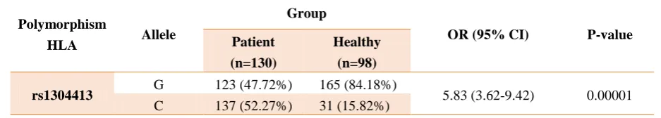
Table3. Allelic frequencies of rs1304413 for the case and control groups
Polymorphism
HLA Allele
Group
OR (95% CI) P-value Patient
(n=130)
Healthy (n=98)
rs1304413 G 123 (47.72%) 165 (84.18%) 5.83 (3.62-9.42) 0.00001 C 137 (52.27%) 31 (15.82%)
Discussion
More than 50 genes have been identified to
influence the risk of T1D, with HLA class II
genes having the greatest impact on the
people's susceptibility [13]. Other loci bear
minor impact on the risk for T1D; however,
the combination of HLA genotypes and
non-HLA single nucleotide polymorphisms
has been shown to aid disease prediction [14,
15]. Several studies have demonstrated a
fundamental role for the HLA in the
susceptibility of, or protection to, T1D
[16-18]. So far it has been understood that
some polymorphism in HLA genes are related
to diabetes. In this study, the effect of
rs310441 polymorphism in the HLA region on
the development of susceptibility or resistance
to T1D among the people with T1D has been
studied. The rs3104413 polymorphism is
located in the intergenic region between
HLA-DRB1 and HLA-DQA1. Several genetic
studies have been published related to
large-scale polymorphisms and autoimmune
diseases one of which is the polymorphisms
examined in this probe. By studying 263
patients with rheumatoid arthritis and 374
control cases in 2014, it was found that the
rs3104413 in the region between HLA-DRB1
HLA-DQA1 has strong links with rheumatoid
arthritis [19]. Cao Nguyen et al. also examined
the frequency of high-risk HLA haplotypes in
case and control groups. For this, three
polymorphisms of the HLA class II loci
(rs3104413, rs2854275 and rs9273363) were
genotyped in all the samples using custom
TaqMan genotyping assay 20x. A study
showed that these polymorphisms can predict
HLA-DR/DQ haplotypes relevant to T1D
with an accuracy (99%) [13, 20]. Three
single-nucleotide polymorphisms in the major
histocompatibility complex region (rs3104413,
rs2854275, rs9273363) were combined to
identify carriers of the high- and low-risk
HLADR And DQ genotypes known to be
associated with autoimmune diabetes (DR3/4,
DR3/3, DR4/4, DR3/X, DR4/X, DR4-DQ7,
DR4/3-DQ8, DR4-DQ8, DRX/X), where the
greatest risk of T1D was found in subjects
heterozygous for these types [13]. Since the
evaluation of diabetes risk development
depends on the type of peoples’ HLA and also
because the HLA typing method is highly
expensive and time consuming, experimenting
a limited number of SNPs and designing an
algorithm the type of HLA can easily be
determined. Furthermore, it has been found
that the rs3104413 can be employed in
this process. Due to the fact that this
polymorphism has a significant association in
Iranian population, the findings of Cao
Nguyen et al. [13] can be utilized in other
forthcoming studies.
Conclusions
This study revealed that there is a significant
association between the frequency of alleles
and genotypes in the patients compared to
healthy subjects. The C/C and C/G genotypes
were more frequent in patients than controls
and G/G genotype was shown to be protective
for T1D. A significant difference was found
for the G allelic frequency in patients with
T1D and in the control group. The allelic
frequency was significantly different between
the two groups. Our findings indicate that
HLA polymorphism (C/G) and (C/C)
genotypes can be considered as genetic risk
factors associated with susceptibility and
(G/G) genotypes associated with protection
against T1D.
Conflict of Interest
There is no conflict of interest to declare.
Acknowledgements
This work was supported by a grant from the deputy of research and technology, Shahid Sadoughi University of medical sciences and was written based on a data set of a medical biotechnology M.Sc. thesis registered in the faculty of medicine.
References
[1]. Atkinson MA, Eisenbarth GS, Michels AW. Type 1 diabetes. Lancet 2014; 383(9911): 69-82. [2]. WHO. Global report on diabetes: World Health
Organization; 2016.
[3]. Whiting DR, Guariguata L, Weil C, Shaw J. IDF diabetes atlas: global estimates of the prevalence of diabetes for 2011 and 2030. Diabetes Res Clin Pract. 2011; 94(3): 311-21. [4]. Morahan G. Insights into type 1 diabetes
provided by genetic analyses. Curr Opin Endocrinol Diabetes Obes. 2012; 19(4): 263-70. [5]. Steck AK, Rewers MJ. Genetics of type 1
diabetes. Clinic Chem. 2011; 57(2): 176-85. [6]. Abbas AK, Lichtman AH, Pillai S. Basic
immunology: functions and disorders of the immune system: Elsevier Health Sciences; 2012. [7]. Florez JC, Hirschhorn J, Altshuler D. The inherited basis of diabetes mellitus: implications for the genetic analysis of complex traits. Ann Rev Genomics Hum Genet. 2003; 4(1): 257-91. [8]. Varney MD, Valdes AM, Carlson JA, Noble
JA, Tait BD, Bonella P, et al. HLA DPA1, DPB1 Alleles and Haplotypes Contribute to the Risk Associated With Type 1 Diabetes Analysis of the Type 1 Diabetes Genetics Consortium Families. Diabetes 2010; 59(8): 2055- 62. [9]. Pociot F, Lernmark Å. Genetic risk factors for
type 1 diabetes. Lancet 2016; 387(10035): 2331-339.
[10].Visscher PM, Brown MA, McCarthy MI, Yang J. Five years of GWAS discovery. Am J Hum Genet. 2012; 90(1): 7-24.
[11]. Karges B, Rosenbauer J, Kapellen T, Wagner VM, Schober E, Karges W, et al. Hemoglobin A1c levels and risk of severe hypoglycemia in children and young adults with type 1 diabetes from Germany and Austria: a trend analysis in a cohort of 37,539 patients between 1995 and 2012. PLoS Med. 2014; 11(10): e1001742.
[12]. You H, Chen J, Zhou J, Huang H, Pan J, Wang Z, et al. Amplification refractory mutation system polymerase chain reaction versus optimized polymerase chain reaction restriction-fragment length polymorphism for apolipoprotein E genotyping of majorly depressed patients. Mol Med Rep. 2015; 12(5): 6829-834.
[13]. Nguyen C, Varney MD, Harrison LC, Morahan G. Definition of high-risk type 1 diabetes HLA-DR and HLA-DQ types using only three single nucleotide polymorphisms. Diabetes 2013; 62(6): 2135-140.
[14]. Pociot F, Lernmark A. Genetic risk factors for type 1 diabetes. Lancet 2016; 387(10035): 2331-339.
[15]. Winkler C, Krumsiek J, Buettner F, Angermüller C, Giannopoulou EZ, Theis FJ, et al. Feature ranking of type 1 diabetes susceptibility genes improves prediction of type 1 diabetes. Diabetologia 2014; 57(12): 2521-529.
[16]. Raha O, Sarkar B, Lakkakula BV, Pasumarthy V, Godi S, Chowdhury S, et al. HLA class II SNP interactions and the association with type 1 diabetes mellitus in Bengali speaking patients of Eastern India. J Biomed Sci. 2013; 20(1): 12. [17]. Noble JA, Martin A, Valdes AM, Lane JA,
Galgani A, Petrone A, et al. Type 1 diabetes risk for human leukocyte antigen (HLA]-DR3 haplotypes depends on genotypic context: association of DPB1 and HLA class I loci among DR3-and DR4-matched Italian patients and controls. Hum immunol. 2008; 69(4): 291-300. [18]. Alves C, Toralles MBP, Carvalho GC. HLA
class II polymorphism in patients with type 1 diabetes mellitus from a Brazilian racially admixtured population. Ethn Dis. 2009; 19(4): 420-24.
[19]. Govind N, Choudhury A, Hodkinson B, Ickinger C, Frost J, Lee A, et al. Immunochip
identifies novel, and replicates known, genetic risk loci for rheumatoid arthritis in black South Africans. Mol Med. 2014; 20(1): 341.
[20]. Assmann TS, Duarte GC, Brondani LA, de Freitas PH, Martins ÉM, Canani LH, et al.
Polymorphisms in genes encoding miR-155 and miR-146a are associated with protection to type 1 diabetes mellitus. Acta diabetol. 2017; 54(5): 433-41.