*Corresponding author: Dileep Kumar ISSN: 0976-3031
Research Article
ISOLATION AND QUANTIFICATION OF PRIMARY METABOLITES AND
ANTIMICROBIAL ACTIVITY OF
PLUMBAGO CAPENSIS
LINN
Dileep Kumar
1., Pushpa Jagarwal
1and Shrama R.A
21
Department of Botany, St.Wilfred P.G. College, Jaipur,- 302020
2Department of Botany, University of Rajasthan, Jaipur- 302004
DOI: http://dx.doi.org/10.24327/ijrsr.2018.0901.1509
ARTICLE INFO ABSTRACT
Medicinal plants contain some organic compounds which provide definite physiological action on the human body. In the present investigation the antibacterial and antifugal activities of the chloroform and ethanolic extract of leaf and stem of Plumbago capensis L. were studied. Antimicrobial activity was determined by using well diffusion method. The extracts exhibited antimicrobial activities with zones of inhibition ranging from 8 to 16 mm. All the extracts exhibited appreciable activity against all the clinically important bacterial and fungal species clinically investigated. Maximum Inhibition zone (16mm) was observed in stem against P.funicullosum and minimum in leaves (8mm) against E. coli. Primary metamolites screening revealed the presence of carbohydrates, proteins, lipids and phenols in the extracts. The antimicrobial activity of the extract was compared with the standard drugs. The ability of the crude extracts of P.capensis plant parts to inhibit the growth of various bacteria and fungi showed its broad spectrum antimicrobial potential, which may be employed in the management of microbial infections.The aim of study was to evaluate the role of Plumbago capensis extraction on antimicrobial activity. Leaves and stem extracts of this plant showed admirable in vitro activity.
INTRODUCTION
Medicinal plants and plant derived metamolites are rich sources of antimicrobial agents. A wide range of plants are used medicinally in different countries and are the source of potential and powerful drugs [1]. The substances that can either inhibit the growth of pathogens or kill them and have no or least toxicity to host cells are considered candidates for developing new antibacterial drugs. In recent years antimicrobial properties of medicinal plants are being increasingly reported from different parts of world [2]. Many efforts have been made to discover new antimicrobial compounds from various kinds of sources such as microorganisms, animals, and plants. One of such resources is folk medicines. Systematic screening of them may result in the discovery of novel effective compounds [3].
Description
Plumbago capensis Thunb. Syn. P.auriculata (Plumbaginaceae) is a small sub-scandent shrub with oblong spathulate leaves and pale blue flowers, indigenous to South
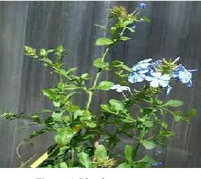
Africa and grown in gardens in India.Mainly leaves,stem and roots are used for the medicinal purpose Figure 1.
Figure 1 Plumbago capensis
The phytochemical evaluation revealed the presence of glycosides, saponins, phytosterols, phenolic compounds, flavanoids, tannins, carbohydrates and triterpenoids.The diversity of phytochemicals found suggests that aqueous and methanolic solvent extracts of these tested plants contain medicinally important bioactive compounds and it justifies
International Journal of
Recent Scientific
Research
International Journal of Recent Scientific Research
Vol. 9, Issue, 1(K), pp. 23649-23653, January, 2018
Copyright © Dileep Kumar., Pushpa Jagarwal and Shrama R.A, 2018, this is an open-access article distributed under the terms of the Creative Commons Attribution License, which permits unrestricted use, distribution and reproduction in any medium, provided the original work is properly cited.
DOI: 10.24327/IJRSR
CODEN: IJRSFP (USA)
Article History:
Received 17th October, 2017 Received in revised form 21st November, 2017
Accepted 05th December, 2017 Published online 28th January, 2018
Key Words:
their use in the traditional medicines for the treatment of different diseases[4]
Literature on plumbago capensis and all species of genus plumbago was survayed, concerning their uses in folk medicine, the different phytoconstituents reported on them as well as pharmacological and toxicological reports. The taxonomy of order plumbaginales, family Plumbaginaceae and genus plumbago was also survayed in addition to chemotaxonomic features of the family. Phytochemical screening of various secondary matabolites from different extracts of plumbago capensis (steroids, alkaloids, phenolic, cardiac glycosides, flavanoids etc) were carried out according to the standard method [5].The roots of Plumbago species are the main source of plumbagin (2–Methyl-5-hydroxy-1, 4 naphthoquinone) which is the commercially important for its broad range of pharmacological activities eg. Anti- helicobacter pylori [6] anti-tumor [7] antiparasitic [8], insect antifeedant [9], anti-hepatoma, anti-intestinal carcinogenesis[10]. [11] studied after phytochamical analysis on plumbago species using Harbone method shows presence of secondary metabolites in these species. The TLC profiling of extract of the Plumbago species showed the presence of Phenol and steroids in different range of band formation.
The Plumbago capensis is used to treat inflammatory disorders, skin diseases. The roots of the Plumbago capensis is used for treatment of warts, broken bones headache. A Decoction of the plant is taken as a remedy for black water fever. The powdered root is taken as a snuff to relive headache. It is also used for prevention of nightmares and warding off lightning. Roots used for curing of various diseases like piles, epilepsy, influenza and jaundice.
The flowers of Plumbago capensis are useful for skin disorder and it can treat bruises, soothing, sun burn, burns, spots and rashes. The studies showed that this medicinal plants contain some organic compounds which provide definite physiological action on the human body. The diversity of phytochemicals found suggests that aqueous and methanolic solvent extracts of these tested plants contain medicinally important bioactive compounds and it justifies their use in the traditional medicines for the treatment of different diseases.
MATERIALS AND METHODS
Collection of Plant Material
The plant leaves and stem used in the study was collected from the fields at Jaipur and authenticated. The voucher (RUBL* No.211571) of experimental plant was Deposited in the Herbarium of Department of Botany, University Of Rajasthan, Jaipur. The plant materials were washed thoroughly with running tab water and chopped into small pieces and dried under shade for a period 10 to 15 days. The dried plant materials were then ground into fine powders using a grinding machine. The powders were placed in sealed airtight bottles, well labeled and stored in the dark room temperature until extraction.
Quantification of Primary Metabolites
Quantification of Primary Metabolites were carried out by using following methods of carbohydrates, proteins, lipids and phenols.
Extraction and Quantification of Carbohydrates
Total Soluble Sugars
The dried experimental plant material (50 mg each) was homogenized in pestle and mortar with 20 mL of 80% ethanol separately and left overnight. Each sample was centrifuged at 1200 rpm for 15 minutes; the supernatants were collected separately and concentrated on a water bath using the method of [12].
Starch
The residual mass obtained after extraction of total soluble sugars of each of the test samples was suspended in 5 mL of 52% perchloric acid [13]. Later, 6.5 mL of water was added to each sample and the mixture was shaken vigorously for 5 minutes.1mL of aliquot of each sample was used for the estimation of carbohydrates using the phenol sulphuric acid [14].
Extraction and Quantification of Proteins
The test sample (50mg each) were separately homogenized in 10 mL of cold 10% trichloroacetic acid (TCA) for 30 min and kept at 40C for 24 hours. These mixtures were centrifuged separately and supernatants were discarded. Each of the residues was again suspended in 10 mL of 5% TCA and heated at 800 on a water bath for 30 minutes. The samples were cooled, centrifuged and supernatants of each were discarded. The residue was then washed with distilled water, dissolved in 10 ml of 1N NaOH, and left overnight at room temperature [15].Each of the above samples (1 mL) was taken and the total protein content was estimated using the spectrophotometer [16].
Extraction and Quantification of Lipids
The test sample were dried, powdered and 100mg was macerated with 10 mL distilled water, transferred to a conical flask containing 30 mL of chloroform and methanol (2/1:v/v) [17]. The mixture was thoroughly mixed and left overnight at room temperature in dark for complete extraction. Later, 20 mL of chloroform mixed with 2 mL of water were added and centrifuged. Two layers were separated, the lower layer of chloroform, which contained all the lipids, was carefully collected in the preweighed glass vials and the colored aqueous layer of methanol which contained all the water soluble substances and thick interface layer were discarded in each test sample. The chloroform layers dried in vacuo and weighed. Each treatment was repeated thrice and their mean values were calculated.
Extraction and Quantification of Phenols
The deproteinized test materials (200mg each) were macerated with 10 mL of 80% ethanol for 2 hours, and left overnight at room temperature. The mixtures were centrifuged and the supernatants were collected separately and maintained up to 40 mL by adding 80% ethanol[18].Total phenol content in each sample was estimated by spectrophotometer [19].
Phytochemical analysis of the plant extract
Antimicrobial Activity
Chloroform and ethnolic extracts was used for determination of antimicrobial activity of P. capensis. Four bacterial and four fungal were selected for the antimicrobial screening.
Microorganisms Used
Clinical laboratory isolates of bacteria viz Streptococcus viridians, Staphylococcus aureus, Escherichia coli, Bacillus and fungi viz Tricoderma ressie, F.fuserium, Pennicilium funicellosum and Aspergillus niger were procured from the Microbiology Laboratory, SMS Medical College, Jaipur.
Preparation of Extract
The chloroform and ethnolic extracts was obtained by macerating 100 g of dried powder of different plant parts in 95% ethanol and kept on a rotary shaker for 24 h, separately. Each of the extract was filtered, centrifuged at 5000rpm for 15 min, dried under reduced pressure and stored at 4 ºC in airtight bottles.
Culture and Maintenance of Bacteria
Aspergillus niger were Above mentioned pure cultures of Streptococcus viridians, Staphylococcus aureus, Escherichia coli, Bacillus and fungal isolates Tricoderma ressie, F.fuserium, Pennicilium funicellosum and used as indicator organisms. These bacteria were grown in nutrient agar medium prepared by autoclaving 8% Nutrient Agar (Difco-Laboratories, Detroit, USA) in distilled water at 15 lbs psi for 25-30 min and incubating at 37°C for 48 h. Each bacterial culture was maintained on the same medium after every 48 h of sub-culturing. A fresh suspension of test organism in saline solution was prepared from a freshly grown agar slant before every antimicrobial assay.
Determination of Antibacterial Assay
In vitro antibacterial activity of the crude chloroform and ethanol extracts were studied against gram +ve and --ve bacterial strains by the agar well diffusion method [20]. Mueller Hinton Agar No.2 (Hi Media, India) was used as the bacteriological medium. The extracts were diluted in 100% dimethylsulphoxide at the concentrations of 5 mg mL-1. The Mueller Hinton agar was melted and cooled to 48-50 ºC and a standardized inoculum (1.5×10⁸ CFU mL-1, 0.5 McFarland) was then added aseptically to the molten agar and poured into sterile petridishes to give a solid plate. Wells were prepared in the seeded agar plates. The test compound (40 µl) was introduced in the well (6 mm). The plates were incubated overnight at 37°C. The antimicrobial spectrum of the extract was determined for the bacterial species in terms of zone sizes around each well. The diameters of zone of inhibition produced by the agent were compared with those produced by the commercial control antibiotic streptomycin and ampicillin. For each bacterial and fungal strain, controls were maintained where pure solvents were used instead of the extract. The control zones were subtracted from the test zones and the resulting zone diameter was measured with antibiotic zone reader to nearest mm. The experiment was performed in triplicate to minimize the error and the mean values are presented.
Determination of Antifungal Assay
Antifungal activity of the experimental plant was investigated by agar well diffusion method [21]. The yeasts and saprophytic fungi were subcultured on Sabouraud’s Dextrose Agar (SDA; Merck, Germany) medium and respectively incubated at 37°C for 24 h and 25°C for 2 - 5 days. Suspensions of fungal spores were prepared in sterile PBS (phosphate buffered saline) and adjusted to a concentration of 106 cells mL-1. Dipping a sterile swab into the fungal suspension was rolled on the surface of the agar medium. The plates were dried at room temperature for 15 min. Wells of 10 mm in diameter and about 7 mm apart were punctured in the culture media using sterile glass tube. 0.1 mL of several dilutions of fresh extracts was administered to fullness for each well. Plateswere incubated at 37°C. After incubation of 24 h, bioactivities were determined by measuring the diameter of inhibition zone (mm). The diameters of zone of inhibition produced were with those of standard clotrimazole used as standard antifungal agent. All the experiments were performed in triplicate and mean values were taken.
RESULTS
Phytochemical screening Investigations on the phytochemical screening of P. capensis extracts revealed the presence of carbohydrates, lipids, proteins and phenols, which are known to be biologically active. These metabolites can exert antimicrobial activity through different mechanisms (Table 1).
Table 1 Phytochemical evaluation from different plant parts of P. capensis
Phytochemical Name Stem
(mg/gdwt)
Leaf (mg/gdwt)
Proteins 150±1.29 135±8.20
Lipids 124±0.02 14±0.08
Phenols 3.90±0.02 4.05±0.02
Carbohydrates
TSS 7.2±0.009 8.0±0.07
Starch 4.3±0.04 10.0±0.03
Values are the mean ± SEM (n = 3 replicates in each group). *P < 0.05; **P < 0.001 compared with the control; P < 0. 001
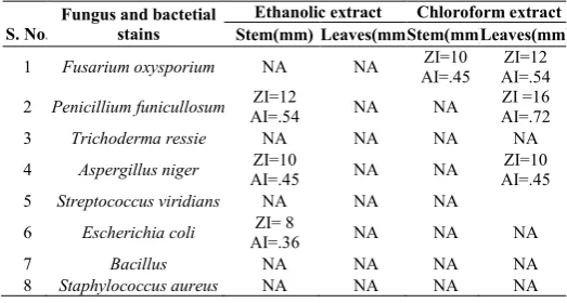
The antimicrobial activity of chloroform and ethanolic extracts of different plant parts of P. capensis were tested against 4 bacterial strains (S. aureus, Bacillus, E. coli and S. viridians) and 4 fungal strains (A. niger, F. fuserium P. funicellosum and T.ressie).
Table 2 Antimicrobial activities of Ethanolic and Chloroform extracts of P.capensis
S. No.
Fungus and bactetial stains
Ethanolic extract Chloroform extract
Stem(mm) Leaves(mm)Stem(mm)Leaves(mm)
1 Fusarium oxysporium NA NA ZI=10
AI=.45
ZI=12 AI=.54
2 Penicillium funicullosum ZI=12
AI=.54 NA NA
ZI =16 AI=.72
3 Trichoderma ressie NA NA NA NA
4 Aspergillus niger AI=.45 ZI=10 NA NA AI=.45 ZI=10
5 Streptococcus viridians NA NA NA
6 Escherichia coli ZI= 8
AI=.36 NA NA NA
7 Bacillus NA NA NA NA
8 Staphylococcus aureus NA NA NA NA
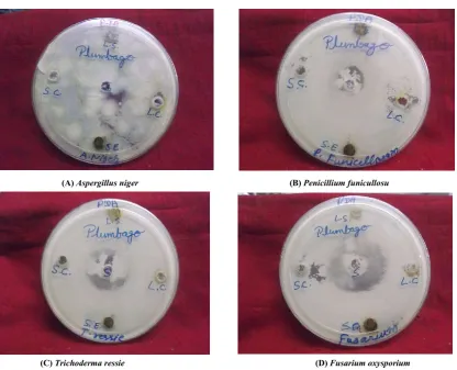
The Inhibition Zone (IZ) was measured by antibiotic zone reader (Table 2). Individually against i maximum IZ was in extract of leaves(16mm), which was at par with that of stem (12mm) and minimum was in stem. In case of P. funicellosum maximum IZ was observed in leaves chloroform (16mm) and minimum in stem (8mm), in F. fuserium maximum IZ was in leaves (12mm) and maximum in stem ethnolic (10mm) against F.fuserium and A.niger also leaves and stem gave maximum IZ (10mm and 10mm, respectively) and Among the bacterial strains against E.coli it was observed that only stem ethnolic (8mm) and leaves did not show any activity.
DISCUSSION
Plants synthesize variety of phytochemicals as part of their normal metabolic activities. Chemical profile of a single plant may vary over a time, as it reacts to changing conditions. Plant scientists and natural products chemists are combing the flora for the phytochemicals and lead compounds, which could be developed for treatment of various diseases. In 2010 a survey of 1000 plants was done out of which, 156 clinical trials for evaluation of their pharmacological activities and therapeutic applications gave encouraging results [22]. This led to the new search for drugs and dietary supplements derived from plants. During the last 10 years pace of development of new antimicrobial drugs has slowed down, while prevalence of resistance has increased multifold [23]. The problem of microbial resistance of growing and outlook for the use of antimicrobial drugs in future is still uncertain therefore,
action must be taken to reduce this problem, such as controlling the use of antibiotics and carrying out research for better understanding of genetic mechanism of resistance. This prompted to evaluate plants as source of potential chemotherapeutic and antimicrobial agent along with their ethnomedicinal use [24]. In the present investigation initial screenings of the experimental plant for possible antimicrobial activities was done using crude ethanolic and chloroform extracts. Nearly all of the identified components from plants that are active against microorganisms are aromatic or saturated organic compounds and most often obtained through ethanol or chloroform extractive. In the present study P. capensis showed antimicrobial potent activity against fungal strains as compared to bacterial strain.
CONCLUSION
In the study, there is necessity to introduce new, biologically safe and active drugs. Naturally the plants possess biologically effective antimicrobial agents. The chloform leaf extract of Plumbago cpensis L. showed good activity against the fungal strains of namely Penicillium funicullosuman and Aspergillus niger . It indicates that the plant leaf contains phytochemical (medicinal) compounds for curing the different human diseases and further investigation should be needed to screen the phytochemicals which are useful for pharmacological studies. The present findings can be of commercial interest to both pharmaceutical companies and research institutes in the production of new antimicrobial drugs. More importantly, there have been no side effects or toxicity reports from many years on this plant.
(A) Aspergillus niger (B) Penicillium funicullosu
(C) Trichoderma ressie (D) Fusarium oxysporium
Acknowledgments
Authors are thankful to University Grant Commission, New Delhi, India for providing fellowship to one of the author Mrs.Pushpa Jagarwal.
References
1. Sivastava, J., Lamhart J and Viatmeyer.1996. Medicinal Plants, an expanding role in development word bank technical paper No.320.
2. Saxena, K.1997. Antimicrobial Screening of Selected Medicinal Plants from India. Journal of Ethano pharmacology, 58(2):75-83.
3. Tomoko. N., Takashi. A., Hiromu. T., Yuka. I., Hiroko. M., Munekazu. I., Totshiyuki. T., Tetsuro. I., Fujio. A., Iriya. I., Tsutomu. N., Kazuhito. W. (2002): J. Health Sci., 48: 273–276.
4. G.H. Urmila, Ganga Rao and T. Satyanarayana, Phytochem. 2013,2(3): 221324.
5. J.B.Harborne, Phytoche. Met. 1998, 3(2): 49-188. 6. CC Wang, YM Chiang, SC Sung, YL Hsu, JK
Chang, PL Kuo, Cancer Lett, 2008, 259, 82–98. 7. Lin Lie-Chwen, Yang Ling-Ling,Chou. Cheng-Jen,
Cytotoxic, Phytochem. Volume 62, 2003, Pages 619-622
8. M. J.Chan-Bacab, L M. Peña-Rodríguez L M., Nat Prod Rep. 2001, 18, 674-688.
9. M A. Villavicencio & B E. Perez-Escandon, Folia Entomol Mex.199286: 191-198.
10. E.Sheeja, S.B. Joshi, D.C. Jain, Ind.J. of Pharm. 2011,41, 273-277.
11. Johnson M., Raja Usha, Nanthini A., Renisheya Joy, Jeba Malar T. (2010) Isozyme Variation and Genetic Relationships among Three Plumbago Species. Journal of Ecobiotechnology. 2 (5): 54-59.
12. Loomis W. E. and Shull C.A. (1973). Methods in Plant Physiology. McGraw Hill Book Co., New York, USA. 13. McCready R. M., Guggoiz J., Silviera V. and Owens
H.S. (1950). Determination of starch and amylase in vegetables. Anal. Chem. 22:1156-1158.
14. Dubois M., Gills K. A, Hamilton J. K., Rebers P. A. and Smith F. (1951). Colorimetric method for determination of sugars and related substances. Anal. Chem. 28: 350-356.
15. Osborne DJ. (1962). Effect of kinetin on protein and nucleic acid metabolism in Xanthium leaves during senescence. Plant .Physiol.37: 595-602.
16. Lowry O. H., Rose H. N, Broug J., Farr A. L. and Randall R. J. (1951). Protein measurement with the Folin-phenol reagent. J. Biol. Chem. 193:265-275. 17. Jayaraman J. (1981). Laboratory Manual in
Biochemistry. Wiley Eastern Limited, New Delhi. 96-97.
18. Bray H.G. and Thorpe W.V. (1954). Analysis of phenolic compounds of interest in metabolism. Meth. Biochem. Anal. 1:27-52
19. M. Johnson, Usha Raja, A. Nanthini, Joy Renisheya, Malar Jeba, J. Ecobio. 2010, 2 (5): 54-59.
20. C.Perez, M. Paul and P. Bazerque, Acta. Biol. Med. Exp. 1990, 15:113-115.
21. Shadidi Bonjar, S. Aghighi and N.A. Karimi, Iran. J. Biol. Sci.2005, 4: 405-412.
22. M.D. Cravotto , PhD. Boffa, M.D. Genzini and M.D. Garella, J. Clin. Pharm Therapeu. 2010, 35: 11- 48 23. D.A. Akinpelu and T.M. Onakaya, African Jour.
Biotechnol. 2006, 5 (11): 1078- 1081
24. K.N. Prashanth, S. Neelam, S. Chauhan, B. Harishpadhi and M. Ranjani, J. Ethnopharmacol. 2006, 107: 182-188.
*******
How to cite this article:Dileep Kumar., Pushpa Jagarwal and Shrama R.A.2018, Isolation And Quantification of Primary Metabolites And Antimicrobial Activity Of Plumbago Capensis Linn. Int J Recent Sci Res. 9(1), pp.23649-23653.