Safety Precautions
Scalpels, dissecting scissors, pins, and needles are all sharp instruments; use caution when handling. Always c u t a w a y from yourself and others. Rotate the dissection pan, i f neces sary, for better control while dissecting. Secure the specimen in the dissection pan before cutting. Wear safety glasses, chemical-resistant gloves, and a chemical-resistant apron. Wash hands thoroughly with soap and water before leaving the laboratory. Please follow all laboratory safety guidelines.
Procedure
1. Carefully follow each step, referring to Figures 1-11 as needed.
2. Read each section completely before continuing to dissect
3. Make careful observations o f each organ and body system.
External Anatomy
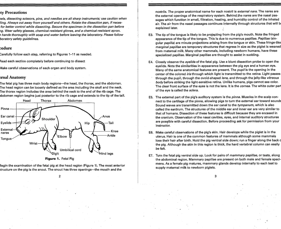
E1. The fetal pig has three main body regions—the head, the thorax, and the abdomen. The head region can be loosely defined as the area including the skull and the neck. The thorax region includes the area behind the neck to the end o f the rib cage. The abdomen region begins just posterior to the rib cage and extends to the tip of the tail.
Head Thorax Abdomen
Pinna
Ear canal
Eyelids
External nares
Tongue
Knee
^Digit
Figure 1. Fetal Pig
Ankle
^ H i n d legs
E2. Begin the examination o f the fetal pig at the head region (Figure 1). The most anterior structure on the pig is the snout. The snout has three openings—the mouth and the
nostrils. The proper anatomical name for each nostril is external nare. The nares are the external openings of the respiratory system. Behind,the nares are the nasal pas sages which function in smell, filtration, heating, and humidity control o f the inhaled air. The air from the nasal passages continues internally through structures that will be
explained later.
-E3. The tip o f the tongue is likely to be projepting from the pig's mouth. Note the fringed appearance of the tip of the tongue. This is due to numerous papillae. Papillae (sin gular papilla) are minute projections arising from the tongue or skin. These fringe-like marginal papillae are temporary structures that regress in size as the piglet is weaned from maternal milk. Many other mammals, including newborn humans, have these specialized papillae. Marginal papillae are thought to assist in suckling.
E4. Closely observe the eyelids of the fetal pig. Use a blunt dissection probe to open the eyelids. Note the similarities in appearance between the pig eye and a human eye. Many o f the same anatomical features are present. The pupil is the opening in the center of the colored iris through which light is transmitted to the retina. Light passes through the pupil, through the ovoid-shaped lens, and through the jelly-like vitreous body before striking the light-sensitive retina. Unlike humans, pigs cannot see color. The clear front surface of the eyes is not the lens. It is the cornea. The white outer part of the eye is called the sclera.
E5. The external part of the pig's auditory system is the pinna. Muscles in the scalp con nect to the cartilage of the pinna, allowing pigs to turn the external ear toward sounds. Sound waves are transmitted down the ear canal to the tympanum, which is also called the eardrum. The structures of the middle ear and inner ear are very similar to that of humans. Dissection o f these features is difficult because they are encased in the cranium. Observation o f the nasal cavities, eyes, and internal auditory structures are possible with careful dissection. Before proceeding ask for permission from your instructor.
E6. Make careful observations of the pig's skin. Hair develops while the piglet is in the uterus. Hair is one of the common features o f mammals although some mammals lose their hair after birth. Hold the pig ventral side down; run a finger along the back of the pig. Although the skin in this region is thick, the hard vertebral column can easily be felt.
Allantoic duct
Umbilical arteries
Umbilical vein
Figure 2. Umbilical Cord E8. Remnants o f the umbilical cord are located on
the ventral abdominal region o f the fetal pig. Observe the cut end (Figure 2). The umbilical cord links the fetal pig to the placenta inside the sow's uterus. Oxygen and nutrients are trans ported to the piglet while deoxygenated blood and other metabolic wastes f l o w back to the sow from the piglet via vessels in the umbilical cord. The largest vessel in the umbilical cord is the umbilical vein. The umbilical vein trans ports oxygenated, nutrient-rich blood from the
placenta to veins near the liver o f the fetal pig. This vessel is called the umbilical vein because it transports fetal blood toward the fetal heart. A pair of umbilical arteries transports blood and wastes back to the placenta (away from the fetal heart). Fetal and maternal blood do not mix in the placenta. A single layer o f cells separates the t w o vascular systems. The fourth vessel is the allantoic duct which links the fetal uro genital sinus (fetal bladder) to the allantoic sac o f the placenta. Many physiological changes occur inside the fetal pig when it is born and the umbilical cord stops func tioning. After birth the urogenital sinus becomes the bladder and part of the reproduc tive organs, the umbilical arteries become bladder ligaments, and the umbilical vein becomes a liver ligament.
E9. Determine the sex of the pig by locating the urogenital opening. In a male pig, the urogenital opening is located immediately posterior to the umbilical cord (Figure 3a). Note the lack o f any external sex organs in the fetal pig. The penis is an interior structure even in the mature male pig. It only extends during copulation. The testes descend into the thin skinned scrotum just before birth. The scrotum is located ven tral to the anus (Figure 3a). In a female, the urogenital openings are located behind the genital papillum near the anus and tail (Figure 3b). The vaginal orifice is located just ventral to the genital papillum. The vaginal orifice is the opening to the internal reproductive organs. Adjacent but ventral to the vaginal orifice is the urethral opening. Urine is excreted through the urethral opening in the female and through the urogeni tal opening in the male.
Umbilical cord
Anus
Scrotum
Urogenital opening
Figure 3a. Male
Umbilical cord
Anus
Urogenital openings
Figure 3b. Female
Internal Anatomy
Oral Cavity
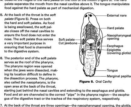
Dissecting pin posterior to j a w
Figure 4 . Head M1. Caution: Always cut away from yourself and
others. Rotate the dissection pan, if necessary, for better control while dissecting. Place the fetal pig onto its left side. Palpate (feel) along the lower jaw, called the mandible, to locate the hinge joint o f the jaw. Insert a dissecting
pin just posterior to the joint (Figure 4). Make an incision from the corner of the mouth back to the dissecting pin. It is better to make several cuts along the same incision when cutting through the strong cheek muscles. Do not attempt to use the scalpel t o cut through the jawbone. Turn the fetal pig onto its right side and repeat.
M2. Carefully place bone-cutting shears into the mouth, aligning the shears with the scal pel incision. Cut through the hinge joint of the mandible (jaw) and the skull. Repeat on the right side. Pry the mouth open exposing the back of the throat. Caution: Fetal pigs may have small teeth that are very sharp.
M3. Closely observe the surface o f the tongue (Figure 5). Recall the marginal papillae on the tip of the tongue. Other types o f papillae are also present—filiform papillae, fungiform papillae, and vallate papillae. Filiform papillae are long and thin projections that do not contain taste buds. They are thought to function in holding food in place during oral digestion. Fungiform papillae are mushroom-shaped projections that con tain taste buds. Vallate papillae are dome-shaped projections toward the back o f the tongue. Salivary glands empty near these papillae.
M4. Observe the ridges of the hard palate on the roof o f the mouth (Figure 5). The hard palate separates the mouth from the nasal cavities above it. The tongue manipulates food against the hard palate as part o f mechanical digestion.
M5. At the back of the throat is the soft palate (Figure 5). Press on both the hard and soft palates. As food is being swallowed, the soft pal ate closes off the nasal cavities t o ensure the food does not enter the
nose. The soft palate thus serves Soft palate a very important purpose in
ensuring that food is channeled to the digestive system.
Cut jawbone
External nare
Hard palate
Nasopharyngeal opening
Esophagus Epiglottis (covering glotis)
Tongue
Marginal papillae
Figure 5. Oral Cavity M6. The posterior end of the soft palate
serves as the roof of the pharynx. The pharynx region was opened when the jaws were dissected, mak ing its location difficult t o define in the dissection process. The pharynx, also called the nasopharynx, is the open area at the back o f the throat,
starting just behind the nasal cavities and extending t o the esophagus and glottis. Food and air are channeled to the correct "pipe" in the pharynx region—the esopha gus of the digestive tract or the trachea o f the respiratory system, respectively.
M7. At the back of the throat are three openings—the nasopharyngeal opening, the glottis, and the esophagus (Figure 5). The nasopharyngeal opening is the opening between the pharynx region and the nasal cavities. Air from the external nares is hydrated and either heated or cooled in the moist nasal cavities before flowing from the nasopha ryngeal opening into the pharynx and into the glottis and the remainder of the respira tory tract.
6
M8. Locate a flap o f cartilage at the back o f the pharynx. This flap, called the epiglot tis, (Figure 5) moves to close off the respiratory system during swallowing. Food is diverted toward the esophagus. Food "going down the wrong pipe" is due to a mal function o f the epiglottis. The epiglottis also functions as one of the seven cartilages forming the larynx or voice box. Use the blunt probe to move the epiglottis out o f the way. The opening behind the epiglottis is the1 glottis. The glottis is the gateway to the trachea and the respiratory tract. :
M9. The middle opening at the back of the throat is the gap f o r the esophagus. The tube for the digestive tract is thus dorsal to that of the respiratory tract. Observe the size and muscle structure around the esophageal opening compared to the glottis. Large pieces of food can lodge the epiglottis over the glottis thus preventing respiration. The relative position o f the esophagus and glottis is also possible in humans.
M10. Once you have completed your observations, close the snout, wrap a piece of string around it, and tie it closed. The string will keep the mouth from drying out while the dissection proceeds.
T h o r a x and Abdominal Regions
T1. Securely tie a twelve-inch piece of string to one wrist o f the fetal pig. Thread the string under the dissection pan and tie the free end to the other wrist. The string should be taut, opening the chest for easier dissection. Repeat with a second string around the ankles.
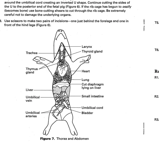
T2. Caution: Always cut away from yourself and others. Rotate the dissection pan, if nec essary, for better control while dissecting the pig in the following steps. Using heavy duty dissection scissors, cut a small hole at Point A on Figure 6. Slip the scissors just under the skin and muscles. Lift the skin and muscles away from the underlying
T3.
organs and make a midline incision, stopping just before the umbilical cord. Cut around the umbilical cord creating an inverted U shape. Continue cutting the sides of the U to the posterior end o f the fetal pig (Figure 6). If the rib cage has begun to ossify (becomes bone) use bone-cutting shears to cut through the rib cage. Be extremely careful not to damage the underlying organs.
Use scissors to make two pairs o f incisions—one just behind the forelegs and one in front o f the hind legs (Figure 6).
Trachea
Thymus gland
Liver
Umbilical vein
Umbilical arteries
Larynx
Thyroid gland
Lung
Cut diaphragm lying on liver
Small intestine
Umbilical cord
Bladder
T4.
Figure 7. Thorax and Abdomen
Use the scalpel to detach the diaphragm and any connecting membranes from the flaps of skin and muscle. Pin open the flaps of skin and muscle. Insert the dissection pins at a 45-degree angle so they do not interfere with the observation of any internal organs. Run a gloved finger along the inside o f the flap of skin and muscle. The pleural
membranes line the internal thoracic cavity and the exterior of the lungs. The pericar dial membrane surrounds the heart. These membranes are very smooth and slick to allow the organs to shift inside the body. The intrapleural space is filled with fluid so the membranes can slide past each other. An infection of the membranes or within the fluid can be very painful as the slipperiness is decreased.
T5. To free the umbilical cord flap, tie a small piece of string around the umbilical vein just beneath the pleural membrane. Tie a second piece of string a small distance below the first one (Figure 7). Use scissors to cut between the t w o pieces of string. The umbilical vein will be discussed in greater detail as part o f the circulatory system so, for now, just lift the umbilical cord flap toward the posterior end o f the fetal pig. There is no need to pin the flap down to the dissection pan.
T6. Closely observe the internal organs o f the fetal pig that are now visible. Observe the heart and lungs visible anterior to the diaphragm and the large liver and coiled intes tines posterior to the diaphragm. The heart and lungs are contained within the tho racic cavity while the liver and intestines are within the abdominal cavity.
Respiratory System
R1. The epiglottis and glottis were located as part o f the Oral Cavity dissection. A i r passes from the external nares through the nasal cavities and past the nasopharyngeal open ing into the pharynx. Next the air travels past the epiglottis into the glottis, the larynx, and the trachea before branching into the t w o bronchi and the lungs.
R2. The respiratory system is easier to study "backwards," that is from the lungs to the external nares. Locate the thin diaphragm posterior to the lungs (Figure 7). Contraction o f the diaphragm enlarges the thoracic cavity and pulls air into the lungs. Since fetal pigs do not breathe in-utero, the diaphragm is not as muscular as it would be in an adult pig.
R3. The lungs in a fetal pig are also very dense compared to that of an adult since they do not breathe in-utero. The right lung consists o f four lobes while the left lung has three lobes. Lobes are named according to the division o f the bronchi, not according to the grooves (fissures) visible on the outside of the lung. Inside the lobes, each bronchus (plural bronchi) divides into smaller and smaller bronchioles. The bronchioles termi nate in alveoli or air sacs, where gas exchange takes place. Each air sac is only one cell thick, allowing for oxygen to diffuse into the oxygen-deficient blood while carbon dioxide diffuses out into the air sac for exhalation.
R4. Use the blunt probe to gently move the lobes of the lungs. Be careful not to damage the heart, the thymus gland, and numerous blood vessels found in the area. Locate the bronchus as it enters one o f the lungs. Trace the bronchus toward the neck t o locate the cartilaginous trachea (Figure 7). It may be necessary t o move the large thy mus gland to see the trachea. The trachea, or "windpipe," is composed of a series of U-shaped cartilages. The trachea is lined with ciliated, mucus-secreting cells. Mucus and moisture are very important for the cells o f the respiratory tract. Dry air would dehydrate the cells and cause cell damage. Mucus traps particles that made it past the hairs and mucus lining o f the nasal cavities.
R5. Follow the trachea to the more bulbous larynx (Figure 7). The larynx is high in the throat. It may be necessary to carefully dissect the neck to the tip of the chin in order t o find the larynx. The larynx is a hard-walled cartilaginous chamber. Sound is pro duced when vocal cords attached to the walls of the larynx vibrate.
Circulatory System
C1. Many fetal pigs have had latex injected into the circulatory system. Single-injected specimens will have red latex, which has been injected into the arteries via the umbili cal arteries or the carotid artery in the neck. Double-injected specimens additionally have blue latex injected into the veins through the jugular or the umbilical vein. The pulmonary circulation system is not fully functional in the fetal pig so it will contain little or no latex. A few large specimens may have lived for a short time after birth and latex may be found in the lungs and pulmonary arteries and veins.
C2. Arteries carry blood away from the heart while veins transport blood toward the heart. Closely observe the area around heart. Locate the heart. Do not confuse the thymus gland, a lymphatic system gland, with the heart. This large endocrine gland lies adja cent to the heart. The thymus gland is discussed in the Endocrine System.
C3. The shiny membrane that surrounds the heart is called the pericardium. The pericar dium acts to keep the heart in its correct location, limit the friction between the heart and the surrounding area, and provide an area o f subatmospheric pressure which assists in cardiac function. Use a blunt probe to open the pericardium.
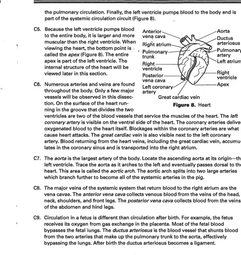
C4. Pigs have a four-chambered heart. The top two chambers are the atria. The bottom t w o chambers are the ventricles. The four chambers are not equal in size. The right atrium receives blood from the various veins of the body and is part of the systemic circulation circuit. The left atrium receives blood from the lungs and is part of the pul monary circulation circuit. The right ventricle pumps blood to the lungs and is part of
the pulmonary circulation. Finally, the left ventricle pumps blood to the body and is part o f the systemic circulation circuit (Figure 8). _v
Anterior vena cava
Right atrium Pulmonary trunk Right ventricle Posterior vena cava Left coronary-artery
Great cardiac vein
Figure 8. Heart
Aorta Ductus arteriosus Pulmonary artery Left atrium
Right ventricle Apex C5. Because the left ventricle pumps blood
to the entire body, it is larger and more muscular than the right ventricle. When viewing the heart, the bottom point is . called the apex (Figure 8). The entire apex is part o f the left ventricle. The internal structure o f the heart will be viewed later in this section.
C6. Numerous arteries and veins are found throughout the body. Only a few major vessels will be observed in this dissec tion. On the surface of the heart run ning in the groove that divides the t w o
ventricles are t w o of the blood vessels that service the muscles of the heart. The left coronary artery is visible on the ventral side of the heart. The coronary arteries deliver oxygenated blood to the heart itself. Blockages within the coronary arteries are what cause heart attacks. The great cardiac vein is also visible next to the left coronary artery. Blood returning from the heart veins, including the great cardiac vein, accumu lates in the coronary sinus and is transported into the right atrium.
C7. The aorta is the largest artery o f the body. Locate the ascending aorta at its origin—the left ventricle. Trace the aorta as it arches to the left and eventually passes dorsal to the heart. This area is called the aortic arch. The aortic arch splits into two large arteries which branch further t o become all o f the systemic arteries in the pig.
C8. The major veins o f the systemic system that return blood t o the right atrium are the vena cavae. The anterior vena cava collects venous blood from the veins o f the head, neck, shoulders, and front legs. The posterior vena cava collects blood from the veins o f the abdomen and hind legs.
C9. Circulation in a fetus is different than circulation after birth. For example, the fetus receives its oxygen from gas exchange in the placenta. Most of the fetal blood bypasses the fetal lungs. The ductus arteriosus is the blood vessel that shunts blood from the two arteries that make up the pulmonary trunk to the aorta, effectively bypassing the lungs. After birth the ductus arteriosus becomes a ligament.
C10. The ductus venosus allows blood from the umbilical vein to bypass the liver. It con nects the umbilical vein t o the posterior vena cava. After birth it becomes ligament in the liver.
C11. Another difference between fetal and newborn circulation is the foramen ovale. The foramen ovale is a valve between the right and left atria. It also functions in the bypass o f blood that would normally travel through the lungs. After birth the hole closes up. Surgery is sometimes performed on human babies to close a foramen ovale that has not seated properly.
C12. The final difference is a major change in the hemoglobin of the fetus compared to hemoglobin after birth. Fetal hemoglobin is able to pick up oxygen at lower partial pressures than that of typical hemoglobin. This physiological change is very impor tant for all placental fetuses as the mother's blood must deliver oxygen to the mother and to the placenta. Therefore the amount o f oxygen in the placenta blood is less than that found in the air.
C13. Use scissors to cut each blood vessel that connects the heart to the rest o f the circu latory system. Holding the ventral and dorsal surfaces of the heart, use a scalpel to carefully bisect the heart. Remove any latex present inside the heart to see the inner chambers and the valves between the atria and the ventricles. Note the thick wall of muscle that separates the two ventricles o f the heart. This is the interventricular septum. Also note the difference in the thickness o f the right and left halves of the heart. Recall that the right half of the heart is responsible for moving blood through the lungs while the left half pumps blood to the rest o f the body.
C14. Valves prevent blood from flowing backward into the atria as the ventricle contracts. Other valves prevent backflow into the ventricles from the blood vessels beyond. The tricuspid valve separates the right atrium from the right ventricle. It has three cusps. The bicuspid or mitral valve separates the left atrium from the left ventricle. It has t w o cusps.
C15. Semilunar valves separate the ventricles from their attached blood vessels. The pul monary semilunar valve is located between the pulmonary trunk (arteries) and the right ventricle. The aortic semilunar valve is between the aorta and the left ventricle.
C16. The chordae tendinae are ligaments that attach the cuspid valves to the muscles that control the valves.
C17.The final organ associated with the circulatory system is the spleen. This long reddish-brown organ is located adjacent to the stomach (Figure 9.on page 14). The spleen stores and releases new red blood cells into the bloodstream, filters out old red blood cells for recycling in the liver, and is also part of the immune system.
Digestive System
D1. Locate the diaphragm once more. Gently push aside the heart (if still present) and lungs to locate the esophagus where it passes through the diaphragm. Notice that the esophagus is a muscular tube. Muscle contraction and body movements push the food down the esophagus to the stomach located in the abdomen.
D2. Below the diaphragm is the very large, brownish liver (Figure 7 on page 8). The human liver has four lobes. The pig liver has five lobes. The lobes are not all the same size. The liver is part of the digestive system because it functions as a digestive gland. It is also part of the excretory system and the endocrine system. A partial list of tasks includes— bile synthesis, synthesis of cellular lipids, cholesterol, and plasma proteins, vitamin and iron storage, recycling o f hemoglobin, detoxification of ammonia to urea, plus detoxifi cation of many chemicals. In a fetal animal, like the fetal pig, red blood cells are synthe sized within the liver as well as in the spleen and eventually in the bone marrow.
D3. Gently move the liver to one side to locate the stomach (Figure 9 on page 14). The esophagus empties into the sac-like stomach. The first valve in the stomach is called the cardiac valve. Located at the anterior end of the stomach, therefore it is the valve closest to the heart. The cardiac valve ensures food and digestive enzymes do not return to the esophagus. Notice the shape o f the stomach. It is not a round balloon shape. Hydrochloric acid and digestive enzymes are secreted by specialized cells that line regions of the stomach. Some nutrients are absorbed by other cells in the stomach lining. Food that has been partially digested passes through the posterior gastric valve, the pyloric sphincter, into the small intestine.
D5. Use scissors to carefully remove the t w o largest lobes of the liver. On the right side of the pig, beneath the liver, locate the small sac-like gallbladder (Figure 9). The gallblad der stores bile secreted by the liver. Bile is secreted into the small intestine to aid in the digestion of fats.
D6. Move the stomach to one side to find the long, flat pancreas (Figure 9). The pan creas is another digestive gland that secretes enzymes used in digestion. Pancreatic enzymes are transported through the pancreatic duct to the small intestine where they aid in the digestion of fats, carbohydrates, and proteins.
Gall bladder—
Duodenum Pancreas Posterior vena cava
Rectum
Diaphragm
Liver (partially removed)
Stomach Spleen Adrenal gland Abdominal aorta Kidney
Ureter
Figure 9 . Abdomen (Liver and Intestines Removed)
D7. Locate the pyloric sphincter once again. The pyloric sphincter acts as the valve between the stomach and the small intestine. Observe the coiled small intestine (Figure 7 on page 8). As in a human, the pig's small intestine can be divided into three parts based on their roles in digestion. The entire small intestine is held together by the mesentery membrane. There are numerous blood vessels and nerves throughout the mesentery.
D8. The first region of the small intestine is the duodenum. The duodenum functions in enzymatic digestion. Enzymes from the liver and pancreas as well as enzymes from the cells lining the duodenum, empty into the internal cavity o f the small intestine. The majority of the chemical digestion o f food occurs in the duodenum, rather than in the stomach.
D9. The last t w o regions o f the small intestine are the jejunum and ileum. Absorption of nutrients occurs in these last two sections. It is difficult to distinguish these t w o regions of the small intestine. Numerous villi, which are tiny projections shaped like fingers, line these last two sections of small intestine. Villi increase the surface area across which absorption occurs. Remove a small portion of the small intestine. Cut the intestine so that it can be laid flat and view the interior surface of the small intestine.
D10. Follow the winding small intestine t o locate the ileocolic valve located where the small intestine and the large intestine join.
D11. Like the small intestine, the large intestine may be subdivided. Just past the ileocolic valve notice a sac-like projection o f the large intestine. This is the cecum. The cecum is thought to house bacterial symbionts that help break down cellulose.
D12. The remainder o f the large intestine, also called the colon, can be divided into three regions—ascending, coiled, and descending. The basic shape of the colon is that of an upside-down U. The main function o f the colon is water reabsorption prior to the elimination o f all undigested material and wastes.
D13. Follow the large intestine from the cecum to the anal sphincter and the anus. Notice that the posterior end of the colon is straight compared to the rest o f the colon. This section is called the rectum. Remove a small portion of the rectum. Meconium may be present. This green matter consists of the remnants o f cells in the internal digestive system and cells swallowed from the amniotic fluid. Clean off the section of rectum if necessary before viewing it with a stereoscope. Compare the internal surface of the rectum to that of the small intestine viewed previously. Use scissors to remove both the small and large intestine from the abdominal cavity. Be very careful to only remove these t w o organs.
Excretory System
EX1. Recall that the liver is also part of the excretory system. Urea excreted by the liver enters the blood system where it travels through the renal arteries to the kidneys for elimination from the body.
EX2. Locate the bean-shaped kidneys along the dorsal body wall (Figure 9). The kidneys do not "float" in the abdominal cavity. They are held in place behind the parietal perito neum membrane. Carefully tease the parietal peritoneum away from the kidneys. Near each kidney is a small adrenal gland that is discussed further in the Endocrine System.
EX4. Trace the ureter to the urinary bladder. Notice the urinary bladder is located between the two umbilical arteries. After birth the umbilical arteries will become ligaments that support the urinary bladder. Urine is stored in the bladder for elimination through the urethra. Trace the urethra to the urogenital opening. When you observe another group's fetal pig to trace the reproductive organs of the other gender, make sure you also trace the urethra to the urogenital opening.
EX5. Also visible near the kidney are the renal artery and renal vein. The kidneys filter waste from the blood and regulate the amount o f water in the blood.
EX6. Carefully remove one o f the kidneys f r o m the body cavity. Slice the kidney in half horizontally to view the basic internal structure o f the kidney. View the kidney with a stereoscope. See Figure 10 while locating the following areas o f the kidney.
EX7. The region where the ureter and renal blood vessels enter or exit the kidney is called the renal hilus.
Blood vessels
Renal hilus Ureter
Renal medulla
Renal cortex
Renal capsule
Figure 10. Kidney Cross-section
EX8. The slightly darker, inner portion of the kidney is called the renal medulla. The outer portion o f the kidney is called the renal cortex. The basic filtration units of the kid ney are microscopic in size and therefore cannot be seen using a stereomicroscope. These kidney tubules or nephrons act to regulate the water and mineral composition of the blood. Part of these filtration tubules lie within the renal cortex while other parts lie within the renal medulla. Different parts o f the nephrons give rise to the visual difference between the renal cortex and renal medulla.
Reproductive System
M a l e
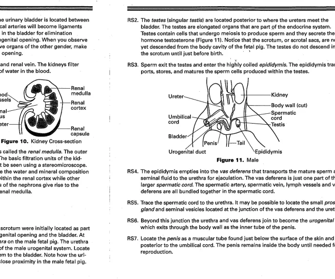
RS1. Recall that the male urogenital opening and the scrotum were initially located as part of the External Anatomy section. Locate the urogenital opening and the bladder. A t the posterior end o f the bladder, locate the urethra on the male fetal pig. The urethra
is a small tube that links the bladder to the rest o f the male urogenital system. Locate the ureters as they exit the kidneys and trace them to the bladder. Note how the uri nary system and the reproductive organs lie in close proximity in the male fetal pig.
RS2. The testes (singular testis) are located posterior to where the ureters meet the bladder. The testes are elongated organs that are part of the endocrine system. Testes contain cells that undergo meiosis to produce sperm and they secrete the hormone testosterone (Figure 11). Notice that the scrotum, or scrotal sacs, are not yet descended from the body cavity o f the fetal pig. The testes do not descend into the scrotum until just before birth. '
RS3. Sperm exit the testes and enter the highly coiled epididymis. The epididymis trans ports, stores, and matures the sperm cells produced within the testes.
Ureter
Umbilical cord
Kidney
Body wall (cut) Spermatic cord Testis
Bladder'
[Penis'
Urogenital duct E p i d i d y m i s
Figure 11. Male
RS4. The epididymis empties into the vas deferens that transports the mature sperm and seminal fluid to the urethra for ejaculation. The vas deferens is just one part of the larger spermatic cord. The spermatic artery, spermatic vein, lymph vessels and vas deferens are all bundled together in the spermatic cord.
RS5. Trace the spermatic cord to the urethra. It may be possible to locate the small prostate gland and seminal vesicles located at the junction of the vas deferens and the urethra.
RS6. Beyond this junction the urethra and vas deferens join t o become the urogenital duct which exits through the body wall as the inner tube of the penis.
RS7. Locate the penis as a muscular tube found just below the surface of the skin and posterior to the umbilical cord. The penis remains inside the body until needed for reproduction.
Female
RS8.
RS9.
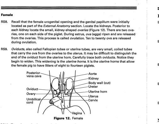
Recall that the female urogenital opening and the genital papillum were initially located as part of the External Anatomy section. Locate the kidneys. Posterior t o each kidney locate the small, kidney-shaped ovaries (Figure 12). There are two ova ries, one on each side of the piglet. During estrus, ova (eggs) ripen and are released from the ovaries. This process is called ovulation. Ten to twenty ova are released during ovulation.
Oviducts, also called Fallopian tubes or uterine tubes, are very small, coiled tubes that carry the ova from the ovaries to the uterus. It may be difficult t o distinguish the end of the oviduct from the uterine horn. Carefully trace both oviducts. Notice they begin to widen. This widening is the uterine horns. It is the uterine horns that allow the female pig to have litters o f eight to fourteen piglets.
Posterior vena cava
Oviduct Ovary
Umbilical cord
Aorta Kidney
Body wall (cut) Ureter
Uterine horn Uterus Cervix
^Vagina
Figure 12. Female
RS10. Trace the uterine horns t o the larger muscular uterus.
RS11. The uterus ends at the cervix, is a muscular constriction that must widen for birth.
RS12. During copulation, sperm is delivered t o the first chamber o f the female reproduc tive system, the vagina. The vagina and the urethra of the excretory system join t o become the urogenital sinus which is located just inside the body of the female pig. Recall that a sinus is any cavity—therefore, the urogenital sinus is a cavity that is part of the urinary and reproductive systems.
RS13. The opening of the urogenital sinus is called the urogenital opening. The urogenital opening lies dorsal to the genital papillum.
Endocrine System
EN1. Endocrine glands located throughout the body secrete hormones into the blood for delivery to specific organs or systems. In addition to hormones secreted by discrete glands, specific hormones are also secreted by the brain, skin, digestive tract, and reproductive system. »
EN2. Locate the thymus gland, which is fourfd anterior to the heart extending along the trachea toward the larynx (Figure 7 on page 8). The thymus gland in the fetal pig is very large as it plays an important role in the development and maintenance o f the immune system. The thymus gland is where white blood cells mature into antibody-producing T-lymphocytes.
ENS. Locate the small, oval, reddish-brown thyroid gland dorsal to the thymus gland and near the larynx (Figure 7 on page 8). The thyroid gland secretes t w o hormones that increase the metabolic rate of cells, which in turn influences growth and development.
EN4. Two small parathyroid glands are located on each lobe o f the thyroid gland.
Hormones excreted by the parathyroid gland cause absorption of calcium from bone, thus increasing the level o f calcium in the blood. The hormone also increases the excretion o f phosphate through the kidneys.
EN5. The adrenal glands are small, bean-shaped organs located anterior to the kidneys (Figure 9 on page 14). The adrenal glands excrete several important hormones including epinephrine, norepinephrine, Cortisol, and testosterone.
Nervous System
NS1. The nervous system of the pig is as complex as that o f a human. Begin the nervous system dissection with the pig dorsal side up. Use a scalpel to cut on either side of the backbone. Lift the skin and muscles off the backbone. Note the nerves extending from each vertebra out to the muscles o f the back.
NS2. Dissection of the cranium can be difficult depending upon the degree o f calcification of the skull. Use a scalpel to remove the skin from the top and back o f the skull. Insert scissors just under the back of the skull. Be very careful not to damage the soft brain under the skull. Make small snips over the top crest of the skull to the arch above the eyes. Cut around the skull back to the original starting point. Gently lift the cranium with forceps. It may be necessary to break or cut the cranium into smaller sections in order to see the top of the brain.
NS3. Three layers of membranes surround the spinal cord and brain in mammals. These are the meninges. The innermost membrane, the pia mater, adheres to the brain. Observe the pattern o f blood vessels visible within the meninges. The brain requires a lot of nutrients and oxygen.
NS4. The line that appears to separate the t w o halves of the brain is the longitudinal fissure. The large halves, or hemispheres, o f the brain form the cerebrum. Notice the convoluted ridges o f the cerebrum. The cerebrum controls voluntary muscle move ments, thinking, memory, judgment, and the senses.
NS5. The cerebellum is also ridged. The cerebellum is the motor coordination center for the brain. It lies under the back portion o f the cerebrum.
NS6. The enlarged area between the cerebellum and the spinal cord is the medulla oblongata. The medulla oblongata controls respiration, heart rate, blood pressure, and regulation of the endocrine system.
Disposal
Place all pieces o f the fetal pig into a resealable bag and return to the teacher. Rinse the dissection supplies with water and carefully dry each item with paper towels.
Connecting t o the National Standards
This laboratory activity relates t o the following National Science Education Standards (1996):
Unifying Concepts and Processes: Grades K-12
Evolution and equilibrium Form and function
Content Standards: Grades 9-12
Content Standard C: Life Science, biological evolution, matter, energy, and organiza tion in living systems
T h e
Fetal Pig Dissection Guide
is available f r o m Flinn Scientific, Inc.
Catalog No. Description
FB1961 Fetal Pig Dissection Guide, pkg. of 10 FB1962 Fetal Pig Dissection Photo Guide, pkg. of 5
PM4070 Fetal Pig, Formaldehyde-free, Medium, Double Injected PM5000 Fetal Pig, Formaldehyde-free, Large, Double Injected