R E S E A R C H A R T I C L E
Open Access
Management of patients with flail chest by
surgical fixation using claw-type titanium
plate
Xufeng Zhang, Zhiqiang Guo, Chuncheng Zhao, Chenyuan Xu and Zheng Wang
*Abstract
Background:The goal of the study was to compare surgical rib fixation using claw-type titanium plate with conservative treatment in the management of patients with flail chest.
Methods:The study retrospectively studied 23 patients suffering from flail chest injury, who admitted to our hospital from October, 2010 to February, 2014. The patients received surgical fixation by using claw-type titanium plate (surgical fixation group). A age and sex-matched cohort of 29 patients received conservative treatment and defined as
conservative treatment group. Outcome variables included number of cases undergoing mechanical ventilation, ventilation time, time of hospital stay, incidence of respiratory complications, incidence of thoracic deformity and postoperative forced expiratory volume in the first second (FEV1).
Results:Compared with conservative treatment group, surgical fixation group had fewer cases undergoing mechanical ventilation, shorter ventilation time, shorter hospital stay, lower incidence of respiratory complications and thoracic deformity and improved pulmonary function. Patients undergoing surgery earlier had shorter time of mechanical ventilation.
Conclusions:Surgical rib fixation with claw-type titanium plate is a reliable and efficient method in the management of patients with flail chest.
Keywords:Flail chest, Surgical rib fixation, Mechanical ventilation, Pulmonary function, Multiple rib fracture
Background
Flail chest is a life-threatening injury and usually de-fined as the presence of three or more consecutive rib fractures in 2 or more places characterized by paradox-ical motion of flailing chest wall [1]. The flail chest-related mortality ranges from 20 % to 30 % [2,3]. In addition to acute morbidities, flail chest also leads to chronic pain and disabilities.
Management of flail chest varies according to the injury severity. Conservative treatment is not sufficient to achieve a good outcome. There is growing evidence that surgical stabilization is a preferred option due to its advantages of shorter mechanical ventilation time and reduced complica-tions caused by mechanical ventilation [4,5]. Despite its cost, surgical fixation should be recommended for treating
appropriate flail chest patients [6]. Moreover, it has been reported that open reduction internal fixation using claw-type plate is an operable and reliable method for treating multiple rib fractures [7]. The study aimed to compare the surgical fixation with claw-type titanium plate with the conservative treatment in the management of patients with flail chest. This study would add weight to evidence in sup-port of the clinical application of claw-type plate.
Methods
Patients
A retrospective study was performed on the patients who were diagnosed with flail chest injury [1, 8] and admitted to our hospital from October, 2010 to February, 2014. Written informed consent was received from each partici-pant and the study protocol was approved by local ethics committee. Indications for surgical fixation included: flail chest with≥3 consecutive rib fractures in≥2 locations; * Correspondence:[email protected]
The Department of Thoracic Surgery, Putuo Hospital, Shanghai University of Traditional Chinese Medicine, NO.164 Lan Xi Road, Shanghai 200062, China
severe paradoxical breathing. Exclusion criteria included: age <20 years or > 80 years; severe associated trauma to head or spinal cord; severe extra-thoracic injuries that was like to cause death during the follow-up; pregnancy. A total of 23 patients received surgical fixation with claw-type ti-tanium plate and were defined surgical fixation group. Of the 23 cases, 17 were due to car accident, 4 due to fall in-jury and 2 due to crush inin-jury. A age and sex matched co-hort of patients with flail chest, who received conservative treatment in our hospital from October, 2010 to February, 2014, was also included by the study, and defined as con-servative treatment group. In the group, 21 cases were due to car accident, 5 cases due to fall injury, 1 case due to blow injury and 2 cases due to crush injury. The patients in the conservative treatment group did not choose sur-gical internal fixation because of economic difficulty or personal reasons.
In accordance with a previous study [9], the flail seg-ments were categorized into two types based on the chest radiographs: posterolateral (PL) flail segment and anterolat-eral (AL) flail segment. The PL flail segment referred to the posterior fractures that influenced the line of posterior rib angle; the AL flail segment referred to anterior fractures lo-cated in the area of anterior rib angle. The lung contusion was graded based on preoperative chest radiographs and computed tomographic (CT) images according to a lung contusion grading system proposed by Balci et al. [10, 11].
Treatment method
Immediately after admission, the patients in the conserva-tive treatment group underwent thorax stabilization using chest straps to reduce paradoxical movement of flailing chest wall. Patients without breathing difficulties were given oxygen, conventional hemostasis, pain medications, mucolytic drugs, necessary fluid resuscitation and hor-mone therapy in the management of lung contusion. Com-bined administration of antibiotics was also taken to prevent pulmonary infection. For patients with serious breathing difficulties or in coma, endotracheal intubation or tracheostomy was performed to provide mechanical ventilator support: synchronization gap mandatory ventila-tion (SIMV) + pressure support ventilaventila-tion (PSV) + positive end-expiratory pressure (PEEP). Patients combined with shock were given fluid and blood infusion therapy. Patients combined with pneumothorax received closed thoracic drainage treatment. Treatment for multiple injuries re-quired consultation with surgeons of relevant departments.
Patients in the surgical internal fixation group also under-went the above conservative treatment. Prior to the surgical fixation, preoperative preparation was made for patients under general anesthesia and endotracheal intubation. Pa-tients were in contralateral supine position. The position of the incision was chosen according to different rib fracture sites. Posterolateral incision was preferred in most patients. The surgery procedure was shown in Fig. 1a-e. An incision
was drawn on the skin of the patient. Special care was taken to preserve the periosteum, intercostal muscle, nerves and blood vessels. Periosteum striping was avoided as much as possible. Anatomical reduction of the rib fracture edges was obtained by using reduction clamp. The fractured ribs were stabilized and fixed by using claw-type titanium plate (Changzhou Waston Medical Appliance Company, Changzhou, China) that was cut to the desired length. The plate was attached to the rib diaphysis. Steel clamp was used to adduct the claw feet of the plate as to tightly hold the rib.
Then, thoracic exploratory surgery was performed in pa-tients requiring this surgery. A small incision was made away from the fixation site so as to prevent postoperative infection. The “coagulating” electrocautery was employed to control active bleeding. Patients with lung laceration were treated differently according to the size and depth of laceration with wedge resection as an option. After closed thoracic drainage, a layered muscular closure was per-formed with absorbable suture. When necessary, other sur-geries were performed in patients with multiple injuries. Prior to surgical internal fixation, the patients combined with shock, subarachnoid hemorrhage or myocardial injury received other symptomatic therapy. The surgical internal fixation was implemented as soon as the patients’ con-dition was improved and their vital signs were stable. All surgeries were completed within 10 days following admission. Patients received routine supportive treat-ment postoperatively.
Statistical analysis
SPSS 11.0 software was used for data processing in the study. The measurement data was expressed as mean ± standard deviation (x ± s). Comparisons be-tween groups were performed using Student's t test or chi-square test or Fisher’s exact probability test if appropriate. P <0.05 represents a statistically signifi-cant difference.
Result
Comparison of conservative treatment and surgical fixation groups
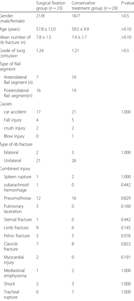
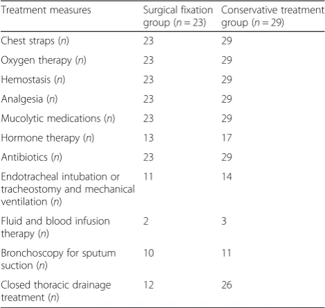
Baseline characteristics of the patients in conservative treatment and surgical fixation groups were summarized in Table 1. There was no significant difference in age, sex, mean number of rib fractures, lung contusion sever-ity, type of flail segment, causes and type of flail chest in-jury, and concomitant injuries between conservative treatment and surgical fixation groups (P> 0.05). Patients in the conservative treatment group received conservative therapies, while patients in the surgical fixation group re-ceived conservative therapies and surgical fixation using claw-type titanium plate. The common conservative mea-sures of the two groups were described in Table 2.
Difference of the conservative measures was insignificant between the two groups (P> 0.05).
Treatment outcomes of the patients in the two groups were compared in Table 3. It showed that surgical fix-ation group had significantly fewer cases taking mechan-ical ventilation (P< 0.01), shorter ventilation time and
shorter ICU stay compared to the conservative
Table 1Baseline data of patients before treatment
Surgical fixation group (n= 23)
Conservative
treatment group (n= 29) P -value
Gender (male/female)
21/8 16/7 >0.5
Age (years) 57.8 ± 12.0 59.5 ± 9.9 >0.10
Mean number of rib fracture (n)
7.8 ± 1.5 7.4 ± 1.7 >0.10
Grade of lung contusion
1.24 1.21 >0.5
Type of flail segment
Anterolateral flail segment (n)
7 10
Posterolateral flail segment(n)
16 19
Causes
car accident 17 21 1.000
Fall injury 4 5
crush injury 2 2
Blow injury 0 1
Type of rib fracture
bilateral 2 3 1.000
Unilateral 21 26
Combined injury
Spleen rupture 1 2 1.000
subarachnoid hemorrhage
1 0 0.442
Pneumothorax 12 16 0.829
Pulmonary laceration
3 0 0.160
Sternal fracture 1 0 0.442
Limb fracture 9 6 0.145
Pelvic fracture 3 5 0.976
Clavicle fracture
7 8 0.822
Myocardial injury
2 0 0.191
Mediastinal emphysema
1 2 1.000
Shock 2 3 1.000
Tracheal rupture
treatment group (P< 0.01;P< 0.05). Moreover, incidence of respiratory complications and thoracic deformity was significantly greater in the conservative treatment group than that in the surgical fixation group (P< 0.005). Rep-resentative CT images of a typical case undergoing sur-gical fixation showed that the thoracic collapse was corrected after the surgical fixation (Fig. 2). It would help improve lung function.
Surgery-related complications and follow up
Postoperatively, 6 patients reported pain because of claw-compression onto neurovascular bundle. Additionally, 1 patient suffered from infection and 1 patient suffered from rejection reaction in the surgical fixation group. During follow-up, osteomyelitis occurred in the patient with in-fection. During the follow-up, the surgical fixation failed in the patient with infection because osteomyelitis was in-duced. He then received thoracoplasty (Fig. 3a-b). The surgical fixation was successful in other patients (Fig. 3c). Patients of the two groups were followed-up for 13.98 ± 3.57 months (range: 12–30 months). No death or plate fracture was reported.
Lung function assessment
Lung function of the patients in the two groups was evaluated postoperatively (Table 4). The surgical fixation group had significantly higher postoperative forced ex-piratory volume in the first second (FEV1) than the con-servative treatment group (P< 0.001), indicating a better lung function.
Association of time to surgery with mechanical ventilation time
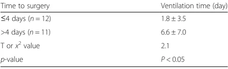
In the surgical fixation group, the association between time to surgery with mechanical ventilation time was fur-ther analyzed. The median time to surgery was 4 days. Subsequently, the 23 cases in the surgical fixation group were subdivided into two subgroups: subgroup A (≤4 days, 12 cases) and subgroup B (>4 days, 11 cases). Subgroup A had significantly shorter mechanical ventilation time than subgroup B (Table 5,P< 0.001).
Discussion
Flail chest is a consequence of multiple adjacent ribs break-ing in multiple places, often accompanied by lung contu-sion. It is observed that fail segment moves in the direction opposite to the rest of chest wall. It has varied patho-physiological consequences, leading ultimately to respira-tory and circularespira-tory failure. The study performed a retrospective study to compare surgical fixation using claw-type plate with conservative treatment in the manage-ment of patients with flail chest. The surgical fixation group had fewer cases undergoing mechanical ventilation, shorter mechanical ventilation time, shorter ICU stay, less respiratory complications, less thoracic deformities and im-proved FEV1 than the conservative method group. More-over, we found that patients with shorter time to surgery appeared to have shorter ventilation time.
The treatment of flail chest focuses on chest wall stabilization, including conservative treatment mea-sures of packing and strapping, external fixation of fractured ribs, internal pneumatic stabilization with mechanical ventilation and surgical fixation [9]. It has been demonstrated that the long-term outcome of non-operative approach is not as optimal as expected due to persistent chest wall pain, and permanent chest wall nonunion and mal-union. Moreover, long-term
Table 2Common conservative measures of two groups
Treatment measures Surgical fixation group (n= 23)
Conservative treatment group (n= 29)
Chest straps (n) 23 29
Oxygen therapy (n) 23 29
Hemostasis (n) 23 29
Analgesia (n) 23 29
Mucolytic medications (n) 23 29
Hormone therapy (n) 13 17
Antibiotics (n) 23 29
Endotracheal intubation or tracheostomy and mechanical ventilation (n)
11 14
Fluid and blood infusion therapy (n)
2 3
Bronchoscopy for sputum suction (n)
10 11
Closed thoracic drainage treatment (n)
12 26
Table 3Treatment outcome of patients in two groups
Category ICU stay
(day)
Mechanical Ventilation (n/%)
Mechanical ventilator time (day)
Respiratory Complications (n/%)
Chest deformity (n/%)
Death (n)
Conservative Treatment (N= 29)
14.2 ± 6.5 24(82.76 %) 13 ± 7.6 22(75.86) 12(41.38 %) 0
Surgical Fixation (N= 23) 5.5 ± 6.4 11(47.83 %) 4.1 ± 6.1 7(30.43) 0 0
T orx2value 4.826 7.113 4.567 10.731 12.372
-P-value <0.05 <0.01 <0.01 <0.005 <0.005
observation on the outcome of patients undergoing surgical fixation for rib fractures reveals that up to 90 % of the involved patients are able to return to work with few limitation at about 2 months postoperatively [12]. The National Institute for Health and Clinical Excellence recommendations in Britain has recom-mended surgical rib fixation in the management of patients with severe fractures [13]. A study suggests potential indications for operative repair of fractured ribs, such as flail chest, painful, movable rib fractures resistant to conservative pain management and rib fracture nonunion [14]. Another recent study regards failure of conservative management and worsening re-spiratory status as indications for surgical intervention
[15]. In contrast, we maintained the belief that severe paradoxical breathing was also a strong indication for surgical fixation of flail chest. In comparison with in-ternal pneumatic stabilization with mechanical ventila-tion, surgical fixation could immediately achieve flail chest stabilization to improve breathing.
The study showed that surgical fixation remarkably re-duced the mechanical ventilation time compared to the conservative treatment (4.1 ± 6.1vs13 ± 7.6). Surgical fix-ation could remarkably reduce the ventilfix-ation time, lead-ing to reduced incidence of respiratory complication and fail chest related mortality [16]. It was similar to previous studies reporting that the ventilation time approximately ranges from 3 to 5 days [15–19]. Nevertheless, it has been
Fig. 2One typical case of rib fracture before and after surgical fixation.a-b, a 3- dimensional reconstruction CT image showing fractured rib before surgery. (c-d), a 3-dimensional reconstruction CT image after the surgery. (e), a chest CT image showing fractured rib before the surgery. (f), a chest CT image showing fractured rib after the surgery. Collapse of the thorax is corrected after the surgical fixation
Fig. 3Follow-up observation.aa chest radiograph of the patient in surgical fixation group, who sufffers from osteomyelitis during the follow up.
reported that the ventilator support time is 6.5 ± 7.0 days for patients receiving operative chest wall stabilization [20]. The incidence of postoperative respiratory complica-tions was 30.43 % in the present study. Previous data shows that the incidence of chest-associated pneumonia is approximate 20 % [16, 18, 21]. Varied flail chest severity and surgical device might be possible causes of these in-consistent results. Different sample size might be another reason. This study showed that surgical fixation using claw-type plate could decrease the incidence of respiratory complications and thoracic deformities compared to con-servative treatment. These observations were evidences supporting the superiority of surgical fixation over conser-vative treatment.
Increasing studies point out that patients following con-servative treatment are at high risk of developing chest de-formity and atelectasis, resulting in working disabilities [22, 23]. They might have persistent chest wall pain and breathing difficulties. In the present study, chest deformity occurred in 12 of 23 (41.38 %) cases of the conservative treatment group, but was not observed in the surgical fix-ation group, suggesting the potent therapeutic efficacy of the surgery for preventing rib cage distortion. Similarly, Granetzny et al. has reported that the chest formality only occurs in one patient with surgical fixation [23].Besides, the patients following surgical fixation displayed a better pulmonary function than those receiving conservative treatment. Likewise, it has been reported that surgical fix-ation achieves better outcome of pulmonary function re-covery [23].
Furthermore, the study also found that in the surgical fixation group, the patients who received surgery in 4 days after injury were weaned from mechanical venti-lation earlier than those who received surgery after 4 days. Similarly, Althausen et al. has reported that the days on ventilator was positively correlated with the time to operation (r = 0.477) in patients undergoing surgical
stabilization of flail chest [24]. It indicated that surgical fixation should be performed as soon as possible. Flail chest injury might be aggravated if it is not treated timely. Once callus is formed, it is difficult to fix the flail chest by using claw-type plate. Thus, the surgery should be performed in 10 days after admission.
The claw-type titanium plate employed in the study was characterized with good ductility and strong corrosion re-sistance. Because of these desirable properties, the claw-type plate form could be adjusted according to the injured rib morphology. The plate bound tightly to the fractured rib with four pairs of claws firmly grasping the rib. The surgical fixation caused complications. In the present study, 6 of 23 patients reported postoperative pain due to compression on blood vessels and nerves. Infection and rejection reaction occurred in 1 patient, respectively. The patient with infection occurred osteomyelitis during the follow-up. The surgical fixation failed in the patient who then received thoracoplasty. Besides, comminuted frac-tures, fractures adjacent to transverse processes of spine or the scapula and rib cartilage fracture were also possible causes of fixation failure of claw-type plate. Additionally, the chest wall adaption might be decreased by application of too many bone plate. What is worse, lung infection might be induced and lung function might be injured in a long term [7].
Collectively, this method was effective and minimally invasive without need for application of drilling or wires or second fixation using kirschner pin. If the pa-tient does not exhibit indications of thoracic explora-tory surgery, rib fixation could be performed outside the pleura. For patients requiring thoracic exploratory surgery, rib fixation should be performed prior to the thoracic exploratory surgery for which a small incision should be made away from the fixation site so as to prevent postoperative infection. Besides, a drainage tube was placed routinely following the surgery. The study had weaknesses. It was a retrospective study with a small sample size. Prospective randomized comparative study enrolling a large number of patients should be performed to verify and extend the results of the study.
Conclusion
Surgical rib fixation using claw-type titanium plate con-fers benefits as evidenced by shorter ICU stay, shorter ventilation time, lower incidence of respiratory compli-cations and chest deformity, and improved pulmonary function in comparison with conservative treatment in the management of patients with flail chest. Patients undergoing surgery earlier have shorter ventilation time. The study suggests that surgical rib fixation with claw-type titanium plate is a operable and effective method.
Table 4Postoperative pulmonary function assessment
Category Postoperative FEV1
Conservative Treatment (N= 29) 1.43 ± 0.06
Surgical Fixation (N= 23) 1.58 ± 0.08
T orx2value 7.73
P-value <0.001
Table 5Association of time to surgery with ventilation time
Time to surgery Ventilation time (day)
≤4 days (n= 12) 1.8 ± 3.5
>4 days (n= 11) 6.6 ± 7.0
T orx2value 2.1
Abbreviations
(CT):computed tomographic; (FEV1): forced expiratory volume in the first second; (PEEP): positive end-expiratory pressure; (PSV): pressure support ventilation; (SIMV): synchronization gap mandatory ventilation; (VALI): ventilator associated lung injury; (VAP): ventilator associated pneumonia.
Competing interests
The authors declare that they have no competing interests.
Authors’contributions
XZ participated in the sequence alignment and drafted the manuscript. ZG carried out the immunoassays. CZ participated in the sequence alignment. CX participated in the design of the study and performed the statistical analysis. ZW conceived of the study, and participated in its design and coordination and helped to draft the manuscript. All authors read and approved the final manuscript.
Received: 21 May 2015 Accepted: 28 October 2015
References
1. Bastos R, Calhoon JH, Baisden CE. Flail chest and pulmonary contusion. Semin Thorac Cardiovasc Surg. 2008;20(1):39–45. doi:10.1053/j.semtcvs.2008.01.004. 2. Jayle CP, Allain G, Ingrand P, Laksiri L, Bonnin E, Hajj-Chahine J, et al. Flail chest in polytraumatized patients: surgical fixation using Stracos reduces ventilator time and hospital stay. Biomed Research International. 2015;2015:1–6.
3. Jayle CP, Allain G, Ingrand P, Laksiri L, Bonnin E, Hajj-Chahine J, et al. Flail chest in polytraumatized patients: surgical fixation using Stracos reduces ventilator time and hospital stay. Biomed Res Int. 2015;2015:624723. doi:10.1155/2015/624723.
4. Nirula R, Allen B, Layman R, Falimirski ME, Somberg LB. Rib fracture stabilization in patients sustaining blunt chest injury. Am Surg. 2006;72(4):307–9. 5. Bottlang M, Long WB, Phelan D, Fielder D, Madey SM. Surgical stabilization
of flail chest injuries with MatrixRIB implants: a prospective observational study. Injury. 2013;44(2):232–8.
6. Bhatnagar A, Mayberry J, Nirula R. Rib fracture fixation for flail chest: what is the benefit? J Am Coll Surg. 2012;215(2):201–5.
7. Chai X, Lin Q, Ruan Z, Zheng J, Zhou J, Zhang J. The clinical application of absorbable intramedullary nail and claw plate on treating multiple rib fractures. Minerva Chir. 2013;68(4):415–20.
8. Bastos R, Calhoon JH, Baisden CE. Flail chest and pulmonary contusion. [J] Semin Thorac Cardiovasc Surg. 2008;20(1):39–45. PMID:18420125. http:// www.ncbi.nlm.nih.gov/pubmed/18420125.
9. Borrelly J, Aazami MH. New insights into the pathophysiology of flail segment: the implications of anterior serratus muscle in parietal failure. Eur J Cardiothorac Surg. 2005;28(5):742–9.
10. Balci AE, Balci TA, Eren S, Ulku R, Cakir O, Eren N. Unilateral post-traumatic pulmonary contusion: findings of a review. Surg Today. 2005;35(3):205–10. doi:10.1007/s00595-004-2925-z.
11. BalcıAE, BalcıTA, ErenŞ, Ülkü R, Çakır Ö, Eren N. Unilateral post-traumatic pulmonary contusion: findings of a review. Surg Today. 2005;35(3):205–10. 12. Sarah M, Quinn C, Granger SR, Vanboerum DH, White TW. Long-term
patient outcomes after surgical stabilization of rib fractures. Am J Surg. 2014;208:88–92.
13. Shaik-Dasthagirisaheb YB, Huang N, Weinberg EO, Shen SS, Genco CA, Gibson 3rd FC. Aging and contribution of MyD88 and TRIF to expression of TLR pathway-associated genes following stimulation with Porphyromonas gingivalis. J Periodontal Res. 2015;50(1):89–102. doi:10.1111/jre.12185. 14. Nirula R, Diaz Jr JJ, Trunkey DD, Mayberry JC. Rib fracture repair: indications,
technical issues, and future directions. World J Surg. 2009;33(1):14–22. 15. Doben AR, Eriksson EA, Denlinger CE, Leon SM, Couillard DJ, Fakhry SM,
et al. Surgical rib fixation for flail chest deformity improves liberation from mechanical ventilation. J Crit Care. 2014;29(1):139–43.
16. Karev D. Operative management of the flail chest. Wiad lek (Warsaw, Poland: 1960). 1996;50:205–8.
17. Balci AE, Eren S, Cakir O, Eren MN. Open fixation in flail chest: review of 64 patients. Asian Cardiovasc Thorac Ann. 2004;12(1):11–5.
18. Ahmed Z, Mohyuddin Z. Management of flail chest injury: internal fixation versus endotracheal intubation and ventilation. J Thorac Cardiovasc Surg. 1995;110(6):1676–80.
19. Nirula R, Allen B, Layman R, Falimirski ME, Somberg LB. Rib fracture stabilization in patients sustaining blunt chest injury. Am Surg. 2006;72:307–9(3). 20. Voggenreiter G, Neudeck F, Aufmkolk M, Obertacke U, Schmit-Neuerburg
K-P. Operative chest wall stabilization in flail chest—outcomes of patients with or without pulmonary contusion. J Am Coll Surg. 1998;187(2):130–8. 21. Tanaka H, Yukioka T, Yamaguti Y, Shimizu S, Goto H, Matsuda H, et al.
Surgical stabilization of internal pneumatic stabilization? A prospective randomized study of management of severe flail chest patients. Journal of Trauma and Acute Care Surgery. 2002;52(4):727–32.
22. Athanassiadi K, Gerazounis M, Theakos N. Management of 150 flail chest injuries: analysis of risk factors affecting outcome. Eur J Cardiothorac Surg. 2004;26(2):373–6.
23. Granetzny A, El-Aal MA, Emam E, Shalaby A, Boseila A. Surgical versus conservative treatment of flail chest. Evaluation of the pulmonary status. Interact Cardiovasc Thorac Surg. 2005;4(6):583–7.
24. Althausen PL, Shannon S, Watts C, Thomas K, Bain MA, Coll D, et al. Early surgical stabilization of flail chest with locked plate fixation. J Orthop Trauma. 2011;25(11):641–7.
Submit your next manuscript to BioMed Central and take full advantage of:
• Convenient online submission
• Thorough peer review
• No space constraints or color figure charges
• Immediate publication on acceptance
• Inclusion in PubMed, CAS, Scopus and Google Scholar
• Research which is freely available for redistribution