RESEARCH
Distribution of Modic changes in patients
with low back pain and its related factors
Yufeng Chen
1,2†, Jie Bao
3†, Qi Yan
2, Cenhao Wu
2, Huilin Yang
2and Jun Zou
2*Abstract
Background: To summarize the clinical distribution of Modic changes in patients with low back pain and explore the related factors.
Methods: A total of 153 patients were enrolled. Gender, age, disk degeneration, herniation, involved segments, lum-bar lordosis angle, and endplate concave angle were recorded, respectively. Patients were divided into two or more groups according to a different classification. The relevant factors were studied with a multivariate logistic regression analysis to analyze their correlation.
Results: A total of 35 patients with type I changes, 110 patients with type II changes, and 8 patients with type III changes. In total, 204 disks were found with Modic changes, L1/2 (10 disks), L2/3 (18 disks), L3/4 (17 disks), L4/5 (76 disks), and L5/S1 (81 disks). Type I changes were distributed mainly under the age of 50. Multivariate regression showed that gender, age, disk degeneration, lumbar lordosis, L4/5 segment lordosis angle, and L5 lower endplate concave angle were related with different types of Modic changes. The regression equation Y = 2.410 − 1.361S − 0.6 33A − 0.654P + 1.106L − 0.990D (Y means type I changes, S means gender, A means age, P means disk degeneration, L means L4/5 segment lordosis angle, and D means L5 upper endplate concave angle). The OR values were S = 0.256, A = 0.531, P = 0.520, L = 3.022, D = 0.372, respectively.
Conclusions: Type II changes are the most common, followed by type I. Modic changes mostly occur in L4/5 and L5/S1; young, male, lower-grade disk degeneration, normal physiological curvature of the lumbar spine, and normal endplate concave angle were associated with type I changes; gender and lumbar curvature were the most relevant factors for different types.
Keywords: Modic changes, Types, Related factors, LBP
© The Author(s) 2019. This article is distributed under the terms of the Creative Commons Attribution 4.0 International License (http://creat iveco mmons .org/licen ses/by/4.0/), which permits unrestricted use, distribution, and reproduction in any medium, provided you give appropriate credit to the original author(s) and the source, provide a link to the Creative Commons license, and indicate if changes were made. The Creative Commons Public Domain Dedication waiver (http://creativecommons.org/ publicdomain/zero/1.0/) applies to the data made available in this article, unless otherwise stated.
Background
Low back pain (LBP) is a common disease, and up to 80% of adults have suffered from it during their lifetime [1]. The lumbar degenerative disease is one of the most important causes for LBP. Endplate locates between the disk and vertebral and intimately connects to the disk. As endplate is very thin, it is difficult to measure through regular imaging methods. With the continuous develop-ment of magnetic resonance imaging (MRI) technology, the relationship between endplate, vertebral, and LBP
has received a lot of attention. Modic changes are a com-mon abnormal signal change in lumbar MRI, reflecting the microscopic changes in tissue biochemistry in the endplate, which is an early manifestation of endplate degeneration. The concept of Modic changes has been in existence for more than 30 years; however, the spe-cific mechanism still remains unknown. The relationship between it and LBP has been widely studied, and most scholars believe that there is a connection [2–6]. Toyone et al. [7] studied 74 LBP patients, 73% (27/37) of patients with type I changes, while only 11% (4/37) with type II changes. They concluded that type I changes are more closely related to LBP. Ohtori et al. [8] found that the number of inflammatory factors such as tumor necrosis factor (TNF) and protein gene product 9.5 (PGP 9.5) was
Open Access
*Correspondence: [email protected]
†Yufeng Chen and Jie Bao contributed equally to this work. 2 Department of Orthopaedic Surgery, The First Affiliated Hospital
significantly increased in patients with type I changes. Different types of Modic changes have different patho-logical changes and therefore have different effects on LBP and surgical success [9–11]. However, no clinical study has been conducted to investigate the relevant fac-tors associated with different types of Modic changes. This study retrospectively analyzed 153 patients with Modic changes and LBP in order to summarize the clini-cal distribution of Modic changes on patients with low back pain and explore the related factors.
Materials and methods General information
Patients who took lumbar MRI for LBP in our hospi-tal from January 2015 to December 2017 and diagnosed with endplate Modic changes were enrolled. According to the following inclusion/exclusion criteria, 153 patients were included in the study.
Inclusion criteria
Patient inclusion criteria are as follows: (1) a typical endplate Modic changes on T1-weighted image and T2-weighted image and (2) clear and complete imaging data.
Exclusion criteria
Exclusion criteria are: (1) fresh compression fracture of the lumbar vertebral; (2) lumbar spondylolisthesis, scoli-osis; (3) tuberculosis, tumor, etc.; (4) Schmorl’s node; and (5) a history of back surgery.
Modic changes
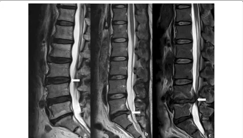
According to the classification criteria proposed by Modic et al. [12], Modic changes were classified into three types: type I—hypointense on T1WI and hyper-intense on T2WI; type II—hyperhyper-intense on T1WI and isointense or mildly hyperintense on T2WI; and type III—hypointense on both T1WI and T2WI (Fig. 1).
Age
According to the classification raised by WHO in 2012, the patients were divided into three groups: young group—under 44 years old; middle-aged group—between 45 and 59 years; elderly group—over 60 years.
Intervertebral disk degeneration
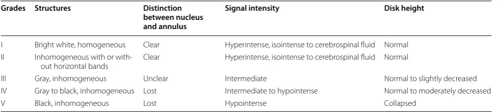
According to the classification raised by Pfirrmann et al. [13], all cases of intervertebral disk degeneration were divided into five grades (Table 1, Fig. 2).
Fig. 1 Typical Modic changes. a Type I changes; b type II changes; c type III changes
Table 1 Classification of intervertebral disk degeneration on MRI (Pfirrman scale)
Grades Structures Distinction
between nucleus and annulus
Signal intensity Disk height
I Bright white, homogeneous Clear Hyperintense, isointense to cerebrospinal fluid Normal II Inhomogeneous with or
with-out horizontal bands Clear Hyperintense, isointense to cerebrospinal fluid Normal
III Gray, inhomogeneous Unclear Intermediate Normal to slightly decreased IV Gray to black, inhomogeneous Lost Intermediate to hypointense Normal to moderately decreased
Disk herniation
According to the classification raised by the American Association of Orthopedic Surgeons, all cases of disk herniation were divided into three types: protruded, extruded, and sequestrated (Fig. 3).
Lumbar curvature
Lumbar lordosis (LL), sacral slope (SS), the lordosis angle of the L4/5 vertebral segment, and the lordosis angle of the L5/S1 vertebral segment were measured (Fig. 4).
Concave angle of vertebral endplate
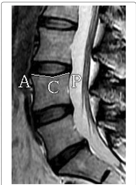
The concave angles (CAVE) of the L4 lower endplate, the L5 upper endplate, and the L5 lower endplate were meas-ured (Fig. 5).
Data analysis
All clinical and imaging data were evaluated by an ortho-pedic surgeon and a radiologist. Data analysis was per-formed by SPSS 20.0 statistical software. Firstly, the univariate Chi-square test was used to study the individ-ual factors, and P < 0.05 indicated statistical significance. Factors with statistical significance were included in a binary logistic regression analysis to study their associa-tions with the different types of Modic changes.
Results
Distribution of different types
There were a total of 35 patients with type I changes, 110 patients with type II changes, and 8 patients with type III changes. The incidence rate was 22.9%, 71.9%, and 5.2%, respectively. In total, 204 disks were found with Modic changes, and the involved segments were: L1/2 (10 disks),
Fig. 2 Typical Pfirrmann classification. a Grade I; b grade II; c grade III;
d grade IV; e grade V
L2/3 (18 disks), L3/4 (17 disks), L4/5 (76 disks), and L5/ S1 (81 disks). Patients under 50 years were more likely to have type I changes, and those over 50 years were more likely to have type II changes.
Relationship between types and age
There were 15 patients in the young group, 14 patients in the middle-aged group, and 6 patients in the elderly group with type I changed; there were 23 patients in the young group, 50 patients in the middle-aged group, and 37 patients in the elderly group with type II changes. There was a statis-tical difference among the three groups (Table 2).
Relationship between types and gender
Patients with type I changes consisted of 23 men and 12 women; patients with type II changes consisted of 49
Fig. 4 The method of measuring lumbar lordosis. a LL; b SS; c L4/5 lordosis; d L5/S1 lordosis
Fig. 5 The method of measuring CAVE. ∠ACP: L4 upper CAVE
Table 2 Distribution of different types of Modic changes with age
*statistically significant difference (p < 0.05)
Groups Modic changes Total χ2 P
Type I Type II
Youth group 15 23 38 7.495 0.024* Middle-age group 14 50 64
Elderly group 6 37 43
men and 61 women. There was a statistical difference between two groups (Table 3).
Relationship between types and affected segments
When two or more segmental were involved, the seg-ment with the largest change area ratio (abnormal sig-nal area/total vertebral body area) was considered as the affected segment. There were 27 L1/4 segments, 55 L4/5 segments, and 71 L5/S1 segments. For patients with type I changes, there were 6 L1/4 segments, 14 L4/5 segments, and 15 L5/S1 segments; for patients with type II changes, there were 13 L1/4 segments, 41 L4/5 segments, and 56 L5/S1 segments. There was no significant difference between the three groups (Table 4).
Relationship between types and intervertebral disk degeneration
When two or more segmental were involved, the seg-ment with the largest change area ratio (abnormal sig-nal area/total vertebral body area) was considered as the affected segment. For patients with type I changes, there were 7 grade II, 10 grade III, 12 grade IV, and 6 grade V; for patients with type II changes, there were 6 grade II, 19 grade III, 31 grade IV, and 54 grade V. There was a statistical difference among all groups (Table 5).
Relationship between types and disk herniation
For patients with type I changes, 23 bulged, 11 pro-truded, and 1 extruded; for patients with type II change,
58 bulged, 46 protruded, and 6 extruded. Patients with type I changes had higher proportions of bulged hernia but lower proportions of protruded and extruded her-nia, but there was no statistical difference among the groups (Table 6).
Relationship between types and lumbar curvature
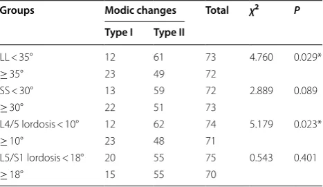
Lumbar lordosis (LL), sacral slope (SS), and the lordosis angles of the L4/5 and L5/S1 vertebral segments were divided into two groups according to the mean degree. The mean degree of LL was 34.7°, SS was 29.7°, and L4/5 and L5/S1 vertebral segment lordosis angles were 9.4° and 17.2°, respectively. Patients with type I changes had Table 3 Distribution of different types of Modic changes
with gender
*statistically significant difference (p < 0.05)
Groups Modic changes Total χ2 P
Type I Type II
Male 23 49 72 4.760 0.029*
Female 12 61 73
Total 35 110 145
Table 4 Distribution of different types of Modic changes with involved segments
Groups Modic changes Total χ2 P
Type I Type II
L1/4 6 13 27 0.978 0.613
L4/5 14 41 55
L5/S1 15 56 71
total 35 110 145
Table 5 Distribution of different types of Modic changes with Pfirrmann classification
*statistically significant difference (p < 0.05)
Groups Modic changes Total χ2 P
Type I Type II
II 7 6 13 14.843 0.002*
III 10 19 29
IV 12 31 34
V 6 54 60
Table 6 Distribution of different types of Modic changes with disk degeneration
Groups Modic changes Total χ2 P
Type I Type II
Protruded 23 58 81 1.902 0.386
Extruded 11 46 57
Sequestrated 1 6 7
Total 35 110 145
Table 7 Distribution of different types of Modic changes with lumbar lordosis angle
*statistically significant difference (p < 0.05)
Groups Modic changes Total χ2 P
Type I Type II
LL < 35° 12 61 73 4.760 0.029* ≥ 35° 23 49 72
SS < 30° 13 59 72 2.889 0.089 ≥ 30° 22 51 73
L4/5 lordosis < 10° 12 62 74 5.179 0.023* ≥ 10° 23 48 71
larger LL and L4/5 vertebral segment lordosis angle, with statistical differences. There was no significant differ-ence for SS and L5/S1 vertebral segment lordosis angle (Table 7).
Correlation between types and CAVE
CAVE was divided into two groups according to the mean degree. The mean degree of L4 lower was 164.8°, that of L5 upper was 168.6°, and that of L5 lower was 165.2°. Patients with type I changes had a smaller degree than type II changes for each CAVE. However, there was a statistical difference only for L5 lower CAVE (Table 8).
Univariate analysis
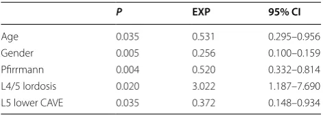
Univariate Chi-square test showed that age, gender, disk degeneration, lumbar lordosis, L4/5 segment lumbar lor-dosis, and L5 lower CAVE were associated with different types. After correlation analysis of the above factors by binary logistic regression analysis, no significant cor-relation was found between lumbar lordosis and types. Binary logistic regression analysis shows that the regres-sion equation is Y= 2.410 −1.361S−0.633A−0.654P+ 1. 106L−0.990D (Y means type I changes, S means gender,
A means age, P means the disk degeneration, L means L4/5 segment lumbar lordosis, and D means L5 upper endplate concave angle. The OR values are S= 0.256,
A= 0.531, P= 0.520, L= 3.022, D= 0.372, respectively (Table 9).
Discussion
Distribution of Modic changes among patients with LBP
Our results showed that type II changes are most com-mon and the proportion is 71.9% for type II, 22.9% for type I, and 5.2% for type III. Modic changes mainly occur in L4/5 and L5/S1 segments, accounting for 77% of all cases. These findings are consistent with previous reports [12, 14]. Many studies had demonstrated that Modic changes mostly occurred in L4/5 and L5/S1 seg-ments, where lumbar suffered maximum stress [9, 15]. Patients under 50 are more likely to have type I changes, and those over 50 are more likely to have type II changes. The incidence of Modic changes in general population is about 6% [6, 16], and the incidence in LBP population is 18% to 62% [7, 9].
Age and types
Our results show that age is one of the factors associ-ated with different types. Modic changes are degenera-tive disease, with a higher incidence rate over 45 [6]. In this study, young patients are closely associated with type I changes, and thus, we hypothesize that there may a transformation between two types, type I can convert to type II with the increasing of age. Braithwaite et al. [17] believed that different types may be different patho-logical processes of the same disease, and all types can be inter-transformable. Modic et al. [12] conducted a 3-year follow-up study and found that 5 of 6 type I changes converted to type II, while 10 type II changes remained unchanged. They, therefore, concluded that type I changes are an unstable state and type II changes are rel-atively stable. A similar study conducted by Mitra et al. [18] found that 48 patients with type I changes, 18 com-pletely transformed into type II, 7 partially transformed into type II, 19 remained unchanged with expanded abnormal signal range, and 4 remained unchanged. They considered that type I changes are a dynamic process, either transform into type II changes or remain type I. However, some paper found that type II changes can also transform into type I changes. Kuisma et al. [14] observed that 70 type II changes, and after 3 years, 10 (14.3%) became type I changes, and demonstrated that type II changes were not as stable as expected. Marsh-man et al. [19] also reported two cases of transforma-tion from type II to type I. With the increase in age, the content of water and collagen in the nucleus pulposus is reduced, the ability to absorb shock and to buffer stress is weakened, and the degeneration of the intervertebral disk is gradually aggravated. Thus, age may be a factor for patients with type II changes.
Table 8 Distribution of different types of Modic changes with endplate concave angle
*statistically significant difference (p < 0.05)
Groups Modic changes Total χ2 P
Type I Type II
L4 lower < 165° 19 47 66 1.430 0.232
≥ 165° 16 63 79
L5 upper < 169° 20 52 72 1.035 0.309
≥ 169° 15 58 73
L5 lower < 166° 21 52 73 5.179 0.023*
≥ 166° 14 58 72
Table 9 Multivariate logistic regression correlation analysis
P EXP 95% CI
Age 0.035 0.531 0.295–0.956
Gender 0.005 0.256 0.100–0.159
Gender and types
Our results show that gender is one of the factors lead-ing to different types of Modic changes. Men patients are associated with type I changes, and women patients are associated with type II changes. Endplate microfrac-tures may be a reason for it. Adams et al. [20] found that microfractures in the endplates and repair of trabecular bone are often seen in spinal cadaver specimens. The process of type I to type II transformation may be the repair process of endplate microfractures. Jensen et al. [21] conducted a prospective study of 100 patients with chronic LBP who had Modic changes, 50 of which went muscle function exercise and the other 50 under-went bed rest treatment. They found LBP relief in both groups, and there was no significant difference in the relief rates between groups. They thought that patients who underwent exercises also increase waist load, which is unbeneficial for the repair of microfracture. Although the muscle strength is enhanced, pain relief is unsatis-factory. It is generally believed that the workload of men patients is greater than women. Hence microfractures of men patients are less well repaired, which then results in persistent microfractures and inflammatory edema.
Disk degeneration and types
Our results show that disk degeneration is one of the fac-tors leading to the different types of Modic changes. We believe this may be related to the biomechanical effects of disk degeneration on the entire lumbar spine. Interver-tebral disk is the largest bloodless tissue in the human body, it largely depends on the nutrients supplied by the cartilaginous endplate; the vascular channels in the car-tilaginous endplate are particularly important for the nucleus pulposus [15]. Holm et al. [22] established a pig model of cartilage endplate injury by perforating the ver-tebral body, after 3 months, the content of water, aggre-can, type II collagen in annulus fibrosus was significantly lower compared to the normal group. Cinotti et al. [23] used the above-mentioned pig endplate injury model to draw a similar conclusion that endplate damage would lead to degeneration, and the degree of degeneration was positively correlated with the extent of endplate damage. Aggrecan and type II collagen are the main components of cartilage endplates, which can increase water content, absorb stress, and buffer shocks. They play an important role in the biomechanics of lumbar vertebrae, and their concentration is the main indicator for evaluating carti-lage endplate degeneration. Modic changes are a mani-festation of endplate biochemical changes on MRI and are early manifestations of endplate degeneration. The damages such as fissures and defects on the endplate also affect the nutrient supply of the intervertebral disk. When the intervertebral disk degenerates, the water
content and type II collagen content in the nucleus posus tissue are reduced, and part of the nucleus pul-posus can protrude to the posterior spinal canal, which weakens the stress absorption and shock buffering effect of the intervertebral disk against axial stress, resulting in a biomechanical change of the entire lumbar vertebrae. The vertebrae, therefore, withstand greater stress and eventually accelerate the damage to the endplate [12]. Severe degeneration of intervertebral disk means severe endplate damage, and hence type II changes often occur. Endplate degeneration and disk degeneration are closely related.
Lumbar lordosis and types
and endplate microfracture was an important factor causing Modic type I changes, which may also be a cause of type II changes in patients with abnormal lumbar curvature.
CAVE and types
Our results show that CAVE is one of the relevant fac-tors leading to the different types of Modic changes. Small CAVE is related to type I changes, and large CAVE is related to the type II changes. We think this may be related to disk degeneration and difference in biomechan-ics. Endplate is a thin layer structure located between the vertebral body and disk, which can be divided into bony endplate and a cartilage endplate. The cartilage endplate is a part of the intervertebral disk, which is directly con-nected with the nucleus pulposus and annulus fibrosus, and serves to isolate the nucleus pulposus from contact with the vertebral cancellous bone. Wang et al. [27] used laser scanning technology to accurately measure the physiological morphological parameters of more than 500 lumbar vertebrae endplates. Endplate is the direct receiver site of disk transfer stress to the vertebral body, so the degree of CAVE plays an important role in the biomechanics of the lumbar spine. Under normal cir-cumstances, the middle part of the endplate is the most stress-bearing area. When a disk degenerates, it will affect the stress distribution of the whole vertebral body, so that the periphery of the endplate will be under more pressure, resulting in the change of endplate morphol-ogy [28]. When the axial pressure of the bony endplate is increased, the body can increase the sagittal diameter of the vertebral body by flattening the endplate, thereby reducing the pressure per unit area. This self-adaptive change can be considered as a self-protection mechanism of the endplate [28]. When endplate degenerates, the content of aggrecan and type II collagen decreases, and endplate loses its elasticity and self-repairing ability. End-plate loses its original concave structure under chronic axial pressure load and flattens. Therefore, the shape of endplate can not only reflect the degeneration of the end-plate itself but also reflect the degree of degeneration of the disk.
Conclusions
In conclusion, type II changes are most common in patients with LBP, followed by type I, and type III is the least common. Modic changes occur mostly in L4/5 and L5/S1 segments. Age, gender, disk degenera-tion, lumbar curvature, VACE was associated with dif-ferent types, among these factors, gender, and lumbar curvature have the strongest correlations. Therefore, for patients with type I changes, we believe that simple
conservative treatment can achieve satisfactory results, and an extended period of conservative treatments is recommended. For patients with type II changes, when conservative treatments cannot achieve satisfactory effects, other aggressive treatment options can be taken to prevent further disease progression. The study is a pre-liminary analysis, and the specific clinical efficacy needs further investigation.
Acknowledgements
We gratefully acknowledge the financial supports by the National Natural Science Foundation of China under Grant Numbers 81472132, 81572183, 81672220, 91849114, as well as Priority Academic Program Development of Jiangsu Higher Education Institutions.
Authors’ contributions
YC and JZ were responsible for designing the study, writing the protocol and report, screening potentially eligible studies. YC and JB were responsible for conducting the search, writing the protocol, and report. HY, YQ and CW con-tributed to data extraction and provided feedback on the report. All authors read and approved the final manuscript.
Funding
This work was supported by the National Natural Science Foundation of China (81472132, 81572183, 81672220, 91849114), Priority Academic Program Devel-opment of Jiangsu Higher Education Institutions (PAPD).
Availability of data and materials
The data used to support the findings of this study were supplied by Jun Zou under license and so cannot be made freely available. Requests for access to these data should be made to Jun Zou, Department of Orthopaedic Surgery, The First Affiliated Hospital of Soochow University 188 Shizi St. Suzhou, China, 215006, Email: [email protected]
Ethics approval and consent to participate
This study was approved by the Ethics Committee of The First Affiliated Hospital of Soochow university and were in accordance with the Helsinki Declaration.
Consent for publication Not applicable.
Competing interests
The authors declare that they have no competing interests.
Author details
1 Department of Orthopaedic Surgery, Jiangsu Taizhou People’s Hospital,
Taizhou 225300, Jiangsu, China. 2 Department of Orthopaedic Surgery, The
First Affiliated Hospital of Soochow University, 188 Shizi St., Suzhou 215006, Jiangsu, China. 3 School of Physical Education and Sports Science, Soochow
Univeristy, Suzhou 215021, Jiangsu, China.
Received: 28 July 2019 Accepted: 28 September 2019
References
1. Deyo RA, Weinstein JN. Low back pain. N Engl J Med. 2001;344(5):363–70. 2. Albert HB, Kjaer P, Jensen TS, et al. Modic changes, possible causes and
relation to low back pain. Med Hypotheses. 2008;70(2):361–8. 3. Luoma K, Vehmas T, Kerttula L, et al. Chronic low back pain in relation
to Modic changes, bony endplate lesions, and disc degeneration in a prospective MRI study. Eur Spine J. 2016;25(9):2873–81.
•fast, convenient online submission •
thorough peer review by experienced researchers in your field • rapid publication on acceptance
• support for research data, including large and complex data types •
gold Open Access which fosters wider collaboration and increased citations maximum visibility for your research: over 100M website views per year •
At BMC, research is always in progress.
Learn more biomedcentral.com/submissions
Ready to submit your research? Choose BMC and benefit from: 5. Kjaer P, Korsholm L, Bendix T, et al. Modic changes and their associations
with clinical findings. Eur Spine J. 2006;15(9):1312–9.
6. Mok FP, Samartzis D, Karppinen J, et al. Modic changes of the lumbar spine: prevalence, risk factors, and association with disc degeneration and low back pain in a large-scale population-based cohort. Spine J. 2016;16(1):32–41.
7. Toyone T, Takahashi K, Kitahara H, et al. Vertebral bone marrow changes in degenerative lumbar disc disease. An MRI study of 74 patients with low back pain. J Bone Join Surg Br. 1994;76(5):757–64.
8. Ohtori S, Inoue G, Ho T, et al. Tumor necrosis factor-immunoreactive cells and PGP 9.5-immunoreactive nerve fibers in vertebral endplates of patients with discogenic low back pain and Modic type 1 or type 2 changes on MRI. Spine. 2006;31(9):1026–31.
9. Rahme R, Moussa R, Bou-Nassif R, et al. What happens to Modic changes following lumbar discectomy? Analysis of a cohort of 41 patients with a 3- to 5-year follow-up period. J Neurosurg Spine. 2010;13(5):562–7. 10. Ohtori S, Yamashita M, Yamauchi K, et al. Low back pain after lumbar
discectomy in patients showing endplate Modic type 1 change. Spine. 2010;35(13):E596–600.
11. Chin KR, Tomlinson D, Deirmengian C, et al. Success of lumbar microdis-cectomy in patients with Modic changes and low-back pain. A Prospec-tive Pilot Study. Spine J. 2006;6(5):139–44.
12. Modic MT, Steinberg PM, Ross JS, et al. Degenerative disk disease: assess-ment of changes in vertebral body marrow with MR imaging. Radiology. 1988;166(1 Pt 1):193–9.
13. Pfirrmann CWA, Metzdorf A, Zanetti M, et al. Magnetic resonance classification of lumbar intervertebral disc degeneration. Spine. 2001;26(17):1873–8.
14. Kuisma M, Karppinen J, Niinimaki J, et al. A three years follow-up of lum-bar spine endplate (Modic) changes. Spine. 2006;31(15):1714–8. 15. Urban JP, Smith S, Falrbank JC, et al. Nutrition of the intervertebral disc.
Spine (Phila Pa 1976). 2004;29:2700–9.
16. Jensen TS, Karppinen J, Sorensen JS, et al. Vertebral endplate signal changes(Modic change): a systematic literature review of preva-lence and association with non-specific low back pain. Eur Spine J. 2008;17(11):1407–22.
17. Braithwaite I, White J, Saifuddin J, et al. Vertebral end-plate (Modic) changes on lumbar spine MRI: correlation with pain reproduction at lumbar discography. Eur Spine J. 1998;7(5):363–8.
18. Kleinstuck E, Dvorak J, Mannion AF. Are “structural abnormalities” on magnetic resonance imaging a contraindication to the successful conservative treatment of chronic nonspecific low back pain? Spine. 2006;31(19):2250–7.
19. Marshman LA, Trewhella M, Friesem T, et al. Reverse transformation of Modic type 2 changes to Modic type 1 changes during sustained chronic low-back pain severity. Report of two cases and review of the literature. J Neurosurg Spine. 2007;6(2):152–5.
20. Adams MA. Biomechanics of back pain. Acupunct Med. 2004;22(4):178–88.
21. Jensen RK, Leboeuf-Yde C, Wedderkopp N, et al. Rest versus exercise as treatment for patients with low back pain and Modic changes. A rand-omized controlled clinical trial. BMC Med. 2012;10:22.
22. Holm S, Holm AK, Ekström L, et al. Experimental disc degeneration due to endplate injury. J Spinal Disord Tech. 2004;17(1):64–71.
23. Cinotti G, Della Rocca C, Romeo S, et al. Degenerative changes of porcine intervertebral disc induced by vertebral endplate injuries. Spine (Phila Pa 1976). 2005;30(2):174–80.
24. Keller TS, Colloca CJ, Harison DE, et al. Influence of spine morphology on intervertebral disc loads and stresses in asymptomatic adults: implica-tions for the ideal spine. Spine J. 2005;5(3):297–309.
25. Tanaka N, An HS, Lim TH, et al. The relationship between disc degenera-tion and flexibility of the lumbar spine. Spine J. 2001;1(1):47–56. 26. Lao LF, Daubs MD, Scott TP, et al. Effect of disc degeneration on lumbar
segmental mobility analyzed by kinetic magnetic resonance imaging. Spine. 2015;40(5):316–22.
27. Wang Y, Battié MC, Videman T. A morphological study of lumbar vertebral endplates: radiographic, visual and digital measurements. Eur Spine J. 2012;21(11):2316–23.
28. Fazzalari NL, Manthey B, Parkinson LH. Intervertebral disc disorganisation and its relationship to age adjusted vertebral body morphometry and vertebral bone architecture. Anat Rec. 2001;262:331–4.
Publisher’s Note