UDC 577.218:577.(112+354.9)+576.38:591.144+57.(085.23+086.164)
Changes in prohB-egF expression aFter FunCtional
aCtivation oF the immune system Cells
T. O. ChUdINa1,2, a. J. LaBINTSeV1, S. I. ROmaNIUk1, d. V. kOLyBO1,2, S. V. kOmISaReNkO1
1Palladin Institute of Biochemistry, National academy of Sciences of Ukraine, kyiv;
2eSC Institute of Biology and medicine, Taras Shevchenko National University of kyiv, Ukraine;
e-mail: [email protected]
The level of proHB-EGF expression on J774, Raji, KG-1 cells derived from different types of human and mouse immune system cells under the standard in vitro culture conditions and during functional activa-tion of these cells was investigated. Changes in the prohB-eGF expression on the cell surface were found to depend on the density of cell population, the content of fetal bovine serum in the culture medium, the presence of mitogenic factors – bacterial lipopolysaccharide and recombinant soluble hB-eGF – rshB-eGF, as well as anti-mitogenic factor – diphtheria toxoid (CRm197). The results obtained are important for the understanding of the functional role of prohB-eGF receptor on the surface of macrophage-like cells and B lymphocytes and indicate the involvement of this receptor in immune response regulation in organism.
k e y w o r d s: prohB-eGF, expression, cell lines, macrophages, B lymphocytes, lipopolysaccharide (LPS), recombinant proteins, shB-eGF, inactive diphtheria toxin CRm197.
H
eparin-binding epidermal growth factor (EGF)-like growth factor (HB-EGF) is ex-pressed on the cell surface as a 208 amino acid transmembrane protein – proHB-EGF, which consists of propeptide and 5 domains: heparin-bin-ding, EGF-like, juxtamembrane, transmembrane and cytoplasmic domains [1]. Processing of proHB-EGF by matrix metalloproteinases (MMPs) and a disin-tegrin and metalloproteinases (ADAMs) results in the formation of 87 amino acid soluble form of HB-EGF (sHB-HB-EGF) which contains heparin-binding and EGF-like domains [2].sHB-EGF, owing to the EGF-like domain, ac-tivates two types of tyrosine kinases receptors – hu-man epidermal growth factor receptors HER1 and HER4 [3, 4]. sHB-EGF is a potent stimulator of cell proliferation and migration, however, its main tar-gets are fibroblasts, smooth muscle cells and epithe -lial cells [5].
proHB-EGF induces proliferation and helps the cell to adhere; it forms complexes with other membrane proteins (such as CD9, DRAP27
(mon-key homologue of CD9), integrin α3β1) essential for cell adhesion and cell migration; and it is involved in signal transmission between cells [6]. In addition to stimulating the cell proliferation and migration, proHB-EGF was shown to be implicated in a variety of normal physiological processes, such as pregnan-cy, wound healing, neuronal recovery after hypoxia and ischemia; as well as in pathological processes, such as tumor growth, smooth muscle cell hyperpla-sia and atherosclerosis [7].
Furthermore, proHB-EGF is a receptor for diphtheria toxin (DT), the main pathogenic factor of Corynebacterium diphtheriae, the pathogenic bacte-rium that causes diphtheria [8]. A toxin can penetrate via an alternative pathway only into immune system cells (macrophages and toxin-specific B cells), owing to phagocytosis and DT-specific immunoglobulin re -ceptors [9]. DT receptor-binding subunit B (SubB) interacts with the EGF-like domain of proHB-EGF allowing the enzymatically active subunit A (SubA) of DT to penetrate the cell. SubA ADP-ribosylates eukaryotic translation elongation factor 2 (eEF2) that
leads to inhibition of protein synthesis and cell death [10]. Cells of different species vary in susceptibili-ty to DT. Thus, cells of most mammalian species (including human and guinea pig) are susceptible to DT, while mouse and rat cells are highly resistant [11].
ProHB-EGF is expressed in the cells of many organs and tissues including vascular endothelial cells, smooth and skeletal muscle cells, keratino-cytes, neurons and tumor cells [12]. ProHB-EGF is also expressed on the immune system cells: mac-rophages [5], CD4+ T cells [13] and B cells [14] that perform totally different functions. The specific fea -tures of proHB-EGF expression on various types of immune system cells and its role in the immune processes remain unexplored. The study of immune cell receptors and their signal transduction pathways is an important area of research, given the prospect of using these fundamental data to develop mecha-nisms to control immune response, as well as to de-velop drugs for the treatment of immunodeficiency, autoimmune diseases, cancer, transplantation com-plications, etc.
The aim of this work was to study changes in proHB-EGF expression on the cell lines derived from macrophage-like cells and B lymphocytes un-der the influence of various activating factors such as: changes in cell density in culture, changes in the content of fetal bovine serum (FBS) in the culture medium, the presence of mitogenic factor – bacte-rial lipopolysaccharide (LPS), as well as previously obtained recombinant proteins: nontoxic form of diphtheria toxin – CRM197 [15] or soluble form of HB-EGF – rsHB-EGF [16].
Previously, we suggested determining proHB-EGF expression on the cell surface using recombi-nant fluorescent DT derivatives, in particular, SubB genetically fused to enhanced green f luorescent protein (EGFP) – EGFP-SubB [17]. This approach allowed determining proHB-EGF expression on the cells not only of susceptible to DT animal species but also resistant to DT [18]. To study the proHB-EGF expression at functional activation of various types of the immune system cells we used the above-mentioned approach with EGFP-SubB-labeled cells and measuring their fluorescence by flow cytometry.
materials and methods
J774, Raji and KG-1 cell lines derived from human and mouse B cells and macrophage-like cells were used in our work. The J774 cell line was
obtained from mouse sarcoma cells (mouse mac-rophage-like cells), the Raji cell line originated from cells of the Burkitt’s lymphoma of the 11-year-old boy (human B cells), and the KG-1 cell line – from bone marrow cells of the 59-year-old patient with acute myelogenous leukemia (human macrophage-like cells). The cell cultures were obtained from the bank of cell lines of RE Kavetsky Institute of Ex-perimental Pathology, Oncology and Radiobiology, National Academy of Sciences of Ukraine.
In the research we also used previously ob-tained in our laboratory recombinant proteins: non-toxic DT analogue – CRM197 [15], HB-EGF soluble form – rsHB-EGF [16] and genetically fused pro-tein EGFP-SubB that consists of subunit B of DT and enhanced green fluorescent protein (EGFP) [17]. CRM197 differs from DT by substitution of Gly52 for Glu within the enzymatically active subunit A that results in a complete loss of toxicity, and slightly alters the conformation of the receptor-binding subu-nit B. Expression and isolation of recombinant pro-teins were performed according to the protocols and recommendations described in the mentioned above publications.
Cell culture. Cells were grown to 70-90% con-fluency in DMEM culture medium containing 10% FBS, streptomycin (100 mg/l), penicillin (10 000 U), and amphotericin В (250 μg/l) at 37 °С, 5% СО2 con-centration.
To study the effect of changing cell density in culture on the proHB-EGF expression, the cells were plated into 24-well plates in a number of 0.75×10⁵,
1.5×10⁵, or 3×10⁵ cells per well, and incubated under
identical conditions.
To study the effect of changes in the medium FBS content on proHB-EGF expression, the cells were plated into 24-well plates of 1.5×10⁵ cells per
well in DMEM medium containing 2, 10, or 20% FBS.
To study the LPS impact on proHB-EGF ex-pression, the cells were plated into 24-well plates in a number of 1.5×10⁵ cells per well in DMEM
me-dium containing 10% FBS. After 6 h of adaptation, a mitogenic bacterial factor LPS (Sigma, USA) was added to a final concentration of 1 μg/ml, and the cells were incubated for 16 h.
To study the influence of the recombinant pro -teins: nontoxic DT analogue – CRM197 or rsHB-EGF on the proHB-rsHB-EGF expression, the cells were plated into 24-well plates in a number of 1.5×10⁵
10% FBS. After 6 h of adaptation, CRM197 to a fi -nal concentration of 1.5 μM or rsHB-EGF to a fi-nal concentration of 0.75 μM was added. The cells were incubated for 20 h.
Cell staining. After incubation, the cells were stained with fluorescent proteins: 1.5 μM EGFP-SubB or 1.5 μM EGFP (control for nonspecific sorption). The optimal number of cells for staining was 0.3-0.5×10⁶ cells per sample. To cells,
prelimi-nary precipitated at 200-300 g for 10 min, we added 1.45 μM fluorescent protein in 200 μl of BSA/PBS solution (0.14 M NaCl, 0.03 M KCl, 0.01 M Na2HPO4 , 0.002 M KH2PO4, 1% BSA; pH 7.2) to each sample. Cells were stained for 15 min at 4 °C. To remove un -bound fluorescent proteins, the cells were gently re -suspended in BSA/PBS solution (added to the volu-me of 1 ml) and again precipitated at 200-300 g for 10 min. The incubation medium was replaced with a similar volu me of BSA/PBS. After this, the suspen-sion of the stained cells was transferred to the test tubes for the flow cytometry analysis.
Flow cytometry. The fluorescence intensity of cells was measured by Coulter Epics XL flow cy -tometer (Beckman Coulter, USA). The results of flow cytometry were normalized to control values of cell autofluorescence, and the output was re-nor -malized to the value of nonspecific sorption (EGFP stained samples).
Parameters employed for the cytometer soft-ware protocol were: small angle forward-scattered light (FS), 90° side-scattered light (SS) and loga -rithmic fluorescence on FL1 channel (515-535 nm). The FS parameters were: voltage (Volts) – 500, am-plification coefficient (Gain) – 1.0. The SS parame-ters were: Volts – 500, Gain – 5.0. FL1 parameparame-ters: Volts – 700, Gain – 10. Flow rate – medium or low depending on the number of cells in the sample. The above settings were determined experimentally.
Based on the data obtained, the cell distribution by forward- and siscattered light that allows de-termining the cell morphology and the fluorescence intensity by the channel FL1 Log were calculated.
Flow cytometry data analysis was carried out using Flowing Software (Cell Imaging Core, Turku Center for Biotechnology, Finland). The number of measurements (events) employed to plot histograms was 10,000. At the histogram analysis, the value of the ratio of the fluorescence intensity in the experi -ment to the fluorescence intensity in the control in the range from 0.5 to 2 was considered to be slightly different from 1, i.e. in these cases, the intensity of
fluorescence in the experiment differed from the in -tensity of fluorescence in the control no more than 2-fold.
results and Discussion
Studying prohB-eGF expression on the surface of immune cells under standard culture conditions in vitro. At the first stage, we studied the proHB-EGF expression on J774, Raji and KG-1 cell lines unexposed to any additional factors. Analysis of cell size and granularity revealed two subpopulations in all studied cell lines: a small granular subpopulation (non-viable cells with damaged cell membrane integ-rity) and a large less granular subpopulation (viab le cells). In further experiments, the proHB-EGF ex-pression on subpopulations of viable cells of J774, Raji and KG-1 lines was studied by measuring the fluorescence of EGFP-SubB ligand.
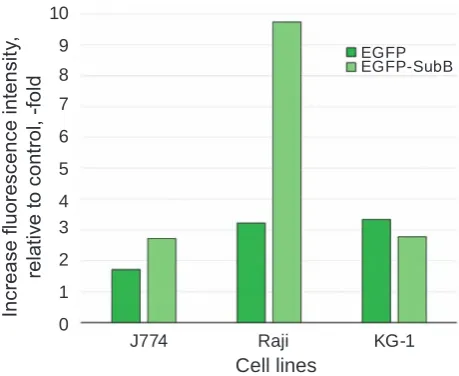
The results of cytofluorіmetric analysis of the cells stained with EGFP-SubB or EGFP (control for nonspecific sorption) are shown in Fig. 1. Autofluo -rescence of unstained cells was taken as a normal signal. The J774 and Raji cells bound proHB-EGF ligand – EGFP-SubB, while the KG-1 cells did not bind EGFP-SubB (the binding was found to be at the level of nonspecific sorption). Raji cells evidently ex -pressed the largest quantity of proHB-EGF on their surfaces (3-fold increase in fluorescence compared to control for non-specific sorption), and J774 cells expressed less proHB-EGF (1.5-2 fold increase in fluorescence compared to control for nonspecific sorption). Further studies were carried out on cells of
Fig. 1. Fluorescence intensity of Raji, J774, kG-1 cells stained with eGFP-SubB or eGFP, relative to control (unstained cells)
Cell lines
J774 Raji KG-1
In
cr
eas
e fl
uo
re
sc
en
ce i
nt
ens
ity
,
re
la
tiv
e t
o c
on
tro
l,
-fo
ld
10
0 1 2 3 4 5 6 7 8
9 eGfp
3 lines derived from immune cells, which possessed different initial levels of proHB-EGF expression: high (Raji), low (J774) and almost zero level (KG-1). KG-1 cells were used as a control, and in further experiments, they did not respond to the activa ting factors by changes in the level of their proHB-EGF expression.
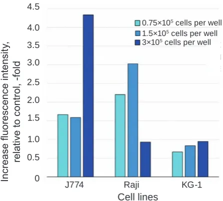
Studying prohB-eGF expression on the cell surface of immune system cells under the influence of activating factors: changes in cell density, chan-ges in FBS concentration, and the presence of LPS, CRm197 or rshB-eGF. The results obtained showed that J774 cells’ ability to interact with proHB-EGF ligand at a high cell density in culture (3×10⁵ cells
per well) increased 2.5-3-fold compared to the con-trol sample (1.5×10⁵ cells per well) (Fig. 2). Raji cells,
on the contrary, better bound proHB-EGF ligand at a lower density. An increase in density up to 3×10⁵
cells per well resulted in a 3-fold decrease in binding proHB-EGF ligand compared to the control sample.
J774 and Raji cells were found to tend to an increased level of proHB-EGF expression with an increase in the medium FBS concentration. J774 cells exhibited approximately 2-fold higher level of proHB-EGF expression at 20% FBS than at 2 and 10% FBS (Fig. 3), while Raji cells exhibited a slight increase in proHB-EGF expression at 10 and 20% FBS (1.3-fold) compared to 2% FBS.
The addition of LPS led to an increase in the level of the proHB-EGF expression by J774 cells Fig. 2. Fluorescence intensity of Raji, J774 and kG-1 cells stained with eGFP-SubB, relative to control (cells stained with eGFP), depending on cell density in plate well
Cell lines
J774 Raji KG-1
In cr eas e fl uo re sc en ce i nt ens ity , re la tiv e t o c on tro l, -fo ld 0 0.5 1.0 1.5 2.0 2.5 3.0 3.5 4.0 4.5
1.5×10⁵ cells per well
3×10⁵ cells per well
0.75×10⁵ cells per well
Fig. 3. Fluorescence intensity of Raji, J774 and kG-1 cells stained with eGFP-SubB, relative to control (cells stained with eGFP), depending on FBS con-centration in culture medium
Cell lines
J774 Raji KG-1
In cr eas e fl uo re sc en ce i nt ens ity , re la tiv e t o c on tro l, -fo ld 0 0.5 1.0 1.5 2.0 2.5 3.0 3.5 4.0 4.5 2% fbs 10% fbs 20% fbs
(more than 2-fold compared to the control) and a slight decrease in the proHB-EGF expression by Raji cells (Fig. 4).
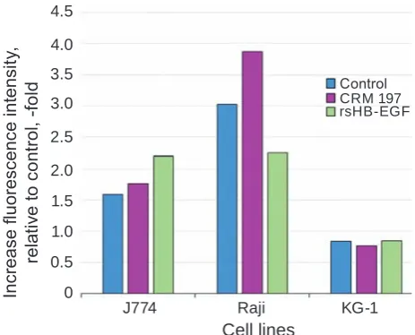
Culturing J774 and Raji cells in the presence of rsHB-EGF resulted in opposite effects (Fig. 5): proHB-EGF expression on J774 cells increased 1.3-fold, and on Raji cells decreased 1.3-fold. Culturing in the presence of a DT analogue (CRM197) had practically no effect on the proHB-EGF expression
Fig. 4. Fluorescence intensity of Raji, J774 and kG-1 cells stained with eGFP-SubB, relative to control (cells stained with eGFP), in the presence of mitogen LPS in culture medium
Cell lines
J774 Raji KG-1
Fig. 5. Fluorescence intensity of Raji, J774 and kG-1 cells stained with eGFP-SubB, relative to control (cells stained with eGFP), in the presence of recombinant proteins: nontoxic analogue of diph-theria toxin CRm197 or analogue of soluble form of hB-eGF – rshB-eGF
Cell lines
J774 Raji KG-1
In
cr
eas
e fl
uo
re
sc
en
ce i
nt
ens
ity
,
re
la
tiv
e t
o c
on
tro
l,
-fo
ld
0 0.5 1.0 1.5 2.0 2.5 3.0 3.5 4.0 4.5
rshb-eGf CRM 197 Сontrol
Changes in prohB-eGF expression on the J774 and Raji cell surface in the presence of activating factors
Cell line J774 Raji
Origin Mouse macrophages Human В cells
Initial level of proHB-EGF low high
Increase in cell density + –
Increase in FBS concentration in medium +
-|-LPS +
--CRM197 +
rsHB-EGF + –
Notes: + increase in proHB-EGF expression; -|- slight increase in proHB-EGF expression; – decrease in proHB-EGF expression; -- slight decrease in proHB-EGF expression; Empty cell – proHB-EGF expression remains unchanged by J774 cells, but enhanced the proHB-EGF
expres-sion (1.3-fold) by Raji cells.
Data on the changes in proHB-EGF expres-sion on the surface of J774 and Raji cells (KG-1 cells were taken as control) under the influence of various activating factors: increasing cell density in culture, increasing FBS concentration, the presence of LPS, CRM197 or rsHB-EGF in culture medium are sum-marized in Table.
Interestingly, the nontoxic DT analogue – re-combinant protein CRM197 – did not alter expres-sion of proHB-EGF by J774 cells but increased proHB-EGF expression by Raji cells. This is
probab-ly due to cell peculiarities across species: human cells (Raji) are susceptible to DT, while mouse cells (J774) are resistant to DT. DT was shown to bind to proHB-EGF on the cell surface of both toxin-suscep-tible and toxin-resistant organisms that led to inter-nalization of the complex [18]. However, in cells of resistant to DT organisms, subA translocation from endosomes to the cytosol did not occur, probably, due to the effect of DT T-domain on the endosomal pH [19]. Mouse cells are characterized by lower DT binding activity of proHB-EGF due to differences in the proHB-EGF amino acid sequences across spe-cies [20] and proHB-EGF-associated transmembrane protein CD9, which in mice is not able to upregulate DT binding activity of proHB-EGF [21]. These dif-ferences are likely to affect signal transmission from proHB-EGF receptor after its binding to CRM197, which increases proHB-EGF expression only on hu-man cells.
Thus, J774 cells derived from mouse mac-rophages and possessed a low initial level of proHB-EGF expression, in most cases responded to activa-ting factors by increasing proHB-EGF expression, while Raji cells derived from human B cells and pos-sessed a high initial level of proHB-EGF expression responded to activating factors mainly by decreasing proHB-EGF expression.
Different types of cells are supposed to employ dif -ferent mechanisms for changing proHB-EGF expres-sion under the influence of activating factors.
The studied J774 and Raji cells derive from different immune system cell types: macrophages and B cells, respectively, which significantly differ in their functional purpose. Macrophages migrate to inflammation area, scavenge and digest pathogens; they are involved in the wound healing process, se-creting a large amount of growth factors. Due to in-tensive sHB-EGF shedding by J774 cells, the level of membrane proHB-EGF expression was low. The main tasks of B cells are recognition of antigen of a pathogenic agent, differentiation into plasma cells, which synthesize specific antibodies. Raji cells are likely to secrete a small amount of sHB-EGF into the medium and therefore, express a large amount of proHB-EGF on their surface.
The functional differences between mac-rophages and B cells underlie different responses of these cells to activating factors. When macrophages, which secrete a significant amount of sHB-EGF in the inflammatory focus, receive signals of an in -crease in their density in the inflammation area, the presence of other mitogen factors and a sufficient amount of sHB-EGF in the medium, they reduce sHB-EGF shedding that lead to an increase in the amount of detectable proHB-EGF on the cell sur-face. This process was observed at the increase in cell density and FBS concentration, as well as un-der the effects of LPS and rsHB-EGF on J774 cells. B cells, possessing a large amount of proHB-EGF on their surface, are always ready to respond to mi-togen agents from other cells by producing their own sHB-EGF. This process is thought to be important for triggering proliferation of some B cell clones that recognize an antigen and interact with T helper cells and to support the formation of a large number of antibody-producing cells and the formation of a strong humoral immune response. Thus, increasing sHB-EGF expression in response to such activating factors as an increase in cell density, the presence of LPS and rsHB-EGF resulted in a decrease in the amount of proHB-EGF on Raji cells.
A slight increase in the amount of proHB-EGF on the Raji cells surface at high FBS concentrations is thought to be caused by activated cell metabolism owing to growth factors present in FBS and an in-crease in proHB-EGF synthesis de novo.
The immune system functions as a well-or-ganized organism owing to a large number of re-ceptors on the cell surface of different immune cell
subpopulations, that are involved in constant signal transmission realized through receptors of neigh-boring cells, binding of cytokines, hormones, neu-ropeptides, growth factors, etc. Recognition of these signals leads to metabolic changes in cell, synthesis or removal of its own receptors. The results obtained showed that proHB-EGF receptor plays an impor-tant role in the regulation of the immune system cell activi ty (at least macrophage-like and B cells), providing (along with other factors) various respon-ses to activating factors and supporting immune sys-tem cells functioning to form full protection of the organism against pathogenic agents.
Зміни в експресії prohB-egF
під час функціональної активації клітин імунної системи
Т. О. Чудіна1,2, А. Ю. Лабинцев1, С. І. Романюк1, Д. В. Колибо1,2, С. В. Комісаренко1
1Інститут біохімії ім. О. В. Палладіна
НАН України, Київ;
2ННЦ «Інститут біології та медицини»,
Київський національний університет імені Тараса Шевченка, Україна;
e-mail: [email protected]
Досліджено рівень експресії proHB-EGF на клітинах ліній J774, Raji, KG-1, що походять з різних типів клітин імунної системи люди -ни та миші, за стандарт-них умов культивуван -ня in vitro та під час функціональної активації цих клітин. Виявлено зміни експресії proHB-EGF на поверхні клітин залежно від щільності клітинної популяції, вмісту фетальної сироват -ки бика у культуральному середовищі, під впли -вом мітогенного чинника – ліпополісахариду бактеріального походження, а також неактивної повнорозмірної форми дифтерійного ток -сину (CRM197) і рекомбінантного анало -га розчинної форми HB-EGF – rsHB-EGF. Одержані результати є важливими для розу-міння функціональної ролі рецептора proHB-EGF на поверхні макрофагоподібних клітин і B-лімфоцитів та свідчать про залучення цього рецептора до регуляції імунних процесів в організмі.
ліпополісахарид, рекомбінантні протеїни, sHB-EGF, неактивний дифтерійний токсин CRM197.
иЗменения
в экспрессии prohB-egF
при функциональной активации клеток иммунной системы
Т. А. Чудина1,2, А. Ю. Лабынцев1, С. И. Романюк1, Д. В. Колибо1,2, С. В. Комисаренко1
1Институт биохимии им. А. В. Палладина
НАН Украины, Киев;
2УНЦ «Институт биологии и медицины»,
Киевский национальный университет имени Тараса Шевченко, Украина;
e-mail: [email protected]
Исследован уровень экспрессии proHB-EGF на клетках линий J774, Raji, KG-1, проис -ходящих от различных типов клеток иммунной системы человека и мыши, при стандартных ус -ловиях культивирования in vitro и при функцио-нальной активации этих клеток. Выявлены из -менения экспрессии proHB-EGF на поверхности клеток в зависимости от плотности клеточной популяции, содержания фетальной сыворотки быка в культуральной среде, под влиянием ми -тогенного фактора – липополисахарида бакте -риального происхождения, а также неактивной полноразмерной формы дифтерийного токсина (CRM197) и рекомбинантного аналога раство -римой формы HB-EGF – rsHB-EGF. Полученные результаты важны для понимания функциональ -ной роли рецептора proHB-EGF на поверхности макрофагоподобных клеток и B-лимфоцитов, и свидетельствуют об участии этого рецептора в регуляции иммунных процессов в организме.
К л ю ч е в ы е с л о в а: proHB-EGF, экс
-прессия, клеточные линии, макрофаги,
B-лимфоциты, липополисахарид, рекомбинант
-ные протеины, sHB-EGF, неактивный дифте
-рийный токсин CRM197.
references
1. Higashiyama S, Abraham JA, Miller J, Fiddes JC, Klagsbrun M. A heparin-binding growth factor secreted by macrophage-like cells that is related to EGF. Science. 1991; 251(4996): 936-939. 2. Yan Y, Shirakabe K, Werb Z. The metalloprotease
Kuzbanian (ADAM10) mediates the transacti-vation of EGF receptor by G protein-coupled receptors. J Cell Biol. 2002; 158(2): 221-226. 3. Jones JT, Akita RW, Sliwkowski MX. Binding
specificities and affinities of EGF domains for ErbB receptors. FeBS Lett. 1999; 447(2-3): 227-231.
4. Auf G, Jabouille A, Delugin M, Guérit S, Pineau R, North S, Platonova N, Maitre M, Favereaux A, Vajkoczy P, Seno M, Bikfalvi A, Minchenko D, Minchenko O. High epiregulin expression in human U87 glioma cells relies on IRE1α and promotes autocrine growth through EGF receptor. BmC Cancer. 2013; 1(1): 597. 5. Besner G, Higashiyama S, Klagsbrun M.
Isolation and characterization of a macrophage-derived heparin-binding growth factor. Cell Regul. 1990; 1(11): 811-819.
6. Nakamura K, Iwamoto R, Mekada E. Membrane-anchored heparin-binding EGF-like growth factor (HB-EGF) and diphtheria toxin receptor-associated protein (DRAP27)/CD9 form a complex with integrin alpha 3 beta 1 at cell-cell contact sites. J Cell Biol. 1995; 129(6): 1691-1705.
7. Raab G, Klagsbrun M. Heparin-binding EGF-like growth factor. Biochim Biophys acta. 1997; 1333(3): F179-F199.
8. Naglich JG, Metherall JE, Russell DW, Eidels L. Expression cloning of a diphtheria toxin receptor: identity with a heparin-binding EGF-like growth factor precursor. Cell. 1992; 69(6): 1051-1061.
10. Van Ness BG, Howard JB, Bodley JW. ADP-ribosylation of elongation factor 2 by diphtheria toxin. Isolation and properties of the novel ribosyl-amino acid and its hydrolysis products. J Biol Chem. 1980; 255(22): 10717-10720.
11. Collier RJ. Diphtheria toxin: mode of action and structure. Bacteriol Rev. 1975; 39(1): 54-85. 12. Vaughan TJ, Pascall JC, Brown KD. Tissue
distribution of mRNA for heparin-binding epidermal growth factor. Biochem J. 1992; 287(Pt 3): 681-684.
13. Blotnick S, Peoples GE, Freeman MR, Eber-lein TJ, Klagsbrun M. T lymphocytes synthesize and export heparin-binding epidermal growth factor-like growth factor and basic fibroblast growth factor, mitogens for vascular cells and fibroblasts: differential production and release by CD4+ and CD8+ T cells. Proc Natl acad Sci USa. 1994; 91(8): 2890-2894.
14. De Vos J, Couderc G, Tarte K, Jourdan M, Requirand G, Delteil MC, Rossi JF, Mechti N, Klein B. Identifying intercellular signaling genes expressed in malignant plasma cells by using complementary DNA arrays. Blood. 2001; 98(3): 771-780.
15. Labyntsev AJ, Korotkevych NV, Manoilov KJ, Kaberniuk AA, Kolybo DV, Komisarenko SV. Recombinant fluorescent models for studying the diphtheria toxin. Russ J Bioorg Chem. 2014; 40(4): 401-409.
16. Korotkevich NV, Kolibo DV, Labyntsev AJ, Romaniuk SI, Komisarenko SV. Obtaining of recombinant human heparin binding EGF-like
growth factor and perspectives of its application in biotechnology. Biotechnology. 2010; 3(4):44-54. (In Ukrainian).
17. Kaberniuk AA, Labyntsev AJ, Kolybo DV, Oliinyk OS, Redchuk TA, Korotkevich NV, Horchev VF, Karakhim SO, Komisarenko SV. Fluorescent derivatives of diphtheria toxin subunit B and their interaction with Vero cells. Ukr Biokhim Zhurn. 2009; 81(1): 67-77. (In Ukrainian).
18. Labyntsev AJ, Korotkevich NV, Kaberniuk AA, Romaniuk SI, Kolybo DV, Komisarenko SV. Interaction of diphtheria toxin B subunit with sensitive and insensitive mammalian cells. Ukr Biokhim Zhurn. 2010; 82(6): 65-75. (In Ukrainian).
19. Labyntsev AJ, Korotkevych NV, Kolybo DV, Komisarenko SV. Effect of diphtheria toxin T-domain on endosomal pH. Ukr Biochem J. 2015; 87(4): 13-23.
20. Abraham JA, Damm D, Bajardi A, Miller J, Klagsbrun M, Ezekowitz RA. Heparin-binding EGF-like growth factor: characterization of rat and mouse cDNA clones, protein domain conservation across species, and transcript expression in tissues. Biochem Biophys Res Commun. 1993; 190(1): 125-133.
21. Hasuwa H, Shishido Y, Yamazaki A, Kobaya-shi T, Yu X, Mekada E. CD9 amino acids critical for upregulation of diphtheria toxin binding. Biochem Biophys Res Commun. 2001; 289(4): 782-790.