Anatomical Sciences Journal 2013, Vol 10, No 2, Pages 87-92
Plastination and Staining of Brain Slices Using Two
Different Dehydration Methods
Mohammad Hossein Asadi, Ph.D.
1, Mohammad-Taghi Joghataei, Ph.D.
2,3,
Abazar Yari, M.Sc.
4, Hossein Bahadoran
Ph.D.
1, Homayoun Naderian,
Ph.D.
5,
Abolfazl Azami-Tameh,
Ph.D.
5*1. Anatomy Department of, Faculty of medicine, Baqiatollah Medical Science University, Tehran, Iran 2. Cellular and Molecular Research Center, Tehran University of Medical Sciences
3. Department of Anatomy, Faculty of medicine, Iran University of Medical Sciences, Tehran, Iran 4. Department of Anatomy, School of Medicine, Alborz University of Medical Sciences, Karaj, Iran.
5. Anatomical Sciences Research Center, Kashan University of Medical Sciences, Kashan, Iran *Corresponding author, E‐mail address: [email protected]
Received: March 2013 Accepted: June 2013
Abolfazl Azami-Tameh has been an Assistant Professor in the Department of Anatomy at Kashan University of Medical Sciences since 2011. His postdoctoral research was conducted in the field of neurodegeneration with Professor Cordian Beyer at the Neuroanatomy Institute of RWTH Aachen University in Germany. His special interest is the study of the role of glutamate receptors in neural survival.
Abstract
Introduction: Unstained formalin-fixed whole brain specimens and brain slices do not give satisfactory results for teaching neuroanatomy. In addition, difficulties in obtaining human brains for dissection have increased the demand for more durable brain specimens that have been obtained by the plastination technique. In the present study brain specimens were sliced, fixed and stained using the Mulligan method.
Materials and Methods: Plastination was performed after two dehydration methods: standard and stepwise. We measured and compared color fading and shrinkage of the specimens between both methods.
Result: There was no color change after dehydration in both methods.
Conclusion: According to the results, stained plastinates that have been dehydrated by the stepwise method are suitable for teaching neuroanatomy.
Keywords: Staining, Acetone, Brain, Anatomical Models, Dehydration
Introduction
Improvement in presenting neuroanatomical structures as a valuable educational tool encourages students and researchers to learn neuroanatomy [1]. Whole and sectioned formalin-fixed brains and spinal cords used in dissection courses have unpleasant smells that are a discouraging factor for students [2]. Numerous fixative solutions, mainly those which contain formaldehyde, are considered toxic and prolonged exposure to them in an indoor environment may be a risk factor for people who handle the specimens or inhale the fumes they give off [3, 4]. There is evidence that medical schools should try to reduce students' exposure to formaldehyde [5].
In recent years plastination has changed the way in which gross anatomy can be presented to students. The plastination process was introduced by Von Hagens [6]. Brain slices plastinated by the silicon10 (S10) technique require macroscopic staining to differentiate between fiber tracts (white matter) and cell bodies (gray matter). In previous studies, brain slices have been stained with different stains followed by the S10 plastination standard method. The slices displayed a fine contrast between the white and grey matter[7].
Although plastination offers a good way of keeping anatomical specimens without the usual problems associated with wet specimens, the cost of its chemicals, in particular acetone, and electricity used for the dehydration process has been considered as a considerable factor that should be reduced [8-10]. The purpose of this work was to compare the shrinkage and color fade of brain plastinates dehydrated under standard conditions in a freezer or by the use of acetone at room temperature.
Materials and Methods
S10, S3 and S6 plastination chemicals used in
this study were purchased from Biodur (Germany). The other chemicals were obtained from Merck (Germany). All study procedures were approved by the Medical Ethics Committee Iran University of Medical Sciences, Tehran, Iran
Fixation
We collected 20 fresh cattle brains. During the dissection process, appropriate care was taken because of the possible risk of transmission of infectious agents from the samples [11]. Vertebral and carotid arteries were clamped with small metal clips and 100 ml buffered fixative solution that contained 5% formalin was injected into the basilar artery. The brains were kept in the fixative for two months and replaced weekly with fresh solution. Then brains were cut into 7 mm thick sagittal or coronal sections with a food slicer. Samples were post-fixed with formalin for an additional week.
Staining
Two hours before staining, sections were washed with water in order to remove the formalin. It is important that sections do not become dry prior to the staining process. The staining procedure was performed according to Tompsett's modification of the Mulligan method [12]. Solutions A, B and C were prepared according to Table 1.
Table 1. Preparation of staining solutions
Solution A
Phenol crystal Copper (II) sulfate Hydrochloric acid Distilled water
50 g 5 g 1.25 ml up to 1000 ml Solution
B Iron (III) chloride Distilled water
1.5 g up to 1000 ml
Solution C
Potassium-
Anat Sci J 2013, Vol 10, No 2
Briefly, brain sections were soaked in pre-warmed solution A (65°C) for two minutes followed by an immediate cool down in ice cold water. Next, samples were immersed in solution B for another two minutes. In this stage the gray matter reacts with iron chloride and is changed into a brown color. After a short wash with water, sections were placed about two minutes in solution C until the color of the gray matter changed to blue. Finally, samples were kept in water overnight.
Dehydration
The ratio of specimen to acetone was 1:5 (v/v). Dehydration was performed either in pure acetone at -25°C freezer (standard method) or at lower concentrations of room temperature acetone (stepwise method) according to Table 2. We measured the purity of acetone using appropriate acetonometers.
Table 2. The process of standard dehydration in cold acetone and stepwise dehydration at room temperature. Acetone purity was measured in each sample group and then replaced either with pure acetone in standard or several degrees of acetone that started from 80% in stepwise dehydration.
Day
2 Day 5 Day 8 Day 11 Day 14 Acetonometry
in standard
dehydration 86% 94% 98% 99% 99.5% Acetonometry
in stepwise
dehydration 71% 83% 90% 96% 99.5% Acetone used
in stepwise
dehydration 80% 90% 98% 99.5% 99.5%
Immersion
S10 and S3 resins were mixed (100:1 ratio) at room temperature and the mixture was vacuumed for 3 hours to remove any air bubbles. Next, samples were placed into S10/S3 resin mixture and maintained for 24 hours at 5°C.
Impregnation
Forced impregnation was done at two weeks and the pressure gradually decreased from 75 mmHg to 5 mmHg, in increments of 10 mmHg.
Curing
At this stage, samples were placed on steel mesh to remove the excess resin. Gas curing was performed with vaporized S6 in a sealed chamber for ten days. In this step, care must be taken to avoid contact between S6 fluid and the specimens. The plastination stages are summarized in Table 3.
Measurement
We measured and compared the areas of the samples before dehydration and after curing. A total of 36 brain slices from the stepwise method and the same number of slices from the freeze substitution method were scanned and measured by Optika Vision 3.9 software (Ponteranica, Italy). Specimen color was measured using
ImageJ 1.440 (National Health Institute, USA) and data analyzed by the student's t-test (SPSS
Table 3. Plastination stages
Stage Process Procedure Time taken
1 Dehydration 1. Dehydrate in ascending concentrations of industrial acetone,
starting at 80% to 100% in room temperature 14 days 2. Dehydrate in absolute [100%] pre-cold acetone using five
successive baths in a -25°C freezer
2 Immersion Dehydrated specimens are immersed in cold S10/S3 resin mixture 1 day 3 Forced impregnation Samples are put in cold plastination polymer and vacuumed 14 days 4 Pre-curing Samples are placed on steel mesh and the excess resin poured out 7 days 5 Gas curing Biodur S10 polymerization with S6 vapor 10 days
Results
Plastination of the unstained brain slices as well as Mulligan-stained sections resulted in specimens that were dry, clear, odorless, and durable.
Color fade
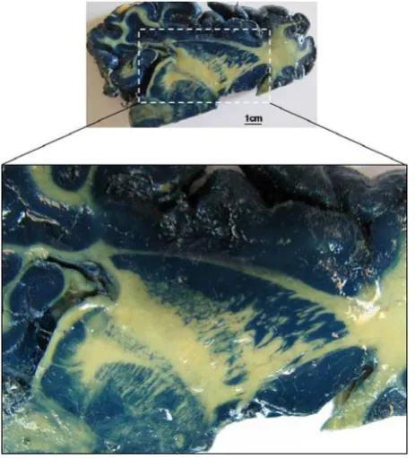
We observed no color fade in stained slices exposed to light. Gray matter was more distinguishable from white matter in Mulligan-stained sections compared to unMulligan-stained slices. Since the white matter areas did not react with the stain in this procedure, there was satis-factory contrast between the blue-stained grey areas and the colorless white matter. Sub-cortical structures and fiber tracts were clearly visible in stained brain cuts (Figure 1).
Figure 1.Sub-cortical nuclei and fiber tracts in a sagittal-stained brain plastinate which can be clearly seen
Shrinkage
The step-wise dehydration method at room temperature caused no significant loss of color in Mulligan-stained slices compared to the standard method (Figure 2). We observed
15.36% surface shrinkage of the samples after plastination with the standard method; 25.54% surface shrinkage was noted with the stepwise dehydration method.
Figure 2. Comparing the colored plastinats in standard and stepwise dehydration methods. Measurement of blue color pixels was done using ImageJ software (P<0.05).
Discussion
Plastination of stained brain slices gives us dry, clear, odorless specimens that are helpful tools in
teaching neuroanatomy. The use of buffered
fixative caused prevention of formalin pigment formation in brain sections.The chemical reaction in Tompsett staining produced a blue color in the gray matter that enabled the observer to clearly visualize the demarcation between gray and white matter, particularly in the sub-cortical structures (Figure 1). Perl's Prussian blue-stained brain sections in anatomy museums that have been exposed to light fade over time [13]. This loss of color could be due to ultraviolet (UV) light in the environment [14]. A thin layer of S10 hardened polymer on the plastinates might protect the color against UV light. Although staining and plastination of brain sections have been studied under freeze substitution conditions [7, 13], there is no data about stepwise dehydration at room temperature.
Anat Sci J 2013, Vol 10, No 2
of the present study was to examine whether dehydration of stained specimens at room temperature affects the solubility of the color. We observed that the blue color of stained brain sections was not changed in room temperature acetone. After six months exposure of the stained sections to light, there was no color loss in both the stepwise and freeze substitution methods. Another aim of the present study was to compare the two conditions of dehydration. In the present study we observed a 10% increase in shrinkage of the samples by the stepwise dehydration method (15.36%) compared to the freeze substitution method, which had a 25.54% increase in shrinkage. This amount of shrinkage was not solely attributed to dehydration but also from staining and impre-gnation. There is some evidence that staining can cause approximately 1% shrinkage in the brain [16]. Sora et al. have reported a surface shrinkage of 4.41% after plastination while using P40 as impregnation resin for 12 hours [17]. A 25% in brain slice surface could be problematic when comparing brain sections with corresponding MRI cuts. Although these plastinats had no severe effect on the morphology and relationship between brain structures, they were still suitable for teaching neuroanatomy. A previous study had approxi-mately the same average amount of shrinkage in
other organs that were dehydrated with pure acetone at cold and room temperatures [18]. Relatively higher shrinkage of stepwise dehyd-ration in our method could be due to the higher amount of fat in brain tissue.
Despite the increased shrinkage we have observed in stepwise dehydration, several brain slices can be dehydrated first with used acetone without the need for a freezer. This method is more cost-effective than the standard method considering the high price of pure acetone and high cost of electrical energy for a freezer.
In conclusion, stepwise dehydration of brain slices at room temperature does not affect color quality. Considering the low cost of stepwise dehydration with used acetone and no need for a freezer, these stained brain slices can be used for educational purposes in anatomy courses. However stepwise dehydration is not suitable for studying the course of nerves and blood vessels and their relationship with each other in the brain.
Acknowledgement
This work was supported by the Deputy of Research at Iran University of Medical Sciences. We express our appreciation to F. Mohammadi and A. Parvanehvar for their excellent technical support.
The authors declare that they have no conflicts of interest in the research.
References
1. Weiglein AH. Plastination in the neurosciences. Acta Anat (Basel) 1997; 158: 6-9.
2. Douglass C, Glover R. Plastination: Preservation technology enhances biology teaching. Am Biol Teach 2003; 65: 503.
3. Gu Y, Fujimiya Y, Kunugita N. Long-term exposure to gaseous formaldehyde promotes allergen-specific IgE-mediated immune responses in a murine model. Hum Exp Toxicol 2008; 27: 37-43.
4. Khaliq F, Tripathi P. Acute effects of formalin on pulmonary functions in gross anatomy laboratory. Indian J Physiol Pharmacol 2009; 53: 93-6.
5. Fleisher JM. Medical students' exposure to formaldehyde in gross anatomy laboratories. N Y State J Med 1987; 87: 385-8.
6. von Hagens G, Tiedemann K, Kriz W. The current potential of plastination. Anat Embryol (Berl) 1987; 175: 411-21.
7. Ulfig N, Wuttke M. Plastination of stained sections of the human brain. Anat Anz 1990; 170: 309-12.
8. O'Sullivan E, Mitchell BS. Plastination for gross anatomy teaching using low cost equipment. Surg Radiol Anat.1995; 17: 277-81.
Reclamation of acetone in plastination laboratories: a simple and inexpensive method. Acta Anat (Basel) 1997; 158: 26-9.
10. Steinke H, Rabi S, Saito T, Sawutti A, Miyaki T, Itoh M. Light-weight plastination. Ann Anat 2008; 190: 428-31.
11. Smith BJ, Holladay SD. Risk factors associated with plastination: Infectious agent considerations. J Int Soc Plast 2001; 15.
12. Tompsett DH. Differential staining of thin brain slices. Ann R Coll Surg Engl 1977; 59: 258.
13. Baeres FM, Moller M.Plastination of dissected brain specimens and Mulligan-stained sections of the human brain. Eur J Morphol 2001; 39: 307-11.
14. Barnett RI, Lyons GW, Driscoll JD, Forrest WJ. Improved sectioning and Berlin blue staining of
whole human brain. Stain Technol 1980; 55: 235-9.
15. John WH, Ralph H. General Chemistry. 2nd ed. New
Jersey, Prentice Hall College Div, 1996.
16.Suriyaprapadilok L, Withyachumnarnkul B. Plastination of stained sections of the human brain: Comparison between different staining methods. J Int Soc Plastination 1997; 12: 27-32.
17. Sora MC, Brugger P, Traxler H. P40 plastination of human brain slices: comparison between different immersion and impregnation conditions. J Int Soc Plastination 1999; 14: 22-4.