Open Access
Research
Impact of contractile reserve on acute response to cardiac
resynchronization therapy
Marie Moonen
1, Mario Senechal
2, Bernard Cosyns
3, Pierre Melon
1,
Eric Nellessen
1, Luc Pierard
1and Patrizio Lancellotti*
1Address: 1Department of Cardiology, CHU Sart Tilman, Liege, Belgium, 22725, chemin Sainte-Foy, Québec, Canada and 3Chirec Hôpital de Braine-l'Alleud Waterloo, rue Wayez 35, 1420 Braine Braine-l'Alleud, Belgium
Email: Marie Moonen - [email protected]; Mario Senechal - [email protected]; Bernard Cosyns - [email protected]; Pierre Melon - [email protected]; Eric Nellessen - [email protected]; Luc Pierard - [email protected];
Patrizio Lancellotti* - [email protected] * Corresponding author
Abstract
Background: Cardiac resynchronization therapy (CRT) provides benefit for congestive heart failure, but still 30% of patients failed to respond to such therapy. This lack of response may be due to the presence of significant amount of scar or fibrotic tissue at myocardial level. This study sought to investigate the potential impact of myocardial contractile reserve as assessed during exercise echocardiography on acute response following CRT implantation.
Methods: Fifty-one consecutive patients with heart failure (LV ejection fraction 27% ± 5%, 67% ischemic cardiomyopathy) underwent exercise Doppler echocardiography before CRT implantation to assess global contractile reserve (improvement in LV ejection fraction) and local contractile reserve in the region of the LV pacing lead (assessed by radial strain using speckle tracking analysis). Responders were defined by an increase in stroke volume ≥ 15% after CRT.
Results: Compared with nonresponders, responders (25 patients) showed a greater exercise-induced increase in LV ejection fraction, a higher degree of mitral regurgitation and a significant extent of LV dyssynchrony. The presence of contractile reserve was directly related to the acute increase in stroke volume (r = 0.48, p < 0.001). Baseline myocardial deformation as well as contractile reserve in the LV pacing lead region was greater in responders during exercise than in nonresponders (p < 0.0001).
Conclusion: The present study showed that response to CRT largely depends not only on the extent of LV dyssynchrony and the severity of mitral regurgitation but also on the presence of contractile reserve.
Background
The number of patients presenting with heart failure is increasing rapidly. Currently, cardiac resynchronization therapy (CRT) has emerged as an adjunctive treatment in
patients with drug-refractory heart failure [1]. The clinical benefit of CRT – evidenced by improvement in symp-toms, quality of life, exercise capacity, and left ventricular (LV) systolic performance – has been largely
demon-Published: 31 December 2008
Cardiovascular Ultrasound 2008, 6:65 doi:10.1186/1476-7120-6-65
Received: 30 October 2008 Accepted: 31 December 2008
This article is available from: http://www.cardiovascularultrasound.com/content/6/1/65
© 2008 Moonen et al; licensee BioMed Central Ltd.
strated [2,3]. Acutely, a CRT-increase in forward stroke volume by ≥ 15% has been shown to predict long-term clinical outcome in both ischemic and non ischemic car-diomyopathy [4]. However, 20% to 30% of patients do not respond to CRT despite application of recommended selection criteria [5]. Therefore, additional selection crite-ria are needed. Response to CRT largely depends on the extent of LV dyssynchrony, the severity of of mitral regur-gitation and the possibility offers to the LV to recruit func-tion [6-9]. CRT response could thus correlate with myocardial viability and inversely be associated with the extent of myocardial fibrosis [10,11]. Furthermore, scar tissue or fibrosis – characterized by lack of contractile reserve – in the LV pacing lead region may prohibit response to CRT. Myocardial contractile reserve – impor-tant prognostic marker in heart failure – can be reliably assessed during exercise echocardiography [12]. This study sought to investigate the potential impact of myo-cardial contractile reserve as assessed during exercise echocardiography on acute response following CRT implantation.
Methods
Patient population
Consecutive patients (n = 51) with severe heart failure, scheduled for implantation of a permanent biventricular pacemaker, were included using established selection cri-teria for CRT: New York Heart Association class III or IV despite optimal medical therapy, LV ejection fraction ≤ 35%, QRS with left bundle branch block configuration or a duration >120 ms. The aetiologies of heart failure were idiopathic dilated cardiomyopathy in 17 patients and ischaemic heart disease in 34. All patients underwent exer-cise Doppler echocardiography before CRT implantation to assess global LV contractile reserve and local contractile reserve in the region of the LV pacing lead (assessed by longitudinal strain using speckle tracking analysis). No patients with ischaemic disease presented inducible ischaemia during test. The protocol was approved by the Human Ethical Committee of our University Hospital and all patients gave informed consent.
Exercise echocardiography
A symptom-limited graded bicycle exercise test was per-formed in a semi-supine position on a tilting exercise table. After an initial workload of 25 W maintained for 2 min, the workload was increased every 2 min by 25 W. Blood pressure and a 12-lead electrocardiogram were recorded every 2 min. Two-dimensional echocardio-graphic recordings were made throughout the test.
Echocardiographic measurements
All echocardiographic parameters were obtained at rest and at peak exercise in the same cycling semi-supine posi-tion (Vivid 7 imaging device, GE Healthcare, United
King-dom) and were obtained in digital format and stored on optical disks for off-line analysis. Two-dimensional gray-scale images (frame rates ≥ 70 s-1) and colour-tissue
Dop-pler imaging (frame rates ≥ 115 s-1) were performed in the
apical views using a narrow sector angle. Left ventricular end-diastolic and end-systolic volumes and ejection frac-tion were measured by the biapical Simpson disk method. Wall motion score analysis was applied to a 16-segment model of the LV with a semiquantitative scoring system (1 = normal, 2 = hypokinesia, 3 = akinesia, 4 = dyskinesia). Quantitation of MR was performed by the proximal isove-locity surface area method as previously described [4]. Intra-LV dyssynchrony was referred to LV dispersion and was calculated as the difference between the longest and the shortest times to peak systolic velocities of 4 opposing basal walls (apical 2-and 4-chamber) [13]. Global LV con-tractile reserve was expressed as the change in LV ejection fraction from rest to peak exercise. Regional contractile reserve in the pacing lead was assessed using speckle track-ing analysis from LV long axis images (mid segments). After tracing the endocardial borders in the end-systolic frame, an automated tracking algorithm outlined the myocardial deformation in the dedicated LV segments [14]. Peak systolic radial strain was only measured in the lateral, posterior and anterior regions, where the LV lead was positioned.
CRT implantation and setting
All patients received a biventricular pacing device for CRT with a right ventricular apical lead and LV pacing elec-trodes positioned through the coronary sinus in a LV epi-cardial vein. This coronary sinus lead was placed in a lateral position in 32 patients, in a postero-lateral posi-tion in 16 and in anterior posiposi-tion in 3. After a successful implant, echocardiography was used to optimize the atrio-ventricular delay in order to maximize LV filling time. Interventricular pacing interval was set to default value (V-V = 0 ms).
CRT response definition
Within the week after implantation, changes in forward stroke volume were assessed. Improvement was defined as an increase in stroke volume ≥ 15% after CRT [2] (see vid-eos).
Statistical analysis
stroke volume after CRT implantation. The optimal improvement in LV ejection fraction and in myocardial strain during exercise to predict response to CRT was determined by receiver operating characteristic curve anal-ysis. To detect independent cofactors associated with response to CRT, a logistic multivariate analysis was per-formed. All significant variables were included in the mul-tivariate model. Reproducibility of echocardiographic measurements has been previously published [15].
Results
Responders versus non responders
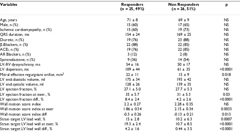
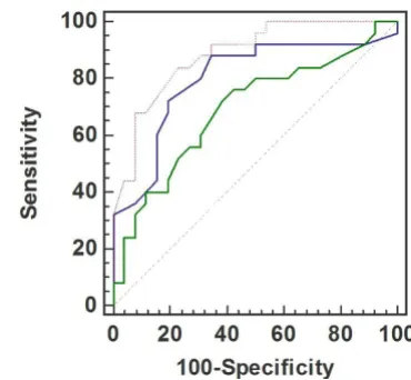
Within the 2 days after CRT implantation, 25 patients (49%) were responders. Baseline clinical and echocardio-graphic parameters between responders and non respond-ers were similar, except for higher degree of mitral regurgitation, greater extent of LV dyssynchrony and bet-ter myocardial deformation by 2D speckle tracking in the LV lead region in responders (Table 1). During exercise, responders showed a greater increase in LV ejection frac-tion. Improvement in LV ejection fraction at peak test was correlated to the improvement in forward stroke volume after CRT (r = 0.48) (Figure 1). By multivariate logistic regression analysis, LV dyssynchrony (p = 0.017), the degree of mitral regurgitation (p = 0.028) and the pres-ence of contractile reserve at exercise (p = 0.006) emerged as independent predictors of response to CRT. Table 2
depicted the area under the curves (ROC-curves) for these 3 independent predictors (Figure 2) (see additional files 1, 2, 3, 4 and 5).
Regional contractile reserve and response to CRT
During exercise, the extent of improvement in regional strain (4.2 ± 1.6% vs 0.44 ± 3.5; p < 0.0001) was greater in patients who presented a significant increase in forward stroke volume. By multivariate logistic regression analysis, the presence of contractile reserve in the LV lead region (OR 0.6: 95% CI 0.39–0.94; p = 0.025) and the global improvement in ejection fraction at exercise (OR 0.59: 95% CI 0.41–0.87; p = 0.008) emerged as independent predictors of response to CRT. An exercise increase in strain values of the target LV lead wall by > 2% (ROC-Curve) yielded a good sensitivity (92%) and specificity (73.1%) for predicting LV reverse remodelling after CRT (AUC 0.89). Among responders, 18 patients had both LV dyssynchrony and regional contractile reserve at exercise (Figure 3). Only 1 out of the 17 patients with no signifi-cant local and global contractile reserve responded to CRT. Figure 4 showed a patient without posterolateral contractile reserve during exercise.
Discussion
The response to CRT is likely modulated by multiple fac-tors. The present study confirms that response to CRT
Table 1: Responders versus non responders
Variables Responders (n = 25, 49%)
Non Responders (n = 26, 51%)
p
Age, years 71 ± 8 69 ± 9 NS
Male, n (%) 15 (60) 17 (65) NS
Ischemic cardiomyopathy, n (%) 15 (60) 19 (73) NS
QRS duration, ms 154 ± 24 169 ± 25 NS
Diuretic, n (%) 19 (76) 23 (88) NS
β-Blockers, n (%) 22 (88) 22 (85) NS
ACEi, n (%) 19 (76) 22 (85) NS
AR Blockers, n (%) 3 (12) 2 (8) NS
Spironolactone, n (%) 9 (36) 14 (54) NS
LV-RV dyssynchrony, ms 54 ± 16 50 ± 17 NS
LV dispersion, ms 109 ± 44 61 ± 35 <0.0001 Mitral effective regurgitant orifice, mm2 22 ± 11 15 ± 9 0.018
LV end-diastolic volume, ml 175 ± 34 193 ± 42 NS LV end-systolic volume, ml 128 ± 26 139 ± 35 NS LV ejection fraction, % 27.1 ± 5.0 27.7 ± 5.3 NS LV ejection fraction at exer., % 35 ± 5.7 31 ± 5.3 0.03 LV ejection fraction diff., % 8.4 ± 2.4 4.2 ± 2.6 <0.0001 Wall motion score index 2.2 ± 0.27 2.28 ± 0.35 NS Wall motion score index at exer 1.86 ± 0.34 2.15 ± 0.34 0.0033 Wall motion score index diff -0.3 ± 0.26 -0.13 ± 0.21 0.013 Strain target LV lead wall, % 15 ± 2.8 10.2 ± 6.3 0.0007 Strain target LV lead wall at exer, % 19.3 ± 2.4 10.7 ± 8.5 <0.0001 Strain target LV lead wall diff., % 4.2 ± 1.6 0.44 ± 3.5 <0.0001
largely depends not only on the extent of LV dyssynchrony and the severity of mitral regurgitation but also on the possibility offers to the LV to recruit function, namely the presence of contractile reserve. The probability to acutely respond to CRT is highly likely in patients with moderate to severe mitral regurgitation, major LV dyssynchrony and significant contractile myocardial recruitment – global and in the LV lead target site – at exercise.
Role of LV dyssynchrony
Several studies have demonstrated that the major predic-tor of responsiveness to CRT is mechanical rather than electrical dyssynchrony [6,13,16,17]. Among available techniques, tissue Doppler imaging has emerged as the most practical method for assessing LV dyssynchrony. As reported previously, we found that a significant mechani-cal delay between the basal segments of the LV, mainly the septum and the lateral wall, on tissue Doppler was highly predictive for response to CRT [6,18]. Patients with a delay ≥ 65 ms showed a significant improvement in for-ward stroke volume under CRT. However, although this beneficial effect was rarely observed in patients with a delay < 65 ms, 34% (9 of 26) of the patients with severe
LV dyssynchrony did not respond to CRT. This empha-sizes that other factors play a role; LV dyssynchrony is nec-essary but not sufficient for CRT response. Indeed, delayed wall motion is mainly a marker of myocardial dysfunction and could be exhibited in viable and scar tis-sue according to loading conditions [19,20].
Effect of mitral regurgitation
Response to CRT might be modulated by the presence of functional mitral regurgitation before implantation. Few authors have shown that patients with severe mitral regur-gitation have less chance of improving with CRT [3]. However, these studies included a limited number of patients. In the CARE-HF study, a randomized trial including a large number of patients, it was, conversely, shown that patients who were not improved were likely to have no significant mitral regurgitation as compared with responders [7]. The results of the present study confirm and extend this observation. Indeed, we found that a mitral effective regurgitant orifice > 15 mm2 was
associ-ated with a higher likelihood of response to CRT. Patients with more severe mitral regurgitation have more LV asyn-chrony (98 ± 49 ms vs 67 ± 36 ms; p = 0.019). The
inter-Table 2: Area under the curves, sensitivity, specificity and optimal cutoff values of predictors for acute CRT response.
Data at inclusion Cutoff values AUC Sensitivity Specificity
LV dyssynchrony > 65 ms 0.80 88% 65.4%
Effective regurgitant orifice > 15 mm2 0.69 76% 57.7%
Changes in LV ejection fraction at exercise ≥ 6.7% 0.89 84% 76.7% Changes in wall motion score index at exercise ≥ 0.23 0.76 80% 69%
Relationship between contractile reserve (improvement in LV ejection fraction during exercise) at inclusion and the per-centage of changes forward stroke volume under CRT
Figure 1
Relationship between contractile reserve (improve-ment in LV ejection fraction during exercise) at inclusion and the percentage of changes forward
stroke volume under CRT.
Receiver operating characteristic curves analysis on various parameters to predict response after CRT
Figure 2
play between LV dyssynchrony and mitral regurgitation is complex [4,21-23]. In fact, the degree of mitral regurgita-tion is directly correlated to the asynchronous activaregurgita-tion of papillary muscles and the decrease in mitral valve clos-ing forces (mainly the consequence of global LV dyssyn-chrony). It could thus be expected that the beneficial effect
of CRT would be greater in patients with significant mitral regurgitation.
Myocardial reserve and response to CRT
In patients with depressed LV function, the identification of contractile reserve during stress echocardiography has been shown to provide important prognostic information in heart failure patients [24,25]. More specifically, in patients referred to CRT, few authors have reported that the presence of residual myocardial viability might mod-ulate the response to CRT [11,14,26-30]. Contractile reserve is a specific marker of underlying myocardial via-bility, which can be reliably assessed during semi-supine exercise echocardiography [12]. To the best of our knowl-edge, this study was the first to examine the value of exer-cise echocardiography for predicting response to CRT. Significant contractile myocardial recruitment – global and in the LV lead target site – characterized patients who presented the greater rise in forward stroke volume. An increase in LV ejection fraction by ≥ 6.7% and/or an increase in local strain by ≥ 2% were found to be predic-tive for response to CRT. Moreover, the extent of LV global contractile reserve was related to the percentage of acute changes in forward stroke volume. These data confirm and extend previous studies by demonstrating that a sub-stantial amount of recruitable myocardium is needed to obtain improvement in LV function after CRT. It could be thus argued that in case of advanced myocardial remode-ling process, fibrosis and loss of contractile material may severely altered myocardial conduction and contractile Number of responders to CRT for 4 different patient
catego-ries based on the presence or absence of global contractile reserve (CR+/CR-) in combination with the presence or absence of contractile reserve in the region of the pacing lead (LEAD+/LEAD-)
Figure 3
Number of responders to CRT for 4 different patient categories based on the presence or absence of glo-bal contractile reserve (CR+/CR-) in combination with the presence or absence of contractile reserve in the region of the pacing lead (LEAD+/LEAD-).
Example of a patient without contractile reserve in the region of the lead pacing
Figure 4
properties, which might in turn impede efficient biven-tricular pacing [31].
Study limitations
Some limitations should be acknowledged. Our data per-tain only to patients with current criteria for CRT implan-tation. The population studied was not completely homogenous since it was composed of patients with myo-cardial dysfunction of ischaemic and non ischaemic ori-gin. However, this represents our daily patients referred for CRT. Moreover, biventricular pacing is still a challeng-ing therapy in both settchalleng-ings.
Conclusion
Heart failure patients referred to CRT have less chance of improving under therapy if they have no significant mitral regurgitation, no LV dyssynchrony and no contractile myocardial recruitment – global and in the LV lead target site – at exercise. It might thus be proposed that the assess-ment of these parameters would be routinely performed before submitting the patients to CRT.
Competing interests
The authors declare that they have no competing interests.
Authors' contributions
All authors participated in the conception and design of the study (MM, MS, BC, PM, EN, LP, PL). The other authors played an important role in the analysis and inter-pretation of data (MM, MS, BC). The last author wrote the paper (PL). All authors revised the manuscript critically for important intellectual content and gave final approval for submission (MM, MS, BC, PM, EN, LP, PL).
Additional material
References
1. Vardas PE, Auricchio A, Blanc JJ, Daubert JC, Drexler H, Ector H, Gasparini M, Linde C, Morgado FB, Oto A, Sutton R, Trusz-Gluza M: Guidelines for cardiac pacing and cardiac resynchronization therapy: The Task Force for Cardiac Pacing and Cardiac Resynchronization Therapy of the European Society of Car-diology. Eur Heart J 2007, 28:2256-2295.
2. Tournoux FB, Alabiad C, Fan D, Chen AA, Chaput M, Heist EK, Mela T, Mansour M, Reddy V, Ruskin JN, Picard MH, Singh JP: Echocardi-ographic measure of acute haemodynamic response after cardiac resynchronization therapy predicts long term clini-cal outcome. Eur Heart J 2007, 28:1143-1148.
3. Cleland JG, Daubert JC, Erdmann E, Freemantle N, Gras D, Kappen-berger L, Tavazzi L, Cardiac Resynchronization-Heart Failure (CARE-HF) Study Investigators: The effect of cardiac resynchronization on morbidity and mortality in heart failure. N Engl J Med 2005, 352:1539-1549.
4. Lancellotti P, Mélon P, Sakalihasan N, Waleffe A, Dubois C, Bertholet M, Piérard LA: Effect of cardiac resynchronization therapy on functional mitral regurgitation in heart failure. Am J Cardiol
2004, 94:1462-5.
5. Abraham WT, Fisher WG, Smith AL, Delurgio DB, Leon AR, Loh E, Kocovic DZ, Packer M, Clavell AL, Hayes DL, Ellestad M, Trupp RJ, Underwood J, Pickering F, Truex C, McAtee P, Messenger J, MIRA-CLE Study Group. Multicenter InSync Randomized Clinical Evaluation: Cardiac resynchronization in chronic heart failure. N Engl J Med 2002, 346:1845-1853.
6. Yu CM, Gorcsan J 3rd, Bleeker GB, Zhang Q, Schalij MJ, Suffoletto MS, Fung JW, Schwartzman D, Chan YS, Tanabe M, Bax JJ: Useful-ness of tissue Doppler velocity and strain dyssynchrony for predicting left ventricular reverse remodeling response after cardiac resynchronization therapy. Am J Cardiol 2007, 00(8):1263-1270.
7. Díaz-Infante E, Mont L, Leal J, García-Bolao I, Fernández-Lozano I, Hernández-Madrid A, Pérez-Castellano N, Sitges M, Pavón-Jiménez R, Barba J, Cavero MA, Moya JL, Pérez-Isla L, Brugada J, SCARS Investi-gators: Predictors of lack of response to resynchronization therapy. Am J Cardiol 2005, 95:1436-40.
8. Reuter S, Garrigue S, Barold SS, Jais P, Hocini M, Haissaguerre M, Clementy J: Comparison of characteristics in responders ver-sus nonresponders with biventricular pacing for drug-resist-ant congestive heart failure. Am J Cardiol 2002, 89:346-50. 9. Richardson M, Freemantle N, Calvert MJ, Cleland JG, Tavazzi L,
CARE-HF Study Steering Committee and Investigators: Predictors and treatment response with cardiac resynchronization
Additional file 1
Case 1: a non-responder with moderate mitral regurgitation, no LV dys-synchrony and transmural necrosis in the posterolateral region. Under CRT, no significant change in mitral regurgitation was observed.
Click here for file
[http://www.biomedcentral.com/content/supplementary/1476-7120-6-65-S1.avi]
Additional file 2
Case 1: a non-responder with moderate mitral regurgitation, no LV dys-synchrony and transmural necrosis in the posterolateral region. Under CRT, no significant change in mitral regurgitation was observed.
Click here for file
[http://www.biomedcentral.com/content/supplementary/1476-7120-6-65-S2.avi]
Additional file 3
Case 2: a responder with severe mitral regurgitation, LV dyssynchrony and viability in the posterolateral region. Under CRT, mitral regurgitation decreased and the aortic TVI increased.
Click here for file
[http://www.biomedcentral.com/content/supplementary/1476-7120-6-65-S3.avi]
Additional file 4
Case 2: a responder with severe mitral regurgitation, LV dyssynchrony and viability in the posterolateral region. Under CRT, mitral regurgitation decreased and the aortic TVI increased.
Click here for file
[http://www.biomedcentral.com/content/supplementary/1476-7120-6-65-S4.avi]
Additional file 5
Cases 1 and 2 responders.Click here for file
Publish with BioMed Central and every scientist can read your work free of charge "BioMed Central will be the most significant development for disseminating the results of biomedical researc h in our lifetime."
Sir Paul Nurse, Cancer Research UK
Your research papers will be:
available free of charge to the entire biomedical community
peer reviewed and published immediately upon acceptance
cited in PubMed and archived on PubMed Central
yours — you keep the copyright
Submit your manuscript here:
http://www.biomedcentral.com/info/publishing_adv.asp
BioMedcentral therapy in patients with heart failure characterized by
dys-synchrony: a pre-defined analysis from the CARE-HF trial.
Eur Heart J 2007, 28:1827-34.
10. Bleeker GB, Kaandorp TA, Lamb HJ, Boersma E, Steendijk P, de Roos A, Wall EE van der, Schalij MJ, Bax JJ: Effect of posterolateral scar tissue on clinical and echocardiographic improvement after cardiac resynchronization therapy. Circulation 2006, 113:969-976.
11. Lim P, Bars C, Mitchell-Heggs L, Roiron C, Elbaz N, Hamdaoui B, Lel-louche N, Dubois-Randé JL, Guéret P: Importance of contractile reserve for CRT. Europace 2007, 9:739-743.
12. Lancellotti P, Hoffer EP, Piérard LA: Detection and clinical useful-ness of a biphasic response during exercise echocardiogra-phy early after myocardial infarction. Am Coll Cardiol 2003, 1(7):1142-7.
13. Bax JJ, Bleeker GB, Marwick TH, Molhoek SG, Boersma E, Steendijk P, Wall EE van der, Schalij MJ: Left ventricular dyssynchrony pre-dicts response and prognosis after cardiac resynchronization therapy. J Am Coll Cardiol 2004, 44:1834-40.
14. Ypenburg C, Sieders A, Bleeker GB, Holman ER, Wall EE van der, Schalij MJ, Bax JJ: Myocardial contractile reserve predicts improvement in left ventricular function after cardiac resyn-chronization therapy. Am Heart J 2007, 154:1160-1165. 15. Lancellotti P, Cosyns B, Piérard LA: Dynamic left ventricular
dys-synchrony contributes to B-type natriuretic peptide release during exercise in patients with systolic heart failure.
Europace 2008, 10:496-501.
16. Pitzalis MV, Iacoviello M, Romito R, Luzzi G, Anaclerio M, Forleo C: Ventricular asynchrony predicts a better outcome in patients with chronic heart failure receiving cardiac resyn-chronization therapy. J Am Coll Cardiol 2005, 45:65-69.
17. Chung ES, Leon AR, Tavazzi L, Sun JP, Nihoyannopoulos P, Merlino J, Abraham WT, Ghio S, Leclercq C, Bax JJ, Yu CM, Gorcsan J 3rd, St John Sutton M, De Sutter J, Murillo J: Results of the Predictors of Response to CRT (PROSPECT) trial. Circulation 2008, 117:2608-16.
18. Notabartolo D, Merlino JD, Smith AL, DeLurgio DB, Vera FV, Easley KA, Martin RP, León AR: Usefulness of the peak velocity differ-ence by tissue Doppler imaging technique as an effective predictor of response to cardiac resynchronization therapy.
Am J Cardiol 2004, 94:817-20.
19. Lancellotti P, Stainier PY, Lebois F, Piérard LA: Effect of dynamic left ventricular dyssynchrony on dynamic mitral regurgita-tion in patients with heart failure due to coronary artery dis-ease. Am J Cardiol 2005, 96:1304-7.
20. Ryf S, Rutz AK, Boesiger P, Schwitter J: Is post-systolic shortening a reliable indicator of myocardial viability? An MR tagging and late-enhancement study. J Cardiovasc Magn Reson 2006, 8:445-51.
21. Breithardt OA, Sinha AM, Schwammenthal E, Bidaoui N, Markus KU, Franke A, Stellbrink C: Acute Effects of Cardiac Resynchroniza-tion Therapy on FuncResynchroniza-tional Mitral RegurgitaResynchroniza-tion in Advanced Systolic Heart Failure. J Am Coll Cardiol 2003, 41:765-770.
22. Kanzaki H, Bazaz R, Schwartzman D, Dohi K, Sade LE, Gorcsan J 3rd: A Mechanism for Immediate Reduction in Mitral Regurgita-tion After Cardiac ResynchronizaRegurgita-tion Therapy. J Am Coll Car-diol 2004, 44:1619-1625.
23. Agricola E, Oppizzi M, Galderisi M, Pisani M, Meris A, Pappone C, Margonato A: Role of regional mechanical dyssynchrony as a determinant of functional mitral regurgitation in patients with left ventricular systolic dysfunction. Heart 2006, 92:1390-1395.
24. Bax JJ, Wijns W, Cornel JH, Visser FC, Boersma E, Fioretti PM: Accu-racy of currently available techniques for prediction of func-tional recovery after revascularization in patients with left ventricular dysfunction due to chronic coronary artery dis-ease: comparison of pooled data. J Am Coll Cardiol 1997, 30:1451-60.
25. Ramahi TM, Longo MD, Cadariu AR, Rohlfs K, Slade M, Carolan S, Vallejo E, Wackers FJ: Dobutamine-induced augmentation of left ventricular ejection fraction predicts survival of heart failure patients with severe non-ischaemic cardiomyopathy.
Eur Heart J 2001, 22:849-56.
26. Hummel JP, Lindner JR, Belcik JT, Ferguson JD, Mangrum JM, Bergin JD, Haines DE, Lake DE, DiMarco JP, Mounsey JP: Extent of
myo-cardial viability predicts response to biventricular pacing in ischemic cardiomyopathy. Heart Rhythm 2005, 2:1211-1217. 27. Da Costa A, Thévenin J, Roche F, Faure E, Roméyer-Bouchard C,
Messier M, Convert G, Barthélemy JC, Isaaz K: Prospective valida-tion of stress echocardiography as an identifier of cardiac resynchronization therapy responders. eart Rhythm 2006, 3:H406-413.
28. Ypenburg C, Schalij MJ, Bleeker GB, Steendijk P, Boersma E, Dibbets-Schneider P, Stokkel MP, Wall EE van der, Bax JJ: Extent of viability to predict response to cardiac resynchronization therapy in ischemic heart failure patients. J Nucl Med 2006, 47:1565-1570. 29. Chalil S, Foley PW, Muyhaldeen SA, Patel KC, Yousef ZR, Smith RE, Frenneaux MP, Leyva F: Late gadolinium enhancement-cardio-vascular magnetic resonance as a predictor of response to cardiac resynchronization therapy in patients with ischaemic cardiomyopathy. Europac 2007, 9:1031-1037.
30. Ypenburg C, Schalij MJ, Bleeker GB, Steendijk P, Boersma E, Dibbets-Schneider P, Stokkel MP, Wall EE van der, Bax JJ: Impact of viability and scar tissue on response to cardiac resynchronization therapy in ischaemic heart failure patients. Eur Heart J 2007, 28:33-41.