R E S E A R C H
Open Access
Right ventricular dysfunction is a predictor of
non-response and clinical outcome following
cardiac resynchronization therapy
Francisco Alpendurada
1, Kaushik Guha
2, Rakesh Sharma
2, Tevfik F Ismail
1, Amy Clifford
1, Winston Banya
3,
Raad H Mohiaddin
1, Dudley J Pennell
1, Martin R Cowie
2, Theresa McDonagh
4and Sanjay K Prasad
1*Abstract
Background:Cardiac resynchronization therapy (CRT) is an established treatment in advanced heart failure (HF). However, an important subset does not derive a significant benefit. Despite an established predictive role in HF, the significance of right ventricular (RV) dysfunction in predicting clinical benefit from CRT remains unclear. We investigated the role of RV function, assessed by cardiovascular magnetic resonance (CMR), in predicting response to and major adverse clinical events in HF patients undergoing CRT.
Methods:Sixty consecutive patients were evaluated with CMR prior to CRT implantation in a tertiary cardiac centre. The primary end-point was a composite of death from any cause or unplanned hospitalization for a major cardiovascular event. The secondary end-point was response to therapy, defined as improvement in left ventricular ejection fraction≥5% on echocardiography at one year.
Results:Eighteen patients (30%) met the primary end-point over a median follow-up period of 26 months, and 27 out of 56 patients (48%) were considered responders to CRT. On time-to-event analysis, only atrial fibrillation (HR 2.6, 95% CI 1.02-6.84, p = 0.047) and RV dysfunction, either by a reduced right ventricular ejection fraction-RVEF (HR 0.96, 95% CI 0.94-0.99, p = 0.006) or tricuspid annular plane systolic excursion-TAPSE (HR 0.88, 95% CI, 0.80-0.96, p = 0.006), were significant predictors of adverse events. On logistic regression analysis, preserved RVEF (OR 1.05, 95% CI 1.01-1.09, p = 0.01) and myocardial scar burden (OR 0.90, 95% CI 0.83-0.96, p = 0.004) were the sole
independent predictors of response to CRT. Patients with marked RV dysfunction (RVEF < 30%) had a particularly low response rate (18.2%) to CRT.
Conclusions:Right ventricular function is an important predictor of both response to CRT and long-term clinical outcome. Routine assessment of the right ventricle should be considered in the evaluation of patients for CRT.
Keywords:heart failure, cardiac resynchronization therapy, right ventricular function, cardiovascular magnetic resonance
Background
Cardiac resynchronisation therapy (CRT) is an estab-lished therapeutic option for selected patients with symptomatic heart failure (HF). Amongst its benefits are reduced mortality, improved exercise tolerance and quality of life [1,2]. However, a proportion of patients do not gain any significant benefit, the reasons for which are unclear. Thus a number of devices are being
implanted with no discernible clinical benefit, which has important healthcare costs implications, as well as exposing patients to unnecessary risks. Our current strategy for assessing benefit with CRT is mainly focused on assessing symptomatic or functional response, but it is increasingly clear that this does not necessarily translate into improved clinical outcomes. It is therefore important to refine the selection criteria for device implantation to better identify those who would benefit-both in terms of response and improved clinical outcomes.
* Correspondence: [email protected]
1CMR Unit. Royal Brompton Hospital. Sydney Street. London, SW3 6NP. UK
Full list of author information is available at the end of the article
Whilst much attention has focused on remodelling of the left ventricle (LV), the role of the right ventricle (RV) in the appropriate selection of patients for CRT remains unclear [3]. Previous studies assessing RV func-tion have utilised echocardiography and radionuclide imaging [4-7]. However, accuracy of RV volumes and function by these techniques may be inaccurate due to the anatomical location and complex geometric struc-ture. Cardiovascular magnetic resonance (CMR) offers superior three dimensional representation of the RV, leading to a more accurate and reproducible assessment of RV function [8]. We therefore sought to assess the impact of RV function on outcomes in HF patients undergoing CRT implantation using CMR.
Methods
Study population
We studied 60 consecutive patients attending the Royal Brompton Hospital heart failure pacing clinic between January 2005 and March 2010 who fulfilled the follow-ing criteria: 1. New York Heart Association (NYHA) class III/IV at the time of CRT implantation; 2. QRS width ≥120 ms; 3. LVEF ≤35% by echocardiography, and; 4. CMR study within 3 months before CRT implantation.
These patients were evaluated for clinical (aetiology of heart failure, symptom status and medication, heart rate, blood pressure) and electrocardiographic (rhythm and QRS width) parameters at the time of device implanta-tion. As this study involved review of local patient medi-cal records, individual consent was not required by our Ethics Committee who approved the study.
Imaging
Cardiovascular magnetic resonance studies were per-formed in 1.5T Sonata or Avanto scanners (Siemens, Erlangen, Germany). A short-axis stack from atrio-ven-tricular level to the apex was acquired using a steady-state free-precession cine sequence (echo time 1.6 ms, repetition time 3.2 ms, flip angle 60°, slice thickness 7 mm with a 3 mm gap, acquisition time of 8-12 cardiac cycles) to quantify left and right ventricular volumes. Long-axis cines were also acquired to define the valve plane throughout the cardiac cycle. An inversion recov-ery gradient echo sequence was used 10 minutes after gadolinium injection (Magnevist® or Gadovist® , 0.1 mmol/kg) to assess myocardial scar. Inversion times were set to null the normal myocardium with images repeated in two stacks of identical short-axis planes but separate phase-encoding directions to exclude artefact.
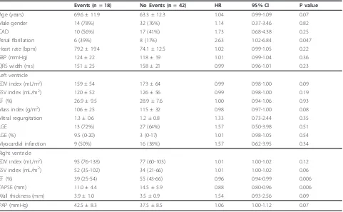
Left and right ventricular volumes were calculated using semi-automated software (CMR tools, Cardiovas-cular Imaging Solutions, London, UK), as previously described (Figure 1) [9,10]. The resulting values were
then indexed to body surface area and compared to reference values from a control population [9,10]. Tri-cuspid annular plane systolic excursion (TAPSE) was measured from the 4-chamber view (Figure 2). RV dys-function was defined as RVEF < 50% or TAPSE < 15 mm; severe RV dysfunction was defined as RVEF < 30% or TAPSE < 10 mm. Peak RV wall thickness was mea-sured from the short-axis slices.
[image:2.595.307.539.88.259.2]Valvular regurgitation was graded as mild (n = 1), moderate (n = 2) or severe (n = 3) by blinded observers, based on the echocardiographic and CMR findings. LVEF was also calculated by echocardiography before and 1 year after device implantation using the Simpson’s method from the 2-chamber and 4-chamber views.
Figure 1 Software used for ventricular volumes and mass measurements. Mid-ventricular short-axis (top) and four-chamber views (bottom) at end-diastole (left) and end-systole (right). Coloured areas represent the left and right ventricular cavities and myocardium. Ventricular volumes are generated from a short-axis stack after being confined by the mitral and tricuspid valve planes (red lines).
[image:2.595.308.539.553.681.2]Pulmonary artery systolic pressure was determined by echocardiography using standard methodology.
Contrast imaging with gadolinium was performed to assess the aetiology of the heart failure. Assessment of late gadolinium enhancement (LGE) in the left or right ventricle was interpreted by blinded observers. When present, the amount of LGE was quantified in a 16-seg-ment model based on the “full width at half maximum” technique by customized analysis software (MRI-MASS, Medis, Leiden, the Netherlands) [11].
Outcomes
All patients were followed-up in a heart failure clinic and standard medications were adjusted and optimized at these appointments. Data was collected from local hospital records, nation-wide summary case records and the Office of National Statistics. The primary end-point was a composite of all-cause mortality or an unplanned hospitalization for a major cardiovascular event; only the first event in each patient was included in this analy-sis [2]. The secondary end-point was echocardiographic response to CRT, defined as an improvement in LVEF by more than 5% by echocardiography 12 months after CRT implantation [12,13].
Statistical analysis
Categorical variables are presented as frequency and percentage. Continuous variables are presented as mean ± standard deviation (SD) if normally distributed, or as median plus inter-quartile range (IQR) as appropriate. Continuous variables were compared with Student’s t-test or the Mann-Whitney for non-parametric data. Cor-relation was assessed with Pearson’s or Spearman’s methods as appropriate.
Time to first event analysis was performed using Cox’s proportional hazard models. Log-rank test was used to compare Kaplan-Meier cumulative event curves. Logistic regression was used to assess response to CRT at 12 months. Multivariate logistic regression analysis using a forward stepwise approach was performed on para-meters that were significant on univariate analysis.
A two-tailed value of p-value < 0.05 was considered significant. All statistical analysis was performed using SPSS 19.0 (IBM, Chicago, Illinois, USA).
Results
Patients
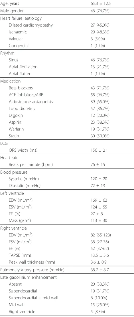
Patient baseline characteristics are presented in table 1. The mean age of the study population was 65.3 ± 12.5 years; most of the patients were male (76.7%). Heart fail-ure was ischaemic in 48.3% of patients, and atrial
fibril-Table 1 Baseline characteristics of the patients
Age, years 65.3 ± 12.5
Male gender 46 (76.7%)
Heart failure, aetiology
Dilated cardiomyopathy 27 (45.0%)
Ischaemic 29 (48.3%)
Valvular 3 (5.0%)
Congenital 1 (1.7%)
Rhythm
Sinus 46 (76.7%)
Atrial fibrillation 13 (21.7%)
Atrial flutter 1 (1.7%)
Medication
Beta-blockers 43 (71.7%)
ACE inhibitors/ARB 58 (96.7%)
Aldosterone antagonists 39 (65.0%)
Loop diuretics 52 (86.7%)
Digoxin 12 (20.0%)
Aspirin 23 (38.3%)
Warfarin 19 (31.7%)
Statin 30 (50.0%)
ECG
QRS width (ms) 156 ± 21
Heart rate
Beats per minute (bpm) 76 ± 15
Blood pressure
Systolic (mmHg) 120 ± 20
Diastolic (mmHg) 72 ± 13
Left ventricle
EDV (mL/m2) 169 ± 62
ESV (mL/m2) 124 ± 55
EF (%) 27 ± 8
Mass (g/m2) 113 ± 30
Right ventricle
EDV (mL/m2) 82 (65-123)
ESV (mL/m2) 38 (27-76)
EF (%) 52 (37-62)
TAPSE (mm) 13.5 ± 5.6
Peak wall thickness (mm) 3.6 ± 0.9
Pulmonary artery pressure (mmHg) 38.7 ± 8.7
Late gadolinium enhancement
Absent 20 (33.3%)
Subendocardial 19 (31.7%)
Subendocardial + mid-wall 6 (10.0%)
Mid-wall 15 (25.0%)
Right ventricle 5 (8.3%)
Data is presented as n (%), mean ± SD, or median (25th
- 75th
[image:3.595.304.539.101.656.2]lation/flutter was found in 23.3% of patients. The mean heart rate was 76 ± 15 bpm, and the mean QRS interval was 156 ± 21 ms (LBBB morphology in 95.0%). The median time between CMR and device implantation was 6 days. Fifty-six out of the 60 CRT (93%) devices were combined with a defibrillator (CRT-D).
Left ventricle
Both mean left ventricular indexed end-diastolic and end-systolic volumes were high (169 ± 62 and 124 ± 55 mL/m2, respectively). The indexed left ventricular end-diastolic volume was increased in 93% of the patients, while the indexed left ventricular end-systolic volume was increased in 99% of the patients compared to normal reference values [9]. The mean LVEF was 27 ± 8%, and the mean mass index was increased at 113 ± 30 g/m2.
Mitral regurgitation was seen in 90% of the patients, and was significant in 25% of patients (18% moderate, 7% severe). Aortic regurgitation was seen in 25% of patients, and was moderate in only one patient.
Right ventricle
Right ventricular indexed volumes and ejection fraction were non-normally distributed, and hence presented as median plus inter-quartile ranges. The median end-dia-stolic and end-syend-dia-stolic volumes were 82 (65-123) mL/m2 and 38 (27-76) mL/m2, respectively. Right ventricular indexed end-diastolic volume was increased in 38% of patients, while the indexed end-systolic volume was increased in 53% of patients [10]. The median RVEF was 52% (IQR 37-62%), with 53% of patients having a RVEF≥50%.
The mean TAPSE was 13.5 ± 5.6 mm, with 40% of patients having values within normal limits when the standard echocardiographic cut-off value of 15 mm was considered. The mean peak RV wall thickness was 3.6 ± 0.9 mm.
In the 52 patients (87%) in whom it could be mea-sured, the mean PASP was 38.7 ± 8.7 mmHg, with 37% of patients having a PAP > 40 mmHg. Tricuspid regur-gitation was seen in 57% of the patients, and was signifi-cant in 14% of patients (12% moderate, 2% severe). No significant pulmonary regurgitation was identified in any patient.
There was a moderately strong correlation between RVEF with TAPSE (r = 0.58, p < 0.01), and a moder-ate correlation between RVEF and LVEF (r = 0.41, p < 0.01). Severity of mitral regurgitation was associated with a higher PAP (p = 0.01) and a lower RVEF (p < 0.01). Modest inverse correlations were observed between RVEF and pulmonary artery pressure (r = -0.37, p < 0.01), RV wall thickness (r = -0.28, p = 0.03) and severity of tricuspid regurgitation (p = 0.03). TAPSE was inversely related with RV wall thickness
(r = -0.29, p = 0.03), but not with pulmonary artery pressure (r = -0.15, p = 0.28) or LVEF (r = 0.09, p = 0.48).
RVEF was similar in patients with an ischaemic and non-ischaemic aetiology (46.9 ± 14.8% vs. 49.5 ± 18.9, p = 0.56), and in patients with AF compared to those in sinus rhythm (45.4 ± 14.9% vs. 49.2 ± 17.8%, p = 0.48).
Myocardial fibrosis
Left ventricular LGE was present in two-thirds of the patients: 19 patients (31.7%) had sub-endocardial enhancement suggesting myocardial infarction, 15 patients (25.0%) had a mid-wall enhancement pattern indicating fibrosis, and the remaining 6 patients (10.0%) had a mixed pattern of myocardial infarction and mid-wall fibrosis. Myocardial infarction was therefore present in 25 patients (41.7%): the septal wall was affected in 11 patients; the inferolateral wall was involved in 15 patients. The median percentage of scar in the myocar-dium was 4% (IQR 0-18%).
Right ventricular LGE was present in 5 patients (8.3%). All these patients had coronary artery disease with infer-ior myocardial infarctions of the LV extending to the inferior free wall of the RV.
Follow-up
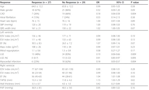
During a median follow-up of 26.1 months (IQR 16.1-39.3 months) after CRT implantation, there were 13 unplanned hospitalizations for a cardiovascular cause (all for heart failure decompensation), and 11 deaths (8 cardiac and 3 non-cardiac deaths). The primary end-point was reached by 18 patients. Atrial fibrillation and RV dysfunction emerged as the only predictors of the primary composite end-point on univariate analysis (table 2). Patients in atrial fibrillation or flutter had a HR of 2.6 (95% CI 1.02-6.84, p = 0.047) for the primary end-point. For each 10% decrease in RVEF, the risk of the primary end-point increased by 40% (HR 0.96, 95% CI 0.94-0.99, p = 0.006); for each 1 mm decrease in TAPSE, the risk of the primary end-point increased by 12% (HR 0.88, 95% CI 0.80-0.96, p = 0.006). Kaplan-Meier curves for RV function assessed by RVEF or TAPSE are displayed on Figure 3.
After documenting AF as a predictor of outcomes, we performed a post-hoc analysis on delivery rate, defined as the percentage of paced left ventricular beats. Data was collected from routine pacing clinic visits during the first year of implantation. Unlike AF, delivery rate was not associated with the primary end-point (HR 0.97, 95% CI 0.88-1.08, p = 0.56).
Response to therapy
increase in LVEF > 5% at one year of follow-up. Thus the response rate using this criterion was 48% (table 3).
Two sets of closely related parameters were associated with non-response to CRT on univariate analysis: (1) coronary artery disease, associated with myocardial infarction and scar burden (myocardial fibrosis plus
[image:5.595.59.540.110.407.2]infarction relative to LV mass), were predictors of non-response to therapy; (2) right ventricular dysfunction (lower RVEF and TAPSE) and hypertrophy (thicker RV free wall). Myocardial scar burden (OR 0.90, 95% CI 0.83-0.96, p = 0.004) and RVEF (OR 1.05, 95% CI 1.01-1.09, p = 0.01) were the only variables to remain signifi-cant on multivariate analysis (table 4).
Table 2 Univariate analysis: Primary end-point (time to death from any cause or an unplanned hospitalization for a major cardiovascular event).
Events (n = 18) No Events (n = 42) HR 95% CI P value
Age (years) 69.6 ± 11.9 63.3 ± 12.3 1.04 0.99-1.09 0.07
Male gender 14 (78%) 32 (76%) 1.14 0.37-3.46 0.82
CAD 10 (56%) 17 (41%) 1.73 0.68-4.38 0.25
Atrial fibrillation 6 (39%) 8 (17%) 2.63 1.02-6.84 0.047
Heart rate (bpm) 79.2 ± 19.4 74.1 ± 12.5 1.02 0.99-1.05 0.22
SBP (mmHg) 124 ± 22 118 ± 19 1.01 0.99-1.04 0.36
QRS width (ms) 151 ± 25 158 ± 21 0.99 0.96-1.01 0.23
Left ventricle
EDV index (mL/m2) 159 ± 54 173 ± 64 0.99 0.98-1.00 0.09
ESV index (mL/m2) 120 ± 52 126 ± 56 0.99 0.98-1.00 0.19
EF (%) 26.9 ± 9.5 28.9 ± 7.6 1.00 0.94-1.06 0.93
Mass index (g/m2) 106 ± 25 115 ± 32 0.98 0.97-1.00 0.08
Mitral regurgitation 1.3 ± 0.6 1.2 ± 0.8 1.33 0.73-2.44 0.35
LGE 13 (72%) 27 (64%) 1.57 0.50-3.98 0.51
LGE (%) 9.5 (0-20) 3 (0-17) 1.01 0.98-1.05 0.54
Myocardial infarction 9 (50%) 16 (38%) 1.57 0.62-3.95 0.34
Right ventricle
EDV index (mL/m2) 95 (76-138) 77 (60-103) 1.01 1.00-1.02 0.12
ESV index (mL/m2) 52 (35-102) 34 (21-66) 1.01 1.00-1.02 0.06
EF (%) 39 (25-54) 55 (43-66) 0.96 0.94-0.99 0.006
TAPSE (mm) 11.0 ± 4.4 14.5 ± 5.9 0.88 0.80-0.96 0.006
Wall thickness (mm) 3.9 ± 1.0 3.5 ± 0.9 1.54 0.93-2.56 0.09
PAP (mmHg) 42.5 ± 8.3 37.5 ± 8.5 1.06 1.00-1.12 0.07
Values are n (%), mean ± SD, or median (25th- 75thpercentile). HR = hazard ratio; CI = confidence interval. CAD = Coronary artery disease; SBP = systolic blood pressure; LGE = late gadolinium enhancement; PAP = Pulmonary artery pressure. Other abbreviations as in table 1.
[image:5.595.57.541.540.715.2]Response to CRT was noted to be lower as RV func-tion deteriorated (Figure 4). Poor RV funcfunc-tion, defined as a RVEF < 30% or a TAPSE < 10 mm, was associated with a particularly low response to CRT (response rates of 18.2% and 26.7%, respectively).
Discussion
This study shows that RV dysfunction is associated with non-response and adverse outcomes in patients on CRT. Despite meeting the standard criteria for CRT implanta-tion, there was a heterogeneous distribution of RV func-tion in our study populafunc-tion. Since LVEF was narrowly distributed by accepted indications for CRT, the wide RVEF distribution observed may indeed explain why RV function was an independent discriminator in this popu-lation with advanced HF. A lower RVEF was associated with a lower LVEF, more significant mitral regurgitation, and a higher PAP. This suggests that the RV
dysfunction may derive from two fundamental mechan-isms, the importance of which may vary from patient to patient: 1) an impairment of global biventricular intrin-sic contractility; 2) pulmonary hypertension secondary to elevated LV filling pressures and mitral regurgitation.
Adverse RV function may reflect more extensive and severe cardiac disease, to which improving myocardial synchrony has little impact. Of note, the response rate of the subgroup of patients with RVEF < 0.3 was less than 20%. Thus, patients with poor RV function were unlikely to benefit from CRT. This may have implica-tions in stratifying patients for device therapy.
Previous work
[image:6.595.56.542.99.394.2]Few studies have investigated RV function and dyssyn-chrony. Previous work has shown that anything up to 20-40% of RV systolic pressure and volume load may originate from contraction from the LV [14]. Table 3 Univariate analysis: Response to cardiac resynchronization therapy (improvement of LVEF≥5% at one year).
Response Response (n = 27) No Response (n = 29) OR 95% CI P value
Age 64.0 ± 12.2 65.8 ± 12.2 0.99 0.95-1.03 0.58
Male gender 18 (67%) 25 (86%) 0.32 0.09-1.20 0.09
CAD 7 (26%) 19 (66%) 0.18 0.06-0.58 0.004
Atrial fibrillation 4 (15%) 7 (24%) 0.55 0.14-2.13 0.38
Heart rate (bpm) 76 ± 15 76 ± 16 1.00 0.97-1.04 0.89
SBP (mmHg) 120 ± 20 119 ± 19 1.00 0.98-1.03 0.76
QRS width (ms) 161 ± 21 150 ± 22 1.02 1.00-1.05 0.07
Left ventricle
EDV index (mL/m2) 156 ± 46 177 ± 71 0.99 0.98-1.00 0.19
ESV index (mL/m2) 111 ± 40 133 ± 63 0.99 0.98-1.00 0.13
EF (%) 30.5 ± 8.5 26.5 ± 7.2 1.07 0.99-1.16 0.07
Mass index (g/m2) 108 ± 26 118 ± 34 0.99 0.97-1.01 0.23
Mitral regurgitation 1.1 ± 0.6 1.3 ± 0.8 0.58 0.27-1.27 0.17
LGE 13 (48%) 24 (83%) 0.19 0.06-0.66 0.009
LGE (%) 0 (0-7) 13 (3-22) 0.91 0.85-0.97 0.005
Myocardial infarction 6 (22%) 18 (62%) 0.18 0.05-0.57 0.004
Right ventricle
EDV index (mL/m2) 77 (67-104) 83 (61-139) 0.99 0.98-1.01 0.28
ESV index (mL/m2) 36 (23-54) 40 (31-96) 0.99 0.98-1.00 0.10
EF (%) 56 (45-63) 44 (28-61) 1.04 1.01-1.08 0.03
TAPSE (mm) 15.3 ± 5.8 11.8 ± 4.2 1.15 1.03-1.29 0.02
Wall thickness (mm) 3.3 ± 0.7 4.0 ± 1.0 0.42 0.21-0.84 0.02
PAP (mmHg) 36.9 ± 8.5 40.5 ± 9.0 0.95 0.89-1.02 0.16
Values are n (%), mean ± SD, or median (25th-75th percentile). OR = odds ratio; CI = confidence interval. Other abbreviations as in tables 1 and 2.
Table 4 Multivariate analysis: Response to cardiac resynchronization therapy (improvement of LVEF≥5% at one year).
Response Response (n = 27) No Response (n = 29) OR 95% CI P value
LGE (%) 0 (0-7) 13 (3-22) 0.90 0.83-0.96 0.004
RVEF (%) 56 (45-63) 44 (28-61) 1.05 1.01-1.09 0.01
[image:6.595.55.538.673.714.2]Conversely, left bundle branch block (LBBB) may inter-fere with ventricular activation and induce mechanical dyssynchrony. Hence LBBB not only affects LV function but may impair RV function as well.
RV functional indices have been previously evaluated in CRT populations. Initial work by Field et al assessed RV function on adverse outcomes in a population of 77 patients undergoing CRT. This study showed that RV dysfunction measured by the Doppler-derived Tei index was a predictor of the composite end-point of death, transplantation and the implantation of left ventricular assist devices [4]. More recent work focused on RV function and response to CRT. In a study of 44 patients undergoing CRT, echocardiographic indices of RV func-tion (TAPSE and RV fracfunc-tional area change) were signif-icantly worse in non-responders versus responders to CRT [5]. The value of TAPSE in predicting reverse remodelling was further validated in the CARE-HF population [6]. A further radionuclide imaging study in 44 patients undergoing CRT showed that those with a low baseline RVEF were less likely to improve in NYHA class, and tended to improve less in functional capacity and LVEF [7].
The results of our study are in line with the above publications. However, we used CMR as the gold-stan-dard technique for assessment of the RV, and RVEF as the reference marker of RV performance. Along with myocardial infarction, RVEF was an independent predic-tor of response to CRT. Among an array of established prognostic markers in HF, RVEF and TAPSE emerged as the strongest predictors of events. Our findings corre-lated response with outcomes over a clinically relevant timeframe, thus supporting the potential of RV function as a predictor of short-term response as well as longer-term outcome following CRT.
Tricuspid annular plane systolic excursion
Although available to all imaging modalities, and despite its validated prognostic value amongst cardiac and pul-monary conditions, RVEF estimation can be challenging as the right ventricle is a complex structure which can-not be modelled with simple geometric assumptions. Therefore, there is some interest in finding simpler ways for assessing RV performance. Of these, tricuspid annu-lar plane systolic excursion (TAPSE) is the most described alternative to RVEF in the literature [15]. This marker of RV longitudinal function is reproducible and easy to obtain, and has shown to be a predictor of adverse outcomes in heart failure, irrespective of NYHA status and LV function [16,17]. In this study, TAPSE not only identified which patients would respond to CRT, but also the patients more likely to have major adverse events. Thus, TAPSE may be used by CMR as an alternative to RVEF for assessing RV function pre CRT when the latter cannot be estimated.
Pulmonary hypertension
[image:7.595.58.556.89.277.2]Increased pulmonary artery pressures have shown to be associated with non-response to CRT [5]. As ejection fraction is influenced by the afterload, it is expected for increasing pulmonary pressures to be associated with decreasing RVEF. A significant negative correlation was indeed observed between PAP and RVEF in our study. However, this correlation was modest and not as strong as other parameters such as LVEF, suggesting that fac-tors other than pulmonary hypertension play a role in RV function. There was a trend for non-response to CRT and adverse outcomes in patients with increased pulmonary pressures, but statistical significance was not reached, probably explained by the smaller study popu-lation compared to other studies [18]. On the other
hand, markers of RV function were significantly asso-ciated with response and adverse events. This suggests that RV function is more important than pulmonary pressure alone for both mechanical response and long-term prognosis in patients on CRT.
Myocardial fibrosis
Late gadolinium enhancement CMR detects myocardial infarction accurately, with superior histological correlation compared to nuclear techniques [19]. This has become an active area of research in the CRT setting, with several subsequent studies documenting the value of scar burden, location and transmurality of myocardial infarctions as a predictor of response to CRT [20-24]. Consistent with this work, our study showed myocardial infarction and scar burden (as determined by percentage of LGE mass) to portend an adverse response to CRT. Although the amount of septal and lateral scar was associated with non-response to therapy (OR 0.99, 95% CI 0.98-1.00, p = 0.02; and OR 0.98, 95% CI 0.97-1.00, p = 0.02, respectively), scar location did not remain significant after including the total amount of scar in a multivariate model.
The lack of response observed with the above para-meters did not translate into poorer outcomes. It is pos-sible that this study was underpowered to detect more adverse events, as larger studies have shown ischaemic heart disease and location of myocardial infarction to portend a worse prognosis in CRT [25-27].
Atrial fibrillation
Atrial fibrillation was the other predictor of outcomes besides RV dysfunction. This mirrors findings of long term studies of patients on CRT [28]. It is recognised that AF with a high ventricular rate may impair the delivery of biventricular pacing, and consequently undermine the clinical benefit of CRT [29]. Despite the paucity of data from randomized control trials in AF populations, recent guidelines support the use of CRT in AF patients, although restricted to a slightly wider QRS duration > 130 ms [30]. A recent European registry suggests that around 20% of patients undergoing CRT are actually in permanent AF, which is in keeping with our findings [31]. Of note, all but one AF patient in this study had QRS > 130 ms. Rate control was satisfactory in our AF cohort, the ventricular delivery rate was high (median 97%, IQR 91-99%), and none of the patients underwent AV nodal ablation. Unlike RV dysfunction, AF was not associated with a lower response rate, sug-gesting that the impact of AF on adverse outcomes was not altered by device implantation.
Response
In addition to the hard clinical endpoints, we assessed the role of RV function on response to CRT. Response is a
contentious topic, with various proposed definitions but no consensus amongst different criteria. The response rate varies widely, depending on the study population and the response criteria used. In a recent paper compar-ing 15 response criteria from the most cited papers, the response rate ranged from 32% to 91%. The same study showed that the agreement between different criteria was poor in 75% of the time, especially between clinical and echocardiographic criteria [32]. For our study, we used LVEF to assess response because it is an objective para-meter and is also one of the selection criteria for CRT. In keeping with previous work, response for this study was an improvement in LVEF≥5% at 12 months [12,13]. The response rate observed was relatively low (48%), but still in accordance with the available literature [32]. Using this criterion, RV dysfunction predicted a failure of response to CRT in terms of LV remodelling, thus sup-porting previous echocardiographic and radionuclide stu-dies [5-7]. It is possible that significant RV dysfunction marks extensive and irreversible adverse remodelling, preventing reverse remodelling and functional recovery after CRT implantation. Improvement in LVEF≥5% was significantly associated with event-free survival in this cohort of patients (likelihood ratio 4.7, p = 0.01). Mechanical remodelling thus appears to be a good surro-gate marker of response to therapy, in line with a recently published analysis of the MADIT-CRT trial, where it was demonstrated that echocardiographic improvement in LV volumes and ejection fraction was associated with improved outcomes [33].
Limitations
This was a retrospective study with a relatively small number of patients and events limiting multivariate ana-lysis on outcomes. Nonetheless, both RVEF and TAPSE emerged as the most significant prognostic markers, suggesting that RV function as assessed by CMR has a powerful role in advanced HF undergoing CRT.
This was a single centre study in a tertiary referral hospital, but it allowed for consistent CMR scanning protocols, and consistent patient selection for CRT and close follow-up of all patients. To ensure a representa-tive cohort, consecurepresenta-tive patients were identified.
The CRT devices implanted during this time period were non-MRI compatible and hence necessitated an alternative imaging modality for consequent follow-up. The development of CMR-compatible CRT devices could overcome this current limitation and may provide new insights on the response of both left and right ven-tricle to CRT.
Conclusions
implantation. As current selection criteria only include patients with poor LV function, it is reasonable to assume that RV function will act as an important discri-minative prognostic marker in patients undergoing CRT. Furthermore, poor RV function appears to identify a subgroup of patients with extensive ventricular remo-delling unlikely to change their natural history. There-fore, CRT may be of no benefit in this subgroup of patients. The presence and amount of myocardial fibro-sis, by contrast, was a predictor of response but did not predict outcomes.
The results of this study suggest that assessment of RV function can provide valuable information pre CRT implantation. Further work in larger cohorts is required to ascertain the precise role of RV evaluation in the selection of patients for CRT as well as the mechanisms for this dysfunction.
List of abbreviations
CMR: cardiovascular magnetic resonance; CRT: cardiac resynchronization therapy; EF: ejection fraction; HF: heart failure; IQR: interquartile range; LBBB: left bundle brunch block; LV: left ventricle; PAP: pulmonary artery systolic pressure; RV: right ventricle; SD: standard deviation; TAPSE: tricuspid annular plane systolic excursion.
Acknowledgements
This project was supported by the NIHR Cardiovascular Biomedical Research Unit of Royal Brompton & Harefield NHS Foundation Trust and Imperial College London. Research support was also received from the BHF and CORDA.
Author details
1CMR Unit. Royal Brompton Hospital. Sydney Street. London, SW3 6NP. UK. 2
Department of Cardiology, Royal Brompton Hospital, Sydney Street, Chelsea, London, SW3 6NP, UK.3Research & Development, Royal Brompton Hospital,
Sydney Street, Chelsea, London, SW3 6NP, UK.4Department of Cardiology. King’s College Hospital, Denmark Hill, London, SE5 9RS, UK.
Authors’contributions
FA, KG, RS and SKP conceived the study. FA, KG, TFI and AC collected the data. WB performed the statistical analysis. All authors drafted, reviewed and approved the manuscript.
Competing interests
KG has received support from Biotronik. RS has received honoraria/research grants from Medtronic, Boston Scientific, Biotronik and Servier. DJP is a consultant to Siemens, and a director in Cardiovascular Imaging Solutions. MRC has received research funding from Medtronic, St. Jude Medical and Boston Scientific.
Received: 6 July 2011 Accepted: 31 October 2011 Published: 31 October 2011
References
1. Cazeau S, Leclercq C, Lavergne T, Walker S, Varma C, Linde C, Garrigue S, Kappenberger L, Haywood GA, Santini M, Bailleul C, Daubert JC:Effects of multisite biventricular pacing in patients with heart failure and intraventricular conduction delay.N Engl J Med2001,344:873-80. 2. Cleland JG, Daubert JC, Erdmann E, Freemantle N, Gras D, Kappenberger L,
Tavazzi L:Cardiac Resynchronization-Heart Failure (CARE-HF) Study Investigators. The effect of cardiac resynchronization on morbidity and mortality in heart failure.N Engl J Med2005,352:1539-1549.
3. Epstein AE, Dimarco JP, Ellenbogen KA,et al:ACC/AHA/HRS 2008 guidelines for Device-Based Therapy of Cardiac Rhythm Abnormalities: executive summary.Heart Rhythm2008,5:934-955.
4. Field ME, Solomon SD, Lewis EF, Kramer DB, Baughman KL, Stevenson LW, Tedrow UB:Right ventricular dysfunction and adverse outcome in patients with advanced heart failure.J Card Fail2006,12:616-620. 5. Scuteri L, Rordorf R, Marsan NA, Landolina M, Magrini G, Klersy C, Frattini F,
Petracci B, Vicentini A, Campana C, Tavazzi L, Ghio S:Relevance of echocardiographic evaluation of right ventricular function in patients undergoing cardiac resynchronization therapy.Pacing Clin Electrophysiol
2009,32:1040-1049.
6. Ghio S, Freemantle N, Scelsi L, Serio A, Magrini G, Pasotti M, Shankar A, Cleland JG, Tavazzi L:Long-term left ventricular reverse remodelling with cardiac resynchronization therapy: results from the CARE-HF trial.Eur J Heart Fail2009,11:480-488.
7. Burri H, Domenichini G, Sunthorn H, Fleury E, Stettler C, Foulkes I, Shah D: Right ventricular systolic function and cardiac resynchronization therapy.
Europace2010,12:389-394.
8. Hudsmith LE, Petersen SE, Francis JM, Robson MD, Neubauer S:Normal human left and right ventricular and left atrial dimensions using steady state free precession magnetic resonance imaging.J Cardiovasc Magn Reson2005,7:775-782.
9. Maceira AM, Prasad SK, Khan M, Pennell DJ:Normalized left ventricular systolic and diastolic function by steady state free precession cardiovascular magnetic resonance.J Cardiovasc Magn Reson2006, 8:417-426.
10. Maceira AM, Prasad SK, Khan M, Pennell DJ:Reference right ventricular systolic and diastolic function normalized to age, gender and body surface area from steady-state free precession cardiovascular magnetic resonance.Eur Heart J2006,27:2879-2888.
11. O’Hanlon R, Grasso A, Roughton M, Moon JC, Clark S, Wage R, Webb J, Kulkarni M, Dawson D, Sulaibeekh L, Chandrasekaran B, Bucciarelli-Ducci C, Pasquale F, Cowie MR, McKenna WJ, Sheppard MN, Elliott PM, Pennell DJ, Prasad SK:Prognostic significance of myocardial fibrosis in hypertrophic cardiomyopathy.J Am Coll Cardiol2010,56:867-74.
12. Bax JJ, Marwick TH, Molhoek SG, Bleeker GB, van Erven L, Boersma E, Steendijk P, van der Wall EE, Schalij MJ:Left ventricular dyssynchrony predicts benefit of cardiac resynchronization therapy in patients with end-stage heart failure before pacemaker implantation.Am J Cardiol
2003,92:1238-1240.
13. Bleeker GB, Bax JJ, Fung JW, van der Wall EE, Zhang Q, Schalij MJ, Chan JY, Yu CM:Clinical versus echocardiographic parameters to assess response to cardiac resynchronization therapy.Am J Cardiol2006,97:260-263. 14. Santamore WP, Dell’Italia LJ:Ventricular interdependence: significant left
ventricular contributions to right ventricular systolic function.Prog Cardiovasc Dis1998,40:289-308.
15. Kaul S, Tei C, Hopkins JM, Shah PM:Assessment of right ventricular function using two-dimensional echocardiography.Am Heart J1984, 107:526-531.
16. Pinedo M, Villacorta E, Tapia C, Arnold R, López J, Revilla A, Gómez I, Fulquet E, San Román JA:Inter- and intra-observer variability in the echocardiographic evaluation of right ventricular function.Rev Esp Cardiol2010,63:802-809.
17. Ghio S, Recusani F, Klersy C, Sebastiani R, Laudisa ML, Campana C, Gavazzi A, Tavazzi L:Prognostic usefulness of the tricuspid annular plane systolic excursion in patients with congestive heart failure secondary to idiopathic or ischemic dilated cardiomyopathy.Am J Cardiol2000, 85:837-842.
18. Tedrow UB, Kramer DB, Stevenson LW, Stevenson WG, Baughman KL, Epstein LM, Lewis EF:Relation of right ventricular peak systolic pressure to major adverse events in patients undergoing cardiac
resynchronization therapy.Am J Cardiol2006,97:1737-40. 19. Wagner A, Mahrholdt H, Holly TA, Elliott MD, Regenfus M, Parker M,
Klocke FJ, Bonow RO, Kim RJ, Judd RM:Contrast-enhanced MRI and routine single photon emission computed tomography (SPECT) perfusion imaging for detection of subendocardial myocardial infarcts: an imaging study.Lancet2003,361:374-9.
clinical and echocardiographic improvement after cardiac resynchronization therapy.Circulation2006,113:969-76.
21. Ypenburg C, Roes SD, Bleeker GB, Kaandorp TA, de Roos A, Schalij MJ, van der Wall EE, Bax JJ:Effect of total scar burden on contrast-enhanced magnetic resonance imaging on response to cardiac resynchronization therapy.Am J Cardiol2007,99:657-660.
22. White JA, Yee R, Yuan X, Krahn A, Skanes A, Parker M, Klein G, Drangova M: Delayed enhancement magnetic resonance imaging predicts response to cardiac resynchronization therapy in patients with intraventricular dyssynchrony.J Am Coll Cardiol2006,48:1953-1960.
23. Chalil S, Foley PW, Muyhaldeen SA, Patel KC, Yousef ZR, Smith RE, Frenneaux MP, Leyva F:Late gadolinium enhancement-cardiovascular magnetic resonance as a predictor of response to cardiac
resynchronization therapy in patients with ischaemic cardiomyopathy.
Europace2007,9:1031-1037.
24. Marsan NA, Westenberg JJ, Ypenburg C, van Bommel RJ, Roes S, Delgado V, Tops LF, van der Geest RJ, Boersma E, de Roos A, Schalij MJ, Bax JJ: Magnetic resonance imaging and response to cardiac resynchronization therapy: relative merits of left ventricular dyssynchrony and scar tissue.
Eur Heart J2009,30:2360-7.
25. Leyva F, Foley PW, Chalil S, Ratib K, Smith RE, Prinzen F, Auricchio A: Cardiac resynchronization therapy guided by late gadolinium-enhancement cardiovascular magnetic resonance.J Cardiovasc Magn Reson2011,13:29.
26. Woo GW, Petersen-Stejskal S, Johnson JW, Conti JB, Aranda JA Jr, Curtis AB: Ventricular reverse remodeling and 6-month outcomes in patients receiving cardiac resynchronization therapy: analysis of the MIRACLE study.J Interv Card Electrophysiol2005,12:107-113.
27. Zhang Q, Fung JW, Chan JY, Yip G, Lam YY, Liang YJ, Yu CM:Difference in long-term clinical outcome after cardiac resynchronisation therapy between ischaemic and non-ischaemic aetiologies of heart failure.Heart
2009,95:113-118.
28. Van Bommel RJ, Borleffs CJ, Ypenburg C, Marsan NA, Delgado V, Bertini M, van der Wall EE, Schalij MJ, Bax JJ:Morbidity and mortality in heart failure patients treated with cardiac resynchronization therapy: influence of pre-implantation characteristics on long-term outcome.Eur Heart J2010, 31:2783-90.
29. Koplan BA, Kaplan AJ, Weiner S, Jones PW, Seth M, Christman SA:Heart failure decompensation and all-cause mortality in relation to percent biventricular pacing in patients with heart failure: is a goal of 100% biventricular pacing necessary?J Am Coll Cardiol2009,53:355-60. 30. Dickstein K, Vardas PE, Auricchio A, Daubert JC, Linde C, McMurray J,
Ponikowski P, Priori SG, Sutton R, van Veldhuisen DJ, ESC Committee for Practice Guidelines, Vahanian A, Auricchio A, Bax J, Ceconi C, Dean V, Filippatos G, Funck-Brentano C, Hobbs R, Kearney P, McDonagh T, Popescu BA, Reiner Z, Sechtem U, Sirnes PA, Tendera M, Vardas P, Widimsky P, Tendera M, Anker SD, Blanc JJ, Gasparini M, Hoes AW, Israel CW, Kalarus Z, Merkely B, Swedberg K, Camm AJ:2010 Focused Update of ESC Guidelines on device therapy in heart failure: an update of the 2008 ESC Guidelines for the diagnosis and treatment of acute and chronic heart failure and the 2007 ESC Guidelines for cardiac and resynchronization therapy.Europace2010,12:1526-36.
31. Dickstein K, Bogale N, Priori S, Auricchio A, Cleland JG, Gitt A, Limbourg T, Linde C, van Veldhuisen DJ, Brugada J:The European cardiac
resynchronization therapy survey.Eur Heart J2009,30:2450-60. 32. Fornwalt BK, Sprague WW, BeDell P, Suever JD, Gerritse B, Merlino JD,
Fyfe DA, León AR, Oshinski JN:Agreement is poor among current criteria used to define response to cardiac resynchronization therapy.Circulation
2010,121:1985-91.
33. Solomon SD, Foster E, Bourgoun M, Shah A, Viloria E, Brown MW, Hall WJ, Pfeffer MA, Moss AJ:Effect of cardiac resynchronization therapy on reverse remodeling and relation to outcome: multicenter automatic defibrillator implantation trial: cardiac resynchronization therapy.
Circulation2010,122:985-92.
doi:10.1186/1532-429X-13-68
Cite this article as:Alpenduradaet al.:Right ventricular dysfunction is a predictor of non-response and clinical outcome following cardiac resynchronization therapy.Journal of Cardiovascular Magnetic Resonance
201113:68.
Submit your next manuscript to BioMed Central and take full advantage of:
• Convenient online submission
• Thorough peer review
• No space constraints or color figure charges
• Immediate publication on acceptance
• Inclusion in PubMed, CAS, Scopus and Google Scholar
• Research which is freely available for redistribution