S H O R T R E P O R T
Open Access
Estimation of the cutoff value of vitamin D: the
Dong-gu study
Seong-Woo Choi
1, Sun-Seog Kweon
2,3, Jin-Su Choi
2, Jung-Ae Rhee
2, Young-Hoon Lee
4, Hae-Sung Nam
5,
Seul-Ki Jeong
6, Kyeong-Soo Park
7, So-Yeon Ryu
1, Hye-Rim Song
8and Min-Ho Shin
2*Abstract
Background:Vitamin D plays an essential role in bone health and growth, but the optimal serum 25-hydroxyvitamin D (25(OH)D) concentration is not known. This study was performed to investigate the optimal 25(OH)D concentration in regard to parathyroid hormone (PTH) concentration in the Korean general population aged 50 years or older.
Findings:The study population consisted of 8,857 subjects (3,545 men and 5,312 women) who participated in the baseline survey of the Dong-gu study, conducted in Korea between 2007 and 2010. Serum 25(OH)D and PTH concentrations were measured by chemiluminescent microparticle immunoassay. The optimal 25(OH)D concentration was estimated by using nonlinear regression model. Our data show that PTH concentration reached a theoretical plateau at 38.2 pg/ml and corresponding 25(OH)D concentration was 21.1 ng/ml in men and PTH concentration at 42.9 pg/ml and 25(OH)D concentration at 13.8 ng/ml in women.
Conclusions:These results indicate that, for Korean general population aged 50 years or older, the optimal 25(OH)D concentration is 21.1 ng/ml in men and 13.8 ng/ml in women.
Keywords:Vitamin D, Parathyroid hormone, 25-hydroxyvitamin D, Cutoff value
Background
Vitamin D deficiency is well known to be a risk factor and an important determinant of osteoporosis [1,2]: 80% to 90% of vitamin D is produced from 7-dehydrocholesterol in the skin after adequate ultraviolet exposure. Only 10% to 20% of vitamin D is derived from dietary sources such as oily fish, milk, butter, eggs, and supplements [3]. Vitamin D main-tains circulating calcium levels by regulating ionized calcium absorption in the bone and intestine, and it indirectly affects the concentrations of parathyroid hormone (PTH) [4].
PTH is a regulator of calcium and phosphate homeostasis [5]. Its secretion increases in response to decreased plasma concentrations of calcium, and it acts to elevate circulating calcium levels by promoting the synthesis of active vitamin D (1,25(OH)D) in the kidney. Additionally, PTH stimulates both calcium release from bone and intestinal calcium absorption and increases the reabsorption of active renal calcium [6].
Based on the hypothesis of hypovitaminosis D and sec-ondary hyperparathyroidism, many researchers have inves-tigated the relationship between vitamin D and PTH to find the optimal vitamin D concentration. However, the results were inconsistent [7-12]. Moreover, evidence from the Ko-rean population is limited [13]. The aim of this study was to investigate the optimal 25-hydroxyvitamin D (25(OH)D) concentration in regard to PTH concentration in 50 years or older urban Koreans.
Methods Subjects
The Dong-gu study is an ongoing prospective population-based study that was designed to investigate the prevalence, incidence, and risk of factors for chronic disease in an urban elderly population [14]. It enrolled 9,260 subjects (3,711 men and 5,549 women) aged 50 years or older between April and July in 2007 to 2010 in the Dong-gu dis-trict of Gwangju Metropolitan City in Korea (35° N). After exclusion of 347 participants who had incomplete data and 56 participants with estimated glomerular filtration rate (eGFR) values of <30 ml/min/1.73 m2, a total of 8,857 sub-jects (3,545 men and 5,312 women) were included in the * Correspondence:[email protected]
2
Department of Preventive Medicine, Chonnam National University Medical School, 160 Baekseo-ro, Dong-gu, Gwangju 501-746, Republic of Korea Full list of author information is available at the end of the article
present analyses. All participants provided informed con-sent, and the study was conducted in accordance with the guidelines in the Declaration of Helsinki. The study was approved by the Institutional Review Board of Chonnam National University Hospital (IRB no. I-2008-05-056).
Measurements
Trained examiners interviewed patients using a standard-ized questionnaire that assessed cigarette use, alcohol con-sumption, physical activity, and menopausal status. Blood was drawn from an antecubital vein after a 12-h overnight fast. Serum was separated within 30 min and stored at −70°C until analysis. All samples were measured using an automated analyzer (model 7600 chemical analyzer, Hitachi Ltd., Tokyo, Japan). The concentrations of serum 25 (OH)D and PTH were measured using an ARCHITECT i2000 chemiluminescent microparticle immunoassay analyzer (Abbott Diagnostics, Abbott Park, IL, USA). The coefficient of variation for the total analytic precision of the assay was ≤10% for 25(OH)D and≤9% for PTH. The lower detection limit of the assay was 3.0 ng/ml for 25(OH)D and 1.0 pg/ml for PTH.
Statistical analysis
Data are presented as the mean ± standard deviation (SD), or as percentages for categorical variables Vitamin D deficiency was defined as 25(OH)D concentration <20 ng/ml. The statistical analysis was conducted using SPSS 18.0 (SPSS, Chicago, IL, USA). The scatterplot and nonlinear regression analysis were used to examine the
association between the serum PTH and 25(OH)D con-centrations for possible thresholds.
Results and discussion
The mean age of the subjects was 65.1 ± 8.1 years, and 60.0% were female. The mean 25(OH)D concentration was 19.2 ± 5.9 ng/ml for males and 15.0 ± 5.1 ng/ml for females (P< 0.001). The mean PTH concentration was 40.6 ± 17.7 pg/ml for males and 44.0 ± 19.4 pg/ml for females (P< 0.001). Male subjects were more likely to be older, have a lower body mass index (BMI), smoke, drink alcohol, and be physically active. The prevalence of vita-min D deficiency ( <20 ng/ml) was 59.8% in men and 86.2% in women (Table 1).
The relationship between serum 25(OH)D and PTH concentrations was studied using the nonlinear regres-sion model [9,15]:
PTH pgð =mlÞ ¼aþb expðc25 OH½ D ng½ =mlÞ:
Our resulting equations were as follows:
PTH pgð =mlÞ ¼34:94 þ 65:24
expð−0:14225 OH½ D ng½ =mlÞðMaleÞ
PTH pgð =mlÞ ¼38:82 þ 86:81
expð−0:21525 OH½ D ng½ =mlÞðFemaleÞ
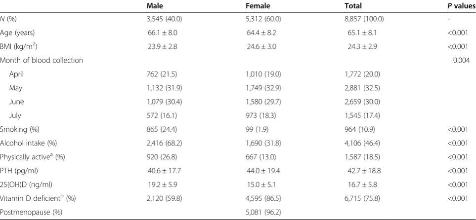
With this approach, the PTH concentration reached a theoretical plateau at 38.2 pg/ml and the corresponding 25(OH)D concentration was 21.1 ng/ml in men (Figure 1)
Table 1 Baseline characteristics of subjects
Male Female Total Pvalues
N(%) 3,545 (40.0) 5,312 (60.0) 8,857 (100.0)
-Age (years) 66.1 ± 8.0 64.4 ± 8.2 65.1 ± 8.1 <0.001
BMI (kg/m2) 23.9 ± 2.8 24.6 ± 3.0 24.3 ± 2.9 <0.001
Month of blood collection 0.004
April 762 (21.5) 1,010 (19.0) 1,772 (20.0)
May 1,132 (31.9) 1,749 (32.9) 2,881 (32.5)
June 1,079 (30.4) 1,580 (29.7) 2,659 (30.0)
July 572 (16.1) 973 (18.3) 1,545 (17.4)
Smoking (%) 865 (24.4) 99 (1.9) 964 (10.9) <0.001
Alcohol intake (%) 2,416 (68.2) 1,690 (31.8) 4,106 (46.4) <0.001
Physically activea(%) 920 (26.8) 667 (13.0) 1,587 (18.5) <0.001
PTH (pg/ml) 40.6 ± 17.7 44.0 ± 19.4 42.7 ± 18.8 <0.001
25(OH)D (ng/ml) 19.2 ± 5.9 15.0 ± 5.1 16.7 ± 5.8 <0.001
Vitamin D deficientb(%) 2,120 (59.8) 4,595 (86.5) 6,715 (75.8) <0.001
Postmenopause (%) 5,081 (96.2)
All values are given asN(%) or mean ± standard deviation. BMI, body mass index; 25(OH)D, 25-hydroxyvitamin D; PTH, parathyroid hormone.a
Subjects who performed 30 min or more of moderate activity at least 5 days a week or 20 min of vigorous physical activity at least 3 days a week were regarded as doing physical activity;
b
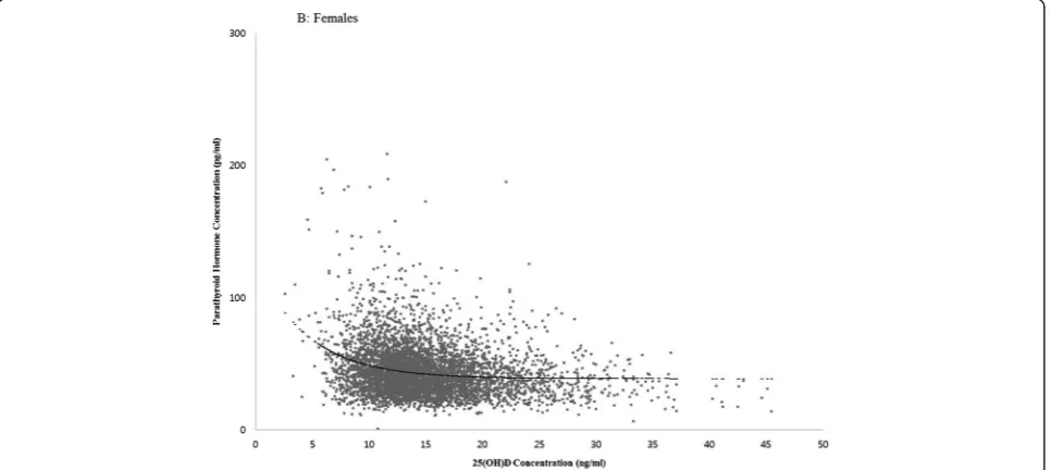
and the PTH concentration at 42.9 pg/ml and the 25(OH) D concentration at 13.8 ng/ml in women (Figure 2).
The optimal 25(OH)D concentration, the threshold value for 25(OH)D at which PTH plateaus, has been sug-gested based on the inverse relation between 25(OH)D and PTH [16]. Because PTH is negatively associated with greater bone loss, maintaining a sufficient concentration of 25(OH)D is believed to have a protective effect on bone health [16]. However, there is a lack of consensus as to what constitutes an optimal 25(OH)D concentration. Some researchers reported that an optimal threshold value was not found [17], while others reported a wide range of
estimates: 8 to 44 ng/ml [7-12], with most clustered at 30 to 44 ng/ml [9-12]. In the present study, PTH reached a plateau at a 25(OH)D concentration of 30 to 40 ng/ml in males, but did not reach a plateau in females. It is possible that ethnic differences may have influenced the relation-ship between 25(OH)D concentrations and PTH. In the NHANES study [18], the optimal concentration was 20 ng/ml in Black Americans but was not found in White or Mexican Americans.
Our data demonstrate the optimal concentration was different according to gender. 25(OH)D is well known to be affected by gender differences. Men tend to spend a
Figure 125(OH)D concentrationversusparathyroid hormone concentration (males).
greater amount of time outdoors than women [19], and the difference in sun exposure may play a role in gender-specific 25(OH)D concentrations. 25(OH)D-binding pro-tein (DBP) may also contribute to gender differences in 25 (OH)D status [20], as DBP levels are significantly higher in women than in men and are positively correlated with overall 25(OH)D concentrations [20].
The ongoing difficulties and controversies associated with the relationship between PTH and 25(OH)D imply that this approach is not the best one to identify vitamin D sufficiency in populations. New approaches to this problem, potentially beyond the hypovitaminosis D, sec-ondary hyperparathyroidism pathway, should be pursued [16].
The main strengths of this study lie in its population-based design and use of a relatively large sample size, which minimized selection bias and provided sufficient statistical power. However, a number of limitations should also be considered. First, the study used a cross-sectional design. Second, it had a comparatively limited ability to explain seasonal changes in 25(OH)D, partly due to a lack of information on sun exposure during the four seasons because the samples were collected April to June during 2007 to 2010. Finally, we performed only a single measurement of the serum 25(OH)D concentra-tions; therefore, the data reflect only a single point in time rather than long-term exposure.
Conclusion
We estimated the optimal 25(OH)D concentration in the Korean general population aged 50 years or older. The optimal 25(OH)D concentration is 21.1 ng/ml at 38.2 pg/ml PTH concentration in men and 13.8 ng/ml 25(OH)D concentration at 42.9 pg/ml PTH concentra-tion in women.
Abbreviations
25(OH)D:25-hydroxyvitamin D; BMI: body mass index; eGFR: estimated glomerular filtration rate; LOWESS: locally weighted estimated scatterplot smoothing; PTH: parathyroid hormone.
Competing interests
The authors declare that they have no competing interests.
Authors’contributions
SWC and MHS contributed to the study design, data acquisition, statistical analysis, interpretation of the results, and manuscript preparation. YHL, HSN, SKJ, KSP, and SYR were involved with the data acquisition and statistical analysis. JSC and JAR conceived and designed the study. SSK participated in the design of the study and carried out the interpretation of data. HRS had an important role in the research and data acquisition. All authors contributed to the preparation and are responsible for the final editing and approval of the manuscript.
Acknowledgment
This study was supported by a research fund from Chosun University, 2014.
Author details
1
Department of Preventive Medicine, Chosun University Medical School, 309, Pilmun-daero, Dong-gu, Gwangju 501-759, Republic of Korea.2Department
of Preventive Medicine, Chonnam National University Medical School, 160 Baekseo-ro, Dong-gu, Gwangju 501-746, Republic of Korea.3Jeonnam Regional Cancer Center, Chonnam National University Hwasun Hospital, 322 Seoyang-ro, Hwasun, Jeollanamdo 519-809, Republic of Korea.4Department of Preventive Medicine & Institute of Wonkwang Medical Science, Wonkwang University School of Medicine, 344-2 Shinyong-dong, Iksan, Jeollabukdo 570-711, Republic of Korea.5Department of Preventive Medicine, Chungnam National University Medical School, Munhwa 1(il)-dong,, Jung-gu, Daejeon 301-747, Republic of Korea.6Department of Neurology & Research Institute of Clinical Medicine, Biomedical Institute of Chonbuk National University Hospital, Chonbuk National University, San 2-20, Geumam-dong, Deokjin-gu, Jeonju, Jeollabukdo 561-180, Republic of Korea.7Department of Preventive Medicine, Seonam University College of Medicine, 439, Chunhyang-ro, Namwon, Jeollabukdo 590-711, Republic of Korea. 8Department of Laboratory Medicine, Chonnam National University Hwasun Hospital, 322 Seoyang-ro, Hwasun, Jeollanamdo 519-809, Republic of Korea.
Received: 18 August 2014 Accepted: 13 February 2015
References
1. Lips P. Vitamin D, deficiency and secondary hyperparathyroidism in the elderly: consequences for bone loss and fractures and therapeutic implications. Endocr Rev. 2001;22:477–501.
2. Holick MF. McCollum Award Lecture, 1994: vitamin D - new horizons for the 21st century. Am J Clin Nutr. 1994;60:619–30.
3. Holick MF. Evolution, biologic functions, and recommended dietary allowances for vitamin D. In: Holick MF, editor. Vitamin D: physiology, molecular biology and clinical applications. Totowa, New Jersey: Humana Press; 1999. p. 1–16.
4. Holick MF. Resurrection of vitamin D deficiency and rickets. J Clin Invest. 2006;116:2062–72.
5. Holick MF. Vitamin D, and bone health. J Nutr. 1996;126:S1159–64. 6. Tomaschitz A, Ritz E, Pieske B, Fahrleitner-Pammer A, Kienreich K, Horina JH,
et al. Aldosterone and parathyroid hormone: a precarious couple for cardiovascular disease. Cardiovasc Res. 2012;94:10–9.
7. Lips P, Wiersinga A, van Ginkel FC, Jongen MJ, Netelenbos JC, Hackeng WH, et al. The effect of vitamin D supplementation on vitamin D status and parathyroid function in elderly subjects. J Clin Endocrinol Metab. 1988;67:644–50.
8. Malabanan A, Veronikis IE, Holick MF. Redefining vitamin D insufficiency. Lancet. 1998;351:805–6.
9. Chapuy MC, Preziosi P, Maamer M, Arnaud S, Galan P, Hercberg S, et al. Prevalence of vitamin D insufficiency in an adult normal population. Osteoporos Int. 1997;7:439–43.
10. Peacock M. Effects of calcium and vitamin D insufficiency on the skeleton. Osteoporos Int. 1998;8:S45–51.
11. Dawson-Hughes B, Harris SS, Dallal GE. Plasma calcidiol, season, and serum parathyroid hormone concentrations in healthy elderly men and women. Am J Clin Nutr. 1997;65:67–71.
12. Krall EA, Sahyoun N, Tannenbaum S, Dallal GE, Dawson-Hughes B. Effect of vitamin D intake on seasonal variations in parathyroid hormone secretion in postmenopausal women. N Engl J Med. 1989;321:1777–83.
13. Kim G, Oh KW, Jang EH, Kim MK, Lim DJ, Kwon HS, et al. Relationship between vitamin D, parathyroid hormone, and bone mineral density in elderly Koreans. J Korean Med Sci. 2012;27:636–43.
14. Kweon SS, Shin MH, Jeong SK, Nam HS, Lee YH, Park KS, et al. Cohort profile: the Namwon study and the Dong-gu study. Int J Epidemiol. 2013;43:558–67.
15. Guillemant J, Taupin P, Le HT, Taright N, Allemandou A, Pérès G, et al. Vitamin D status during puberty in French healthy male adolescents. Osteoporos Int. 1999;10:222–5.
16. Willett AM. Vitamin D, status and its relationship with parathyroid hormone and bone mineral status in older adolescents. Proc Nutr Soc.
2005;64:193–203.
17. Vieth R, Ladak Y, Walfish PG. Age-related changes in the 25-hydroxyvitamin D versus parathyroid hormone relationship suggest a different reason why older adults require more vitamin D. J Clin Endocrinol Metab.
2003;88:185–91.
hormone in the National Health and Nutrition Examination Survey. Osteoporos Int. 2011;22:1745–53.
19. Carnevale V, Modoni S, Pileri M, Di Giorgio A, Chiodini I, Minisola S, et al. Longitudinal evaluation of vitamin D status in healthy subjects from southern Italy: seasonal and gender differences. Osteoporos Int. 2001;12:1026–30.
20. Bolland MJ, Grey AB, Ames RW, Horne AM, Mason BH, Wattie DJ, et al. Age-, gender-, and weight-related effects on levels of 25-hydroxyvitamin D are not mediated by vitamin D binding protein. Clin Endocrinol. 2007;67:259–64.
Submit your next manuscript to BioMed Central and take full advantage of:
• Convenient online submission
• Thorough peer review
• No space constraints or color figure charges
• Immediate publication on acceptance
• Inclusion in PubMed, CAS, Scopus and Google Scholar
• Research which is freely available for redistribution