R E V I E W
Open Access
Immunological challenges associated with
artificial skin grafts: available solutions and
stem cells in future design of synthetic skin
Saurabh Dixit
1,2†, Dieudonné R. Baganizi
1†, Rajnish Sahu
1†, Ejowke Dosunmu
1, Atul Chaudhari
1, Komal Vig
1,
Shreekumar R. Pillai
1, Shree R. Singh
1and Vida A. Dennis
1*Abstract
The repair or replacement of damaged skins is still an important, challenging public health problem. Immune acceptance and long-term survival of skin grafts represent the major problem to overcome in grafting given that in most situations autografts cannot be used. The emergence of artificial skin substitutes provides alternative treatment with the capacity to reduce the dependency on the increasing demand of cadaver skin grafts. Over the years, considerable research efforts have focused on strategies for skin repair or permanent skin graft transplantations. Available skin substitutes include pre- or post-transplantation treatments of donor cells, stem cell-based therapies, and skin equivalents composed of bio-engineered acellular or cellular skin substitutes. However, skin substitutes are still prone to immunological rejection, and as such, there is currently no skin substitute available to overcome this phenomenon. This review focuses on the mechanisms of skin rejection and tolerance induction and outlines in detail current available strategies and alternatives that may allow achieving full-thickness skin replacement and repair.
Keywords:Skin allograft, Immune rejection, Biomaterial, Scaffolds, Stem cells, Skin tissue engineering, Skin substitutes
Background
The skin, a subcomponent of the integumentary system, is a substantial fast-growing organ comprised of the epi-dermis, dermis and hypodermis layers, which in adults weighs about 7–8 pounds, covering 21–22 square feet of surface area (2-m square). The skin is a protective bar-rier for toxins, micro-organisms, radiation, and mechan-ical impacts along with regulating several physiologmechan-ical functions including temperature control, preventing de-hydration and providing sensory detection and immune surveillance [1, 2]. Human skin frequently is damaged/ injured resulting in the loss of its integrity and physio-logical balance, which may result in significant disability and infections. The skin’s natural restorative capacity usually is sufficient to repair and heal itself when dam-aged/injured [3]. However, skin grafts are required for
severe skin injuries to protect the exposed layers of skin and allow the damaged portion to reform. Transplanting autologous skin grafts [4, 5] is the therapeutic approach of choice that successively reform the skin, but extensive injuries and chronic skin wounds could result in an in-sufficient number of autografts, especially in severe burn cases [6] and skin morbidities [7, 8]. When required, in such cases, either allogeneic or xenogeneic, skin grafts are used for transplantation. Despite allogeneic trans-plantations becoming more tolerant with immunosup-pressive treatment, there are still some issues with early rejection. Skin allograft rejection is the recipient’s im-mune response following the recognition of alloantigens leading to the cellular destruction. Allogeneic or xeno-geneic skin grafts may be employed but their short-term graft survival time limits their clinical use [9]. Skin allo-grafts transplantation is employed for severe clinical cases to protect the damaged skin areas, but considering the conundrum of the rejection mechanism, the recipi-ent may require additional transplantation from a differ-ent donor [10]. Alternative strategies are now being developed to overcome skin allograft rejections and * Correspondence:[email protected]
†Equal contributors
1Center for Nanobiotechnology Research and Department of Biological
Sciences, Alabama State University, 1627 Harris Way, Montgomery, AL 36104, USA
Full list of author information is available at the end of the article
allowing adequate skin repair [11, 12]. Novel treatment approaches include the use of stem cell-based therapies, specific immunosuppressive therapies targeting T cells or donor immune cells and skin tissue engineering. Sev-eral tissue-engineered skin substitutes are commercially available and used in clinical settings with negligible risk of immunogenic responses such as the Integra dermal regeneration template [13]. Available engineered skin substitutes are composed of either a cellular or acellular component and a biological (autologous, allogeneic, and xenogeneic) or synthetic (polymer) scaffold [14]. However, available skin alternatives engineered to mimic natural skin still do not provide a permanent so-lution [5, 14, 15]. This review gives an insight into different approaches and innovative advances to allow overcoming skin allografts rejection.
Immunological rejection
Mechanisms of skin graft rejection
Allografts have been used for many years in transplant-ation; however, donor tissue availability remains a critical issue. Cadaver tissues, especially organs, are in high de-mand and harvesting of skin has to be completed rapidly [16] post-death and preserved [17]. Critical issues associ-ated with allografts are availability and rejection. Laboratory-grown artificial tissues are now in develop-ment to help overcome the immunological rejection is-sues [18, 19]. Over the years, synthetic skins comprised mostly of human cell lines with biodegradable materials have been used for transplantation onto burned and wounded skin patients [20, 21]. Even though the artifi-cial skin products are in development and available com-mercially, they are still prone to rejections [7].
Skin autografts transplantation is a well-known med-ical procedure. Grafting between genetmed-ically identmed-ical in-dividuals (syngeneic graft) can be successful without a prolonged immunosuppressive treatment. Even though immunosuppressive treatments for organ transplantation are effective in preventing early rejection, skin tissues whether from both donor or engineered are continu-ously failing [22]. Skin graft successfully placed at the donor site but rejected within 1–2 weeks is consistent and is termed first set rejection. The second set of rejec-tion is even faster if grafted from the same donor. Graft rejection is an intricate mechanism, which involves an array of processes and ultimately potent inflammatory responses initiated by innate immune responses and de-struction of the donor tissue [23]. The rate of rejection of donor tissue at the recipient’s graft site is dependent on the graft volume and antigens incompatibilities be-tween both. The role of T lymphocytes in graft rejection is vital as evidenced from studies in nude mice, which do not reject allogeneic skin grafts because they lack CD4+ and CD8+ functionality [24]; however restoring
this functionality with adoptive transfer of normal T cells initiates rejection of the skin graft [25, 26]. Therefore, a hurdle in allogeneic skin grafting is the triggering of CD4+ and CD8+ T cells immune responses, sometimes involving both for first set rejection, although second set rejection could be facilitated by antibodies [26].
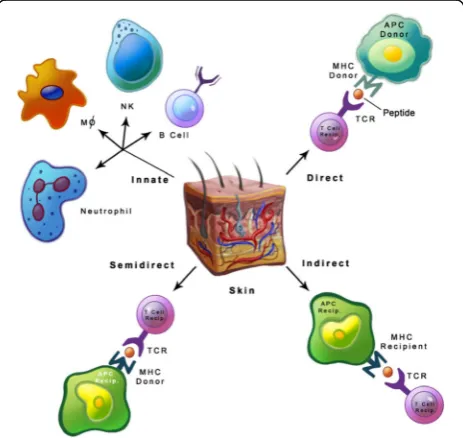
The mechanism of skin graft rejection (Fig. 1) starts with responses from dendritic cells (DCs), macrophages, polymorphonuclear cells, angiogenic mediators, and cy-tokines to promote rejection [22, 23], followed subse-quently by the activation of T cells (CD4+ and CD8+). Further, accumulation of inflammatory cytokines and ef-fector T cells permeate the skin graft to commence re-jection [22, 26, 27]. The event/stimulus that triggers skin graft rejection arises from a mismatch in donor MHC and recipient T cells receptors (TCRs) [28]. Even though matching the MHC type is critical in avoiding skin grafts rejection, a single genetic difference at the loci of MHC molecules can still commence the rejection process by stimulating alloreactive T cells [10]. Additionally, even if the rejection rate is not very high in genetically related donor and recipient [29], it can be controlled by im-munosuppressive drugs. The only scenario where allo-graft transplantation without immunosuppressive drugs is successful when the donor and recipient are identical twins, with true human leukocyte antigen (HLA) match [30], which shows the immunological importance of MHC molecules in rejection of transplants. Thus match-ing the HLA types [31] between non-identical twins
improves the rate of graft transplantation, but HLA typ-ing methods are not precise due to complexity and poly-morphism of MHC molecules [32]. Another reason is differences in minor histocompatibility antigens (MHA) that also vary in individuals for HLA type matching, which is a consideration in assessing graft rejection [33].
Most allografts require MHC class matching for allo-geneic transplantation, and the primary reason for a re-jection response against foreign MHC molecules is TCRs specificity. Foreign MHC antigen recognition be-tween both the recipient and donor tissues initiates the rejection process at the graft site. The repercussion of allorejection is the initiation of adaptive immune re-sponses especially with alloreactive T cells [22]. The allorecognition mechanism for skin grafts is distinguish-able from other tissue transplantations. Apparently, skin graft rejection is potentially a much broader response generated to destroy the donor skin graft. The demon-stration on corneal transplant rejection in mice indicates that only the CD4+ indirect pathway involving minor antigens leads to rejection. The CD8+ T cells pathway has limited functionality but no cytotoxicity [34].
Dendritic cells from donor grafts also have a crucial function in initiating the rejection process by their mi-gration [35] into the donor’s lymphoid organs for antigen presentation to initiate the adaptive immune response [36]. The adaptive alloimmune response from DCs is attributed to Langerhans cells (LCs) from the epidermis and dermis. Langerhans cells are distributed in the epidermis and contribute up to 3% in epidermal cells [22]; they express CD1a in humans and have the capability of microbial lipid antigen presentation to T cells [37].
Adaptive immunity in allorejection
Direct allorecognition
Direct allorecognition pathway (Fig. 1) is the outcome of the interaction between the recipient’s T cells in the lymphatic system with foreign MHC molecule from mi-grating leukocytes of the donor. Direct allorecognition exploits CD4+ and CD8+ subsets of T cells for identifi-cation of MHC class I and II molecules from a donor, but antigen processing by the recipient’s antigen present-ing cells (APCs) is not mandatory. This rapid donor-recipient recognition generates robust T cells responses [22] with alloreactive activated T cells from the recipient migrating to the donor tissue for the direct attack [10]. The donor MHC class II and class I molecules recogni-tion by the recipient’s CD4+ and CD8+ T cells respect-ively generate a robust response. Although recognizing foreign MHC molecules is not precisely the nature of T cells, the observed cross-reactivity between self and for-eign MHC molecule suggests that some non-specific T cells might be involved in this process [22, 28]. This
phenomenon of foreign MHC molecules recognition was proposed with TCRs similarities on donor’s and re-cipient’s T cells to identify the nominal and allogeneic MHC proteins [38] and cross-reactivity [26, 30]. The lack of specificity in the direct pathway proposes the plausible cause for transplant rejection even in highly matched MHC molecules between donor and recipient [30] and corroborate that even a single variation in MHC molecule can promote graft rejection [10, 22]. Leukocytes depletion in donor tissue prolonged allograft survival but remained ineffective in averting the rejec-tion process [10]. In a study using mixed lymphocytes in vitro, where allorecognition points towards the non-specificity of antigen recognition, and in vivo with mice lacking CD4+ T cells, where the indirect pathway trig-gering via antigen presentation is not possible, CD8+ T cells alone were observed to be functional in graft rejection [39–41].
Dendritic cells of donor grafts are the key initiators for direct pathway activation in the graft recipient’s immune system. This premise was proven by depletion of the do-nor’s DCs which halted the immunogenicity, as the later addition of the donor’s DCs reverted the whole process [42]. Diminishing donor APCs via antibody treatment [10] may propose an alternative mechanism in avoiding the direct allorecognition process to prevent rejection. Similarly, alloreactive T cells activation via endothelial cells may pose an alternate process in the allograft rec-ognition [26]. The robust T cell response generated in the direct pathway by itself can cause complete rejection, but alloantibodies production is not the outcome of dir-ect allorecognition pathway [43].
Indirect allorecognition
Allorecognition via the indirect pathway (Fig. 1) is pri-marily contributed by the recipient’s APCs presenting foreign proteins to T cells from grafts and activating CD4+ T cells. The indirect pathway is based solely on CD4+ T cells. The robustness of indirect allorecognition is significantly less than direct allorecognition where CD4+ and CD8+ T-cell phenotypes partake without antigen processing but are adequate for rejection of grafts. The indirect pathway is evidence of the conven-tional antigen processing and presentation by APCs. Even though the indirect pathway represents specificity, the rejection is also very often due to minor H antigen loci differences [10, 33].
However, the overall contributory role of indirect allore-cognition in the immunological graft rejection could not be validated when tested alone [45]. Although the indir-ect pathway is the basis for long-term rejindir-ection, it can also activate macrophages, thus resulting in tissue injury and fibrosis, furthermore developing alloantibodies against allografts [46].
The indirect pathway selectively depends on CD4+ T cells, but the support of CD8+ T cells is also evident; a process termed cross-dressing [22]. CD8+ T cells partici-pation in graft rejection was underscored by in vivo experiments with single MHC class I peptide presenta-tion followed by confirmapresenta-tion of skin allografts rejecpresenta-tion [47, 48]. Allorecognition by CD8+ T cells also initiates the acute allograft rejection, but activation of the CD8+ T cells dependent indirect pathway may require support from CD4+ T cells [41, 49]. Evidently, the indirect path-way remains the assertive mechanism in long-term allor-ecognition as long as the allograft remains on the recipient’s graft site and role of memory T cells remains affirmative in rejection and tolerance [26, 48].
Semi-direct allorecognition
Direct allorecognition is the most dependable pathway in rejection followed by the indirect pathway, but the al-ternate pathway known as cross-dressing has been re-ported [22, 50]. In this semi-direct allorecognition pathway (Fig. 1), the recipient APCs acquires intact MHC molecule from the donor for antigen presentation to T cells; this contrasts to the indirect pathway where the processed peptides of allogeneic MHC molecules on the recipient’s MHC molecules are displayed by the re-cipient’s APCs [22]. This process indicates that the same APCs present MHC class II and I molecules for CD4+ and CD8+ T cells activation [23]. This controversial rep-resentation of both MHC molecules, which are pre-sented by the same DCs and referred to as “three-cell interaction” [51] was supported by staining the MHC molecules and inducing T-cell specific proliferative re-sponses [52]. This sharing of MHC molecules has subse-quently been explored using different DCs subsets and was observed to be a natural phenomenon of DCs for transfer efficiency [53]. Such recognized DCs presenting MHC class I and II molecules was further documented in mice [52]; however, the semi-direct pathway involve-ment in graft allorecognition was not evident in in vivo studies [50]. The mechanism of the MHC transfer ap-parently is via exosomes [54, 55] that are released by multiple cell types and hypothesized to be representing the MHC molecules onto their membranes. The inter-action of MHC class I molecule-deficient DCs, and CD8 + T cells specific to an antigen with exosomes accom-modating MHC molecule revealed the transfer of mole-cules from exosomes to DCs [54]. Substantiation of
allogeneic MHC molecule transfer undoubtedly indicates the semi-direct pathway involvement, but this pathway’s precise role in allograft rejection still warrants further investigations.
B cells
Allograft recognition is dominated mainly by T cells al-though B cells are al-thought to be involved. Pre-existing alloantibodies against blood groups and polymorphic MHC antigens are reasons for antibody-mediated allo-graft rejection, and B cells also have the capability of antigen presentation [23]. Both antibody production and the antigen presentation capability of B cells potentially participate in allograft rejection [56]. Others have pro-posed that B cells can promote T cells activation via co-stimulatory pathways and cytokines release [26, 57]. Alloreactive memory T cells also represent the diverse functionality of T cells in allograft rejection [58]. More-over, recent investigation for comprehending the diverse functionality of B cells in allograft rejection revealed that proliferation, differentiation, and memory T cells func-tionality were increased, which may be due to B cells ability to function as APCs [58]. B-cell deficiency in mice further suggested that the extended graft survival rate resulted from the absence of antigen presentation functionality [59]. Depleting mature B cells with anti-CD20/anti-CD19 monoclonal antibodies in mice acceler-ated the rejection of skin allografts [60, 61] by enhancing the allospecific memory T cell immunity, which may ex-plain B cell participation in allorecognition and survival [56–61].
Innate immunity in allorejection
T cells proliferation [71]. Macrophages do not play a dir-ect role in allorejdir-ection since they are not efficient in priming naïve T cells [72, 73]. Macrophages are hetero-geneous depending on their functions and are in large numbers in human allotransplants. In general, macro-phage activation follows the classically activated M1 and alternatively activated M2 phenotypes. M1 macrophages are activated by Th1 secreted cytokines, i.e., IFN-γ and TNF-α [22, 73, 74]. M2 macrophages are induced by IL-4 produced by Th2-activated T cells as well as baso-phils and mast cells in response to injuries [75]. Macro-phages are frequently observed in acute rejections, which may indicate their defensive functions in necrosis and pro-inflammatory cytokines secretion.
Neutrophils participate in allograft rejection by secret-ing chemokines thus leadsecret-ing to T cells activation and proliferative responses [71]. The role of innate immunity elements in immunological rejection of allografts is not highly evident. However, support of adaptive immune re-sponses via innate immunity cells may be a more logical explanation since they are rapid responders against for-eign molecules. Overall, the process of allograft rejection is by the direct, indirect and semi-direct pathways, but innate immunity components may participate along with adaptive immune responses to boost the allograft rejec-tion process.
Approaches to avoid skin immune rejection
Therapies to escape skin rejection
The use of skin autographs is the most efficient method and the treatment of choice to avoid immunogenicity in reconstructive skin transplantation. However, there are limitations in using skin autografts for patients with deep and/or large wounds or with extensive burns [76, 77]. In such circumstances, transplantation of split-thickness skin allotransplants or full-thickness skin from live donors or in the form of cadaveric skin provides a replacement that reforms the functional skin [22, 76]. Nonetheless, there are limitations to the use of allogeneic skin grafts since in-variably they are rejected due to the triggering of the host immune response that subsequently leads to their short-life span [22, 76, 78]. Furthermore, efficacious immuno-suppressive treatments usually used in organ transplants to prevent early rejection are either less or/not effective in skin transplantation [77]. Treatment of skin allografts be-fore operation allows decreasing the immunogenicity, but it is insufficient over the long-term [78]. Newer treatment procedures have thus been developed to overcome skin grafts rejection to prolong skin graft survival.
Therapies addressing donor-derived DCs
A large number of researchers have reported on effective therapies addressing donor-derived DCs to induce skin graft tolerance. Indeed, as donor-derived DCs are critical
in acute immune responses in skin transplants under-lying allograft rejection, there is every likelihood that their depletion or inhibition results in prolonged survival of skin grafts [78, 79]. Most DC-based strategies aimed at inhibiting the antigen presentation process predomin-antly by targeting donor-derived LCs and the DC-subpo-pulations expressing MHC class II glycoproteins are important in the initiation of allograft rejection [22, 78]. Strategies using chemical agents to modulate the activity of DCs have shown a high improvement of skin grafts survival. For example, treatment of skin grafts with Glio-toxin, an epipolythio dioxopiperazine (ETP) immuno-suppressive mycotoxin, significantly reduced the epidermal density of LCs and altered their function, resulting in the enhancement of skin graft survival and induction of donor-specific tolerance onto MHC-mismatched recipient mice [80]. Gliotoxin apparently immunomodulates the functions of immunocompetent cells and reduces contact hypersensitivity responses through the induction of suppressor cells thus leading to inhibition of graft rejection for prolonged survival with-out altering the skin functions [80]. Treatment of skin grafts with 10-dimethyl-1,2-benzanthracene (DMBA) depleted LCs and therefore the class II MHC antigens from the graft, which enhanced the survival of C57BL/6 skin grafted onto BALB/c recipients [81]. Norcantharidin (NCTD, C8H8O4), a cantharidin that promotes
hematopoiesis extended the survival time of allogeneic skin grafts in mice by modulating the activity of DCs func-tion towards tolerance and inducing immune silencing via inhibiting the activity of calcineurin phosphatase [82].
survival effect of this treatment resulted from β2mAb, which impaired the functions of HLA-class I antigen, and UVC-treatment which reduced the number of skin APCs for an efficiently localized immunosuppression [85]. UVC-treatment inhibits the induction of contact hypersensitivity responses by depleting LCs and limiting their migratory capacity [86–88]. Furthermore, it in-duces the release of epidermal growth factors, promotes proliferation of endothelial cells, restores melanin pro-duction which accelerates wound healing and restoration of skin homeostasis [89, 90]. Also, UVC-treatment of dermal fibroblasts increases the release of fibronectin in the cellular microenvironment for the contraction of fibroblast-populated collagen lattices, thereby resulting in increased healing through wound contraction [90, 91].The loading of donor’s antigens with donor-derived immature DCs and third-party DCs was also reported to partly induce skin transplantation tolerance against rejection in mice [92].
Inactivation and deletion of alloresponsive T cells
Approaches exploiting the inactivation of functional T cells subpopulations have been revealed to induce dur-able tolerance and allograft survival. Activation of T cells by recognition of allogeneic skin grafts is sufficient to initiate acute rejection. Supposedly, this is accomplished by inducing CD4+ and CD8+ phenotypic activation with subsequent production of the Th1 pro-inflammatory cy-tokines, IL-2 and IFN-γ [22]. In mice, the endogenous ligand for FMS-like tyrosine kinase 3 (Flt3 ligand, FL) stimulated robust tolerance of skin grafts in recipients of FL-mobilized donor cells [79]. FL-induced skin graft tol-erance was inherent to the durable macro-chimerism of persistent blood and selective suppression of donor-reactive T cells [79]. Furthermore, treatments based on antibodies with immunosuppressive effects induced tol-erance ultimately leading to the survival of skin grafts. Use of the FN18 antibody specific for the CD3 antigen of rhesus monkey T cells significantly extended the sur-vival of skin grafts in rhesus by modulating or depleting T cell subsets [93]. Anti-Ly49A mAb YE1/48 reactive against the Ly49 receptors expressed on T cells, NK, and NKT, regulate immune responses through inhibition/ac-tivation of MHC class I molecules were shown to delay MHC molecules-mismatched allogeneic skin graft rejec-tion in mice [94]. YE1/48 mAb prolonged the survival of skin grafts by inhibiting only the primary immune re-sponses to allografts [94].
Treatment with an anti-CD80 mAb combined with cyclosporin A, an immunosuppressive drug, suppressed the activation of T cells and triggered alloantigen-specific non-responsiveness resulting in significant in-crease of skin grafts survival in a preclinical rhesus mon-key model [95]. Transfusion of C57BL/6 mice recipients
with donor BALB/c spleen cells and CD154 anti-body also permitted skin grafts acceptance and survival [96, 97]. Prolonged rat skin xenografts survival occurred following transfusion of mice with donor-specific cells and monoclonal an anti-CD154 mAb [97]. The longevity of graft survival provided by this treatment entailed the continuous activation of CD4+ and alloresponsive T cells without IFN-γin the graft [96]. Furthermore, prolonged allografts survival times were observed in recipients treated with donor-specific transfusion and an anti-CD154 mAb essentially by deletion of alloantigen-specific CD8+ T cells, which led to an allotolerant state [98–100]. In addition to CD8+ T cell deletion, the initi-ation of skin allograft survival required CD4+ T cells, but other mechanisms along with different CD4+ T cells subsets can induce skin transplantation tolerance [100].
The success of T cells depletion approaches relies on the generation of stably mixed chimaerism in which host T cells are ablated to achieve tolerance of donor’s MHC mismatched grafts [101]. The clinical application of this approach, however, has been limited by the need for pre-transplant treatment with myeloablative agents, their potential toxicities and split tolerance due to unmatched minor antigens [101–103]. Also, post-depletion of humoral responses and the repopulation of memory T cells without xenogeneic antibody production and/or over-immunosuppression represent a considerable chal-lenge [103]. Even though T cells depletion approaches proved highly effective in animal models, the matching of minor antigens to prevent effector T cells from reject-ing donor skin grafts might not be possible in clinical practices due to the lack of effective in vivo T cell depleting agents [101–103].
Bioengineering
interface of the acellular matrix membrane provides a physical barrier between the recipient and donor tissues for interrupting the effector pathway to protect from the allorecognition pathway underlying the humoral rejec-tion [76]. The network of adhesive molecules of bioengi-neered membranes acts as a barrier to cellular migration while, at the same time, it grants free diffusion of nutri-ents and oxygen [76]. It is still not well understood how bioengineered membranes provide protection that re-sults in prolongation of allograft survival especially when mature complex capillaries, arterioles, and venules have developed [76].
Gene therapy
Gene therapy is also a promising approach to induce tol-erance and effectively extending the survival time of skin allografts. Transduction of hematopoietic fetal liver cells with human IL-10 (hIL-10) gene before transplantation delayed rejection and lengthened the survival time of mouse skin allografts [107, 108]. Overexpression of IL-10 was achieved by inserting the IL-IL-10 plasmid into GPE86 fibroblastic cell line to produce retroviral vectors carrying the hlL-I0 gene [107].This provision by IL-10 is because it is an immune-regulatory cytokine that exerts its immunosuppressive activities by inhibiting the syn-thesis of Th1 cytokines [107, 108]. Therefore, regulating the effects of T cell responses through overexpression of IL-10 in the donor can induce long-term tolerance and improve graft survival. Donor hematopoietic stem cells transduced with hIL-10 extended the survival of donor skin allografts through the continued production of IL-10 and induction of donor cell chimerism and engraft-ment which protected allogeneic grafts from rejection [107, 108]. However, a full tolerance was not attainable with this treatment strategy. Transgenic expression of human CTLA4Ig (cytotoxic T-lymphocyte-associated antigen 4-immunoglobulin) reportedly also lengthen the survival of xenogeneic skin grafts on burn wounds in rats and mice [109, 110]. Cytotoxic
T-lymphocyte-associated antigen 4-immunoglobulin
modulates T cells functions by competitively inhibit-ing the CD28 and B7 co-stimulatory pathways [109, 111]. Hence, CTLA4Ig by down-regulating activated T cells could induce transplantation tolerance and re-duce immune rejections. Transgenic CTLA4Ig locally inhibited human lymphocyte activation and prolifera-tion without significantly affecting the systemic im-mune function which led to prolonging grafts survival of the transgenic skin [109, 110]. Furthermore, trans-fection of allogeneic skin flaps with CTLA4Ig and OX40Ig gene mediated by lentivirus vectors signifi-cantly increased the survival time of tissue allografts in rats [112]. OX40 is a subcomponent of the TNF superfamily of receptors involved in T cell
co-stimulation [78, 112]. Local transfer of OX40Ig and CTLA4Ig genes inhibited the rejection of allografts and expanded survival time by decreasing the quan-tity of CD4+ T cells, increasing the clonal expansion of T helper subset 2 (Th2) subpopulations and down-regulating IL-2 and IFN-γ expressions [112]. Skin gene therapy holds great promise in allotransplanta-tion tolerance and improvement of long-term allo-grafts survival. Gene therapy provides the advantage of a local production of immunosuppressive mole-cules. Cells or organs are treated ex vivo with gene transfer vectors before implantation allowing the pro-duction of immunomodulatory proteins in the donor grafts and resulting in local rather than systemic im-munosuppression [113]. However, some key risks to consider are the fact that the host immune response limits repeated administrations of the vector and safer vectors need to be developed [114, 115]. Moreover, although the gene expression and protein production are transient, the introduced mutagenesis, the im-munogenicity and alloimmune response, and vector stability in the host represent important clinical chal-lenges to avoid endangering patients [115, 116]. Chal-lenges also include the development of more efficient and durable vectors for sustained expression of the desired gene in vivo with minimal toxicity principally in regards to genomic integration and immune re-sponse [113, 115–117]. Noteworthy of mention is the difficulty in achieving a specific and uniform therapeutic transfer to different compartments of the skin that must be addressed [117].
Antioxidant therapy
Antioxidant therapies of donor skins from C57BL/6 mice before transplantation, or BALB/c mice recipient skins with Salen-Manganese (Salen-Mn) complexes were demonstrated to delay allograft rejection [118]. Salen-Mn delayed allograft rejection and increased skin allograft survival by reducing reactive oxygen species (ROS)-mediated graft tissue damage, by reduction of anti-donor cytotoxic responses via the decrease of Th1 alloreactive cells and increase of donor-specific Th2 cells, and by suppression of inflammatory reactions [119]. However, the precise mechanism underlying the promotion of anti-inflammatory T cell responses was not elucidated in these studies.
Skin tissue engineering to overcome rejection
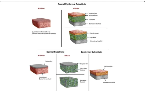
artificial skins incorporating an extracellular matrix of biomaterials and cells (autologous cells, allogeneic cells or xenographic tissues) with minimum risks of rejection (Fig. 2) [12, 120–122]. Bioengineered skin substitutes act first as protective dressings to limit infection and fluid loss and further function as skin equivalents to provide temporary wound covers or permanent skin replace-ments [123, 124]. Bioengineered skins are either acellular or cellular and are composed of epidermal and/or der-mal equivalents enclosed into a matrix scaffold of bio-materials which are further incorporated into the body during the regeneration of new skin (Fig. 2) [121, 125, 126].
Biomaterial components used to make skin substitutes include natural and synthetic materials that provide a matrix scaffold onto which grafted skin cells grow and spread [121, 124]. Some natural materials include fibro-nectin, collagen, chitosan, hyaluronan, and glycosamino-glycans (GAGs) [121]; synthetic materials include synthetic polymers like polyethyleneglycol (PEG), poly lactic-co-glycolic acid (PLGA) and natural polymers like dextran, chitosan, gelatin, fibrin, and hyaluronic acid [127–130]. Biomaterial scaffolds are designed in a solid and porous three-dimensional (3D) form with the aim of performing several functions including promoting
interactions between cells and the biomaterials and the deposition of the extracellular matrix (ECM).). Due to cell size along with cell migration requirements and transport, the optimal scaffold pore size is usually around 100 μm, but pore sizes greater than 300μm are highly recommended to achieve, especially, the forma-tion of capillaries [131]. This porous scaffold permits sufficient transport of nutrients, regulatory and growth factors for proliferation, differentiation, and survival of cells. The scaffold also undergoes controlled biodegrad-ation while supporting tissue regenerbiodegrad-ation with similar physical, mechanical and functional properties; and in-ducing a minimal degree of toxicity, immunogenicity and inflammation [124, 132]. These biomaterials particu-larly enable to overcome limitations of rapid and permanent implementation of the grafted skin while reducing the incidences of infection and rejection [121, 122]. Nevertheless, there are no bioengineered skin sub-stitutes to completely replicate skin or fulfill all its func-tions mentioned above [12, 121].
Immune response to bioengineered skins
The implantation of bioengineered skins elicits a series of host immune reactions, first towards the cellular component and further to the biomaterial component
[133]. However, there is lack of reported studies regard-ing the host immune responses to biologic scaffold ma-terials, and those available studies mostly evaluate the inflammation response. The biomaterial implantation initiates inflammation responses through a series of events, collectively known as foreign body response starting with proteins adsorption from the ECM on the biomaterial surface followed by the attraction, adhesion, and activation of phagocytes such as monocytes and/or macrophages in the implant site [133–135]. Phagocytic cells might also be attracted to the inflammatory site by molecular patterns that mimic pathogen-associated mo-lecular patterns (PAMPs) that may be on the biomate-rial, through innate receptors or the recognition of proteins adsorbed to the biomaterial by APCs [134]. Activated macrophages then secrete a wide range of cytokines (i.e., IL-1, IL-6, IL-10, TGF-β), chemokines (IL-8, MCP-1, and MIP1-α/β), growth factors and ECM enzymes [135, 136]. Depending on the biomaterial and cellular component in the bioengineered skin, these medi-ators could direct the skin tissue repair and inflammatory response to the biomaterial or mediate other variable re-sponses including the migration and proliferation of fibro-blasts and skin tissue regeneration [133, 135, 137].
Moreover, surface contact of complement proteins with biomaterials and the adsorbed protein layer triggers the complement cascade that leads to the activation of inflammatory cells, which subsequently mediate other processes, including maintenance of inflammation, activation, and initiation of tissue repair or promotion of T and B cells development [133, 136]. Hence, selection of biomaterials for bioengineering of artificial skin tis-sues depends, in addition to addressing functional skin requirements, on the potential host responses towards them. However, although biomaterials can cause inflam-mation, they contribute minimally to transplant rejection [133, 135]. Furthermore, the elimination or inactivation of cellular elements that lead to immunogenic responses from the matrix scaffold (i.e., cells and cellular antigens) allows the artificial skin to exhibit minimum early rejec-tion and not to cause a chronic rejecrejec-tion reacrejec-tion after implantation [105, 138].
Skin substitutes with natural biomaterials
Naturally occurring biomaterials capable of reproducing the micro-architecture and physiological functionality of the ECM are more widely used in designing skin substi-tutes, and they include fibronectin, collagen, chitosan, hyaluronan, and glycosaminoglycans (GAGs) [121]. Their main advantages reside in their inherent proper-ties of biological recognition, low antigenicity, bio-degradability, low toxicity and low chronic inflammatory responses [125, 139–141].
Collagen
Collagen-based matrix scaffolds are the most attractive matrix for artificial ECMs. Collagen, an essential struc-tural component of the ECM, comprises more than 70% of the dry weight of the dermis [142, 143]. Over twenty different types of collagens exist of which Type I and III are more abundant in dermal tissues, while Type IV and VII are the major components of the basement mem-brane [121, 124, 142–145]. The advantage of using colla-gen resides in its high biocompatibility, biodegradability, and weak antigenicity while offering support for cell attachment and growth compared with other natural biomaterials [141, 142]. The use of collagen, especially of animal origin is currently widespread for the develop-ment of in vitro three dimensions (3D) full thickness skin equivalent models that exhibit close morphology and metabolic activity of human skin to study skin biol-ogy, wound healing, and skin cancer [146, 147]. The im-provement of culture techniques has led to a successful commercialization of artificial human skins based on collagen as their biomaterial component (Table 1).
Integra® is an artificial skin dermal replacement com-posed of non-living ECM of porous bovine Type I colla-gen and a glycosaminoglycan (chondroitin-6-sulfate) with a disposable silicone epidermal layer [7, 124, 148, 149]. The dermal collagen matrix is incorporated and becomes vascularized while the silicone epidermal mem-brane is temporary and subsequently replaced by a thin layer of autograft [124, 148, 150]. Integra® is used for coverage of burn wounds particularly those requiring partial and full thickness repairs, and also successfully for chronic ulcer treatment of diabetic foot ulcer (DFU) [124, 148]. Integra presents low risks of immunogenic responses and no immunologically significant increase of antibody activity [150].
Alloderm® is an artificial skin of acellular collagen matrix containing an entire basement membrane complex used as a scaffold for dermal tissue remodeling [124, 148]. Alloderm® is composed of human allograft skin tissue processed from fresh cadaver skin to remove the epider-mis cellular material and freeze-dried after processing [124, 148]. It is used for resurfacing of full thickness burn wounds and temporary wound covers [124]. Alloderm® is accepted by the recipient’s immune system since the allo-geneic cells, and antigenic materials are removed render-ing it immunologically inert, and therefore reducrender-ing the antigenic response [123, 124, 148].
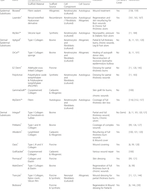
Table 1Some current commercially available bio-engineered skin substitutes
Type Brand Components Indication Permanent
Cover
References
Scaffold Material Scaffold Source
Cell Component
Cell Source
Epidermal Substitutes
Bioseed® Fibrin sealant Allogeneic Keratinocytes (cultured)
Autologous Wound treatment Yes [158,159]
Laserskin® Benzyl-esterified Hyaluronan derivative
Recombinant Keratinocytes / Fibroblasts (cultured)
Autologous/ Allogeneic
Regeneration and skin resurfacing for Burn wounds & Chronic full thickness ulcers
Yes [161,165,167]
MySkin™ Silicone layer Synthetic Keratinocytes (cultured)
Autologous Neuropathic, pressure & Diabetic foot ulcers
Yes [11,184]
Dermal/ Epidermal Substitutes
Apligraf® Type I Collagen Bovine Keratinocytes and fibroblasts (cultured)
Allogeneic Partial & Full thickness burns, chronic wounds, Leg & Foot ulcers
No [6,11,101,120]
OrCel™ Type I Collagen sponge
Bovine Keratinocytes and fibroblasts (cultured)
Allogeneic Healing of autograft donor sites, Reconstruction of recessive dystrophic epidermolysis bullosa
No [6,11,101]
EZ Derm® Aldehyde-cross linked Collagen
Porcine Dressing for partial
thickness burns
No [11,120,145]
PolyActive Polyethylene oxide terephthalate & Polybutylene terephthalate (PEO/PBT)
Synthetic Keratinocytes and fibroblasts (cultured)
Autologous Dressing for partial thickness wounds
No [11,183]
GammaGraft™ Cryopreserved Collagen
Cadaveric & Allogeneic
Skin graft for burns,
chronic wounds
No [100]
MyDerm™ Fibrin Autologous Keratinocytes
and fibroblasts (cultured)
Autologous Coverage of full thickness skin loss
Yes [116] [152,157]
Dermal Substitutes
Integra® Type I Collagen & Chondroitin-6-Sulfate
Bovine Partial and full
thickness wound, burns, Chronic ulcers
No (Semi) [6,11,101,120,121]
PriMatrix® Type I and III Collagen
Bovine Coverage of complex
wounds
Yes [99,126,127]
Alloderm® Lyophilized Collagen
Cadaveric & Allogeneic
Resurfacing of full thickness burn wounds & Wound cover
Yes [100,101,120]
Oasis® Type I, III and V Collagen
Porcine Wound covering Yes [6,99,120]
GraftJacket® Cryopreserved Collagen
Cadaveric & Allogeneic
Various wound repair Yes [100]
Permacol® Collagen and Elastin
Porcine Skin dressing Yes [99,121]
MatriDerm® Type I Collagen & Elastin
Bovine Regeneration of full
thickness burn & chronic wounds
Yes [6,99]
Trancyte® Type I Collagen, Nylon mesh, Silicon film
Porcine & Synthetic
Neonatal fibroblasts (cultured)
Allogeneic Wound dressing for partial thickness burns
Yes [11,121,144]
Biobrane® Porcine
& Synthetic
Regeneration & Wound dressing for Partial &
bovine Type I collagen [148, 151]. Apligraf® is employed as an epidermal substitute in the treatment of partial to full thickness burns, chronic wounds, venous leg ulcers as well as diabetic foot ulcers [7, 124, 148]. It promotes healing by providing in addition to ECM components, essential growth factors, and cytokines including TGFα/ TGFβ, IL-1, IL-6, and IL-8 [151]. Since Apligraf® does not contain any APCs, it does not cause immunological rejection or support any significant humoral or cellular immune responses [151, 152]. Apligraf®is considered im-munologically inert as it does not contain APCs, and thus not cause immunological rejection or support in-duction of significant humoral or cellular immune re-sponses [151, 152]. Studies have shown the absence of humoral or cellular responses to keratinocytes or fibro-blasts of Apligraf®; however, safe and reliable human cell sourcing represents a well-recognized problem [153, 154]. Moreover, in clinical trials performed using Apligraf®, no signs or symptoms of rejection were de-tected in vitro to bovine collagen or alloantigens expressed on keratinocytes or fibroblasts in Apligraf® [155, 156]. However, Apligraf® requires joined grafting with an autologous epithelial supplier because the grafted allogeneic cells are temporary [12, 157].
OrCel™is a bi-layered skin construct where the dermis is composed of cultured neonatal keratinocytes and fibroblasts derived from foreskin tissues that are seeded into a Type I collagen matrix [7, 120]. OrCel™is used for reconstruction in recessive dystrophic epidermolysis bullosa and healing of autografts donor sites [120]. Simi-lar to Apligraf®, OrCel™ promotes healing by mimicking cytokine and growth factor expression in the healing skin (TGF-α, fibroblast growth factor-1, keratinocyte growth factor-1, etc.) [12, 124]. PriMatrix®, Oasis®, and TissueMend™ are other available collagen matrix-based
skin substitutes, which also do not exhibit immuno-logical rejection. PriMatrix® is a fetal bovine dermal substitute comprising extracellular Type I and III colla-gen matrix scaffolds and used for the coverage of com-plex wounds to stimulate vascularization and dermal regeneration [122, 158, 159]. Oasis® is an acellular dermal substitute fabricated from porcine small intestine [7, 122, 148]. It is composed of a matrix of collagen (Types I, III and V) and growth factors (TGF-β and fibroblast growth factor-2) processed to remove cell components [122, 148] and commonly employed for wound covering in lower limb wound treatment. Oasis® is decellularized and therefore does not elicit immuno-logical responses. TissueMend™is an acellular skin sub-stitute with collagen matrix scaffold used for tendon and ligament repair tissue remodeling [160, 161]. Because TissueMend™is depleted of all cellular components and immunogens, it does not elicit inflammation and foreign body reactions [161].
Cross-linked and complexed collagen
Due to the fast biodegradation rate of untreated collagen scaffolds often accompanied by the loss of mechanical strength, various modification techniques have been used to enhance its biodegrading rate, optimize its mechanical property and increase its cellular integration. These include cross-linking treatments or the combin-ation of collagen with other natural or synthetic poly-mers [143, 162, 163]. Various cross-linking methods have been explored including, but not limited to, colla-gen scaffolds cross-linked with glutaraldehyde (GA), (1-ethyl-3-(3-dimethylaminopropyl) carbodiimide (EDC) alone or with N-hydroxysuccinimide (NHS), or electro-spinning [163–166]. These scaffolds are fabricated with collagen or a mixture of collagen and polymer (chitosan,
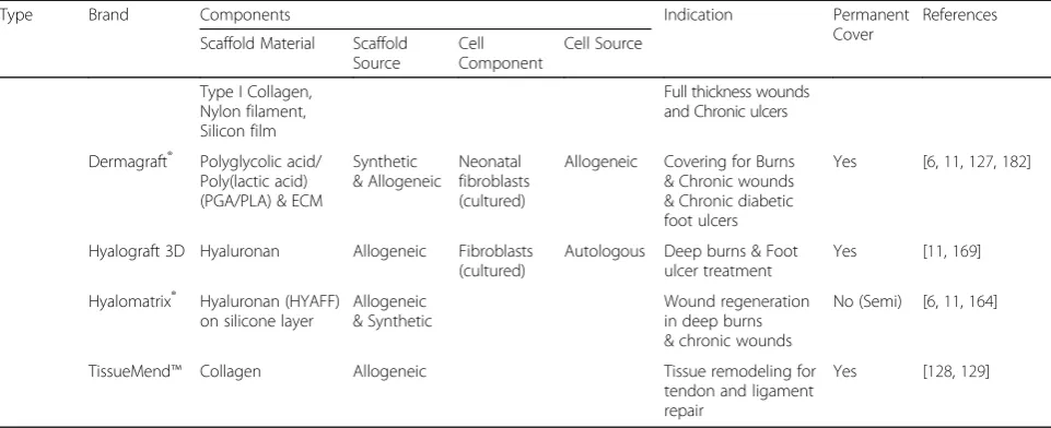
Table 1Some current commercially available bio-engineered skin substitutes(Continued)
Type Brand Components Indication Permanent
Cover
References
Scaffold Material Scaffold Source
Cell Component
Cell Source
Type I Collagen, Nylon filament, Silicon film
Full thickness wounds and Chronic ulcers
Dermagraft® Polyglycolic acid/ Poly(lactic acid) (PGA/PLA) & ECM
Synthetic & Allogeneic
Neonatal fibroblasts (cultured)
Allogeneic Covering for Burns & Chronic wounds & Chronic diabetic foot ulcers
Yes [6,11,127,182]
Hyalograft 3D Hyaluronan Allogeneic Fibroblasts (cultured)
Autologous Deep burns & Foot ulcer treatment
Yes [11,169]
Hyalomatrix® Hyaluronan (HYAFF) on silicone layer
Allogeneic & Synthetic
Wound regeneration in deep burns & chronic wounds
No (Semi) [6,11,164]
TissueMend™ Collagen Allogeneic Tissue remodeling for
tendon and ligament repair
PLGA, PEG) [167–171], elastin protein [166, 172] or other ECM constituents (hyaluronic acid, glycosamino-glycans) [169, 173–175] and are freeze-dried and treated with GA, EDC/NHS or electrospun for cross-linking. Cross-linked scaffolds decrease biodegradation and increase biocompatibility [162, 164]. Moreover, they efficiently accelerate cell infiltration and proliferation and decrease the inflammatory reaction [167, 168, 173]. Biobrane®, TransCyte®, EZ Derm®, Permacol®, and Matriderm®are some commercially available skin substi-tute employing cross-linked or complexed collagen matrix (Table 1).
Biobrane® is a dermal biosynthetic skin substitute which contains Type I porcine collagen packing an inner dermal layer of a 3D nylon filament that is also partially imbedded in an outer epidermal layer of an ultrathin sili-cone film [7, 124, 176]. Biobrane®is used for partial and full thickness burn wound dressing, particularly in the pediatric population as well as for chronic ulcers for which it provides temporary wound repair and regener-ation [7]. TransCyte® is a temporary skin substitute made of a synthetic polymeric epidermal membrane and human neonatal fibroblasts cultured on a scaffold of porcine collagen coated with bio-absorbable polyglactin and containing a silicone covered nylon mesh attached to it [12, 149, 176]. Within the nylon mesh, fibroblasts proliferate, secrete matrix proteins/growth factors and are inactivated by freezing before grafting [148, 176]. TransCyte® is used for temporary wound dressing of partial thickness burns [124, 148, 176]. EZ Derm® is an acellular xenogeneic (porcine) dermal matrix composed of an aldehyde cross-linked collagen matrix [12, 148, 177] used for the temporary dressing of partial thickness
burns [177]. Porcine products do not undergo
vascularization, and the aldehyde cross-linking treatment allows prevention of host immune responses and, conse-quently, no rejection [177]. Matriderm® is composed of an extracellular bovine Type I collagen matrix with elas-tin and used for full thickness burn and chronic wounds [7, 122]. Permacol® is an acellular porcine-derived der-mis with collagen and elastin matrix used as a temporary skin dressing [122, 149].
Fibronectin and fibrin
Fibronectin is a ubiquitous glycoprotein and a major multifunctional constituent of the ECM [144, 178]. Fibronectin has multiple functions including, in particu-lar, promoting the adhesion, proliferation, and contrac-tion of cells (macrophages, fibroblasts, etc.) that participate in wound healing [178–180]. Moreover, fibro-nectin interacts with several growth factors and there-fore regulate their expression and serve as reservoir increasing their local bioavailability [181]. However, since fibronectin is inhibited by mechanical stretching
and fails to promote vascularization, there is a limited number of fibronectin-based biomaterials available [121, 144]. Nevertheless, we can include the use of fibronectin associated with fibrin as a matrix to support skin cell growth (keratinocytes and fibroblast) for skin replace-ment [121, 182]. Fibrin, a fibrous protein derived from soluble plasma fibrinogen, which supports keratinocytes and fibroblast proliferation and migration in wound healing is also a potential source of natural biomaterials for skin substitute [144, 183]. The fact that fibrin is autologous and a potent source of growth factors re-quired for wound healing is a net advantage for using a fibrin matrix [144].
MyDerm™ is a fully autologous bi-layered living engi-neered skin substitute employing fibrin as the scaffold [144, 184]. It is constructed using keratinocytes, and fibroblasts skin biopsied cells and fibrin from the pa-tient’s plasma as biomaterials [144, 184, 185]. MyDerm™ is suitable for coverage of full-thickness skin loss [144, 184] and is assimilated and integrated into the patient’s skin without causing immune rejection and cross-contamination [144, 184]. Fibrin is also used as a sealant in tissue-engineered skin substitutes [182, 186] to mimic the final coagulation cascade step where soluble fibrino-gen is converted into insoluble fibrin by thrombin [186, 187]. The sealant polymerizes in a semi-rigid fibrin clot, thus serving as a sealing barrier to prevent leakage from the skin structure [186, 187]. Fibrin sealant presents sig-nificant advantages including biocompatibility and bio-degradability, without inducing inflammation and foreign body reactions [187]. Fibrin sealant is used with skin grafts to improve the fixation and uptake of the graft and in tissue engineering of skin, substitutes to ac-celerate wound healing [182, 188, 189]. BioSeed® is an autologous skin substitute consisting of cultured autolo-gous keratinocytes re-suspended in a fibrin sealant and is mainly used for wound treatment, e.g., chronic leg ulcers [190, 191].
Hyaluronic acid (HA)
HA is a ubiquitous linear polysaccharide composed of repeating β-1, 4-linked D-glucuronic acid (GlcA) and
advantages, notably, including biocompatibility, bio-degradability, and susceptibility to chemical modification and cross-linking, which have resulted in HA-based bio-material scaffolds and skin tissue bio-constructs exhibit-ing rare adverse effects and antigenic reactions [194, 195]. Some HA-derived materials are thus commercially available mostly for skin replacement in wound healing (Table 1).
Laserskin®is a thin and transparent epidermal substi-tute sheet of benzyl esterified HA derivative [193, 197] whose surface area is cultured with autologous kerati-nocytes and/or allogeneic fibroblasts and applied to the wound in an inverted fashion [193, 197]. Laserskin® is successfully used for dermal regeneration and skin re-surfacing to treat burn wounds or chronic full-thickness ulcers without adverse effects and antigenic reaction [193, 197–199]. Hyalomatrix® is a bi-layered acellular dermal substitute of the hyaluronan-based scaffold with a temporary external layer of silicone, which acts as an epidermal barrier [7]. Hyaluronan-based scaffold incorporates into the wound, delivers hyaluronan and induces the formation of neodermis [196]. Hyalomatrix® is clinically used to stimulate the healing process in deep burns and chronic wounds treatments [7, 200]. Hyalograft 3D is also an acellular dermal skin substitute composed of a bilayer of hyaluronan-based scaffold [105]. Unlike Hyalomatrix®, it lacks the pseudo-epidermal silicone layer but has au-tologous fibroblasts that secrete the necessary growth factors/cytokines to sustain the healing wound [201]. Hyalograft 3D is used mainly in conjunction with Laserskin® for deep burns and foot ulcers treatment [105, 196]. Both Hyalograft 3D and Hyalomatrix® are biocompatible and biodegradable and do not induce any foreign body reactions since their components are acellular [12].
Skin substitutes with synthetic biomaterials
The use of polymers to fabricate hydrogels scaffolds is another promising alternative in skin tissue engineer-ing. Hydrogels matrix scaffolds have been developed and exhibit greater properties including 3D network structure with structural resemblance to ECM, high permeability and diffusion for oxygen and nutrients, precise design and control of mechanical properties, and excellent biocompatibility and biodegradation [13, 127, 129, 202]. Biomaterials used for making hydrogel scaffolds range from synthetic polymers including PEG, PLGA and natural polymers like dextran, chitosan, gel-atin, fibrin, and hyaluronic acid [127–130]. Natural polymers have unique advantages because they are bio-compatible, biodegradable and have crucial biological functions. Nonetheless, their use is limited by their
potential immunogenic reactions and relative inability to form mechanically stable constructs [127, 203]. In contrast, synthetic polymers possess superior mechan-ical properties but often lack natural ligands and/or biological epitopes to interact with cell receptors or sol-uble proteins [203, 204]. Consequently, a combination of natural and synthetic hydrogels is often used, thus producing a cellular responsive hydrogel matrix exhibit-ing excellent mechanical and structural properties with high biocompatibility and bio-functionality [204, 205].
Porous and nanometer-sized fibrous matrix scaffolds have been fabricated to support skin tissue formation for skin wound repair and more importantly for slow release of essential growth factors required for tissue regeneration [206]. Nanofibrous scaffolds are made of natural and synthetic polymer complexes: poly(L-lactic acid)-co-poly(ε-caprolactone) (PLA-PCL) and gelatin; collagen and chitosan; PCL and collagen; PCL and PEG; PCL and collagen; chitosan and polyvinylalcohol (PVA); PEG and fibrinogen and others [203, 206, 207]. Furthermore, nanofibrous scaffolds contain open con-trollable cellular pores allowing an endowed cell adhe-sion and proliferation to form new tissues [206]. The scaffold material is then seeded with skin cells (kerati-nocytes/fibroblasts) and/or functionalized with growth factors or cytokines for their controlled delivery [206]. The scaffold undergoes degradation and absorption [206], and although most of them increase the inflam-matory response [206, 208], no immunogenic reactions or rejection have been reported.
Other forms of hydrogel scaffolds that have devel-oped and tested include bi-layered scaffolds composed of chitosan for reconstructing severe burns which exhibited a reasonable tolerance of chitosan and tissue regeneration [209]. Moreover, several skin substitutes using polymer-based scaffolds are currently available in
clinical practices (Table 1). Examples include
polymer support layer and is used for neuropathic, pressure and diabetic foot ulcers [212].
Stem cells in the development of perfect skin and avoidance of immune rejection
Skin regeneration growth and repair are evolutionary processes, but scarring is an ultimate consequence. In cases of severe skin injuries, a large portion of the skin is damaged, thus rendering it prone to infections and de-void of performing its basic thermoregulation function. Current options available for severely damaged skin re-placements are autologous grafts or allogenic skin grafts where recipients are treated with immune-suppressants to prolong survival of transplant. Nevertheless, immuno-suppressant treatments are toxic to skin recipients with chronic disabling diseases leading to infections and can-cer [213, 214].To avoid immune rejection, tissue biolo-gists now employ cadaver skin therapy capable of surfacing full thickness burns known as Alloderm® (as discussed above). Other commercially available skin options used to avoid immune rejection are Permacol®, which is a porcine-derived acellular matrix, and Apli-graf® (organogenesis) human allogeneic neonatal fore-skin fibroblast [121]. Despite these research endeavors, these skin substitutes are not fully capable of solving the problems of graft rejection. Tissue engineering of artifi-cial skin to mimic natural skin and which is immuno-compatible is emerging as the solution for skin graft re-jection [215]. However, challenges are still eminent in designing tissue-engineered donor skins to match the re-cipient HLA gene complex system, which codes the MHC complex of human responsible for regulation of immune system, or making modifications in the genetic makeup so that there are neutral surface receptors.
Advancement in tissue engineering and cell biology after three decades has resulted in many alternatives to wound healing and tissue regeneration. Ideally, skin re-placement should functionally and physically mimic nat-ural skin, be resistant to infection, have vascularization, hair follicles, sebaceous glands, and more importantly lack of antigenicity [216]. Skin replacements that are commercially available can protect the wound and help to reestablish epidermal and dermal layers, but they lack sweat glands, blood vasculatures, and hair follicles.
The emergence of stem cells with transformation capacities into different tissues and organ systems of the body, make them exceptionally attractive for human biomedical applications, including skin regeneration. Development in cell biology has made mesenchymal and embryonic stem cells technologies bring some surety to complete skin regeneration, mainly by increasing the chances of developing autologous skin grafts with re-duced chances of immune rejection [217].
Mesenchymal stem cells (MSCs)
limited MSCs studies have addressed investigating the secondary immune response. For example, normal and diabetic mouse models administered MSCs via the tail vein or the pancreatic routes exerted low immunogen-icity and immunosuppressive properties during the ini-tial period of transfusion [223]. However, during the later stage, mice receiving MSCs via the pancreatic route produced insulin and expressed MHC II, generating sig-nificant T cell responses. In contrast, mice receiving MSCs by tail vein remained immune-privileged. These results underscored how differences in the transplant-ation routes and microenvironments can influence the immunogenicity of MSCs, thus making them attractive for artificial skin regeneration. MSCs reportedly pro-longed skin grafts survival time also in a nonhuman pri-mate baboon model [224]. MSCs and regulatory T cells function collectively to drive the immune system thereby increasing the probabilities for allograft survival [225].
Umbilical cord Wharton’s Jelly, an excellent source of stem cells (WJ-MSCs), isolated by removal of both arter-ies and vein [226] have been shown to exhibit similar properties as those described above for MSCs. In addition, WJ-MSCs can differentiate into cells of meso-derm, ectoderm and endoderm origins [227]. Stem cells isolated from WJ-MSCs are well tolerated by Severe com-bined immunodeficiency (SCID) mouse and they do not induce adverse reaction after transplantation and in vitro soft agar assays [227]. Moreover, WJ-MSCs treated with inflammatory cytokines exhibited higher activity of immu-nomodulation as compared to treated bone marrow-derived MSCs. Growing WJ-MSCs on de-cellularized am-niotic biological scaffold induced scar free wound healing, hairs and better biomechanical strength after transplant-ation onto SCID mouse than did MSCs alone [226]. Other MSCs such as adipose tissue-derived stem cells (ADSCs) were revealed to be immunosuppressive, thereby making their use appealing for transplantation without employing cytotoxic drugs [228]. Prior treatment of animals with a single dose of ADSCs before skin transplantation pro-longed their skin transplants survival by expansion of CD4+ Tregs, IL-10 production and suppression of Th17 responses [228]. Overall, MSCs are attractive for regener-ation of perfect dermal replacement and have been tested in commercial artificial skin substitutes [229–231].
Embryonic stem cells (ESCs)
ESCs developed from the inner cell mass of mouse blas-tocysts were described in 1981 [232] followed by the first derived human ESCs (hESCs) in 1998 [79]. However, there are lots of ethical questions associated with using human fetus for regeneration of artificial organs. It is also difficult to generate tailored-specific ESCs for treat-ment of specific diseases or patients. We can address this issue by inducing pluripotency in adult stem cells by
direct remodeling. Somatic cells can be remodeled to an embryonic-like status by transfer of nucleus from som-atic stem cells to oocyte. [233–235] or by fusion with ESCs [236]. Researchers cloned mice by injecting nuclei from hair follicle and keratinocytes and showed that skin somatic stem cells can easily differentiate into whole or-ganisms [237]. In addition, stem cells nuclei can be rede-signed to pluripotency by exposing them to unfertilized oocytes cytoplasm as discussed later in the review. ESCs, with its self-renewal and pluripotent capabilities, are an encouragement for tissues/organs regeneration and their ability to differentiate into a variety of cell lineages has stimulated research in generating neurons [238], cardio-myocytes [239], hepatocytes [240], hematopoietic pro-genitor cells [241] and skins [242, 243].
ESCs are believed to be immune privileged cells albeit with conflicting results. Experiments using undifferenti-ated and differentiundifferenti-ated cells in a mixed lymphocyte reac-tion (MLR) showed limited or absence of human peripheral blood mononuclear cells (hPBMCs) and hu-man peripheral blood lymphocytes (hPBLs) proliferative responses, which were attributed to diminished MHC class II expression levels by hESCs [241]. In opposite to this, MLR performed with added CD4+ T cells and DCs mixed with hESCs demonstrated not only that hESCs lacked inhibition of T cells proliferation, but they also in-duced their proliferation [244]. This may be because hESCs express MHC class I, but do not express MHC class II and costimulatory molecules; whereas mature DCs display both MHC class I and II, and costimulatory molecules such as CD80, CD86, and CD40, which confer upon them the potent capacity for T-cell activation.
The pluripotent capability of ESCs highlights their potential applicability for future therapeutics in tissue regeneration to treat numerous severe illnesses. Similarly, the immunogenicity of ESCs represents one of the major obstacles precluding the successful translation of ESCs-based therapies. The immunogenic characteristics of ESCs are dynamic and in constant flux depending on their dif-ferentiation state and the environment surrounding them. When ESCs are undifferentiated, their high proliferation rate and low expression of potentially immunogenic sur-face proteins present an elusive target for the immune sys-tem. However, after differentiating and immunogenic cell surface markers are increased, ESCs are at increased risk of immunologic rejection. hESCs can be best used for re-generative medicine therapy as suggested by Taylor et al. [245] by creating hESCs bank typed with human leuko-cytes antigen to avoid immune rejection.
Induced pluripotent stem cells (iPSCs) to escape immune rejection
expression of somatic cells occurs without modifying DNA into an ESCs stage with multipotent capability. This advancement can resolve ethical and short-coming issues of employing ESCs in regenerative medicine. Vital organs of our body such as brain, skin, bone and skeletal muscles have self-renewal capacity in the form of stem cells, which can regenerate injured tissues and are responsible for normal growth and repair mechanisms [246]. However, their limitations reside in being difficult to culture, lack proliferative capacity, undergo apoptosis after transplantation, inability to develop vascularization and expensive for in vitro maintenance. These limita-tions prevent their application for artificial skin develop-ment and regeneration. Notwithstanding, some of these shortcomings and apprehensions were solved after the discovery of iPSCs in 2006 [247] when Takahashi and Yamanaka introduced four transcription factors (Oct 3/ 4, Nanog, Lin28, and SOX 2) into mouse fibroblasts resulting in ESCs exhibiting continuous proliferative capacity and differentiated into different cell types. iPSCs have characteristics that are well-suited for regeneration repair since cells from a transplant recipient can be modified by reprogramming them into multi lineages and increasing their chances to reduce immune rejec-tion, which can be further exploited for the treatment of genetic disorders [247, 248]. The continuous modifica-tion and progress in iPSCs’reprogramming modification can give new directions to regeneration, particularly arti-ficial skin implants. The evidence is provided that re-veals iPSCs can be differentiated into different cell lineages, which can lead to the formation of fully differ-entiated 3D skin structures with skin appendages and vascularization [14, 249]. Biology of skin makes it easily accessible, from both patient and normal healthy indi-vidual where iPSCs can serve as an ideal platform for re-generation of skin since skin somatic stem cells have an affinity for “Yamanaka factor” than any other system of the body. Somatic stem cells from skin have an affinity towards transcription factors Oct3/4, Sox2, Klf4, and c-Myc that are required for induction of pluripotency in cells with cells other than integumentary origin [249, 250]. Investigators have shown that precursors of mel-anin along with the hair forming units of our body have high basal expression levels of SOX2 and Klf4 transcrip-tion factors, which help to differentiate them easily into iPSCs [251]. Results from another study show that not all‘Yamanaka factors’are required for induction of pluri-potency into somatic skin cells since this process could equally be accomplished with only the Oct4 factor. Reprogramming of somatic stem cells into iPSCs is also less labor intensive [252]. Studies confirming the possi-bility of reprogramming somatic cells showed that indu-cing pluripotency in keratinocytes resulted in regrowth of epidermis after exposure to bone morphogenetic
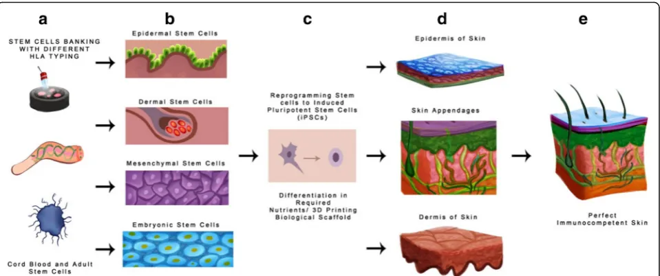
protein 4 and vitamin A metabolite [253, 254]. In a dif-ferent study, mouse fibroblasts were converted into iPSCs; differentiated into melanocytes and then embry-oid bodies when co-cultured with wingless-type 3 and EDN3 stem cell factors [255]. Even though iPSCs show great promise towards organ regeneration and growth, the long-term in vivo compatibility issues are unknown. They display many genetic and epigenetic aberrations that can cause cancerous growth or graft immune rejec-tion. iPSCs are known to induce low levels of immuno-genicity, have decreased T cells infiltration and reduced expression of JCLN1 and NOHA genes that are respon-sible for immunogenicity, and suppressing skin and tera-toma tissues [233]. Qiau Lu et al., [256] reported generation of hypo-immunogenic hiPSCs by exposing them to allogenic hPBMCs. These cells expressed re-duced MHC class II, IFN-γ, TNF-α, and IL-17; moderate MHC class I and HLA-G co-stimulatory molecules and high levels of IL-10 from Tregs in comparison to human skin fibroblast. So far, we have made significant advance-ments in developing strategies for culturing and recon-struction of 3D skin biological constructs that bear similarities to the normal competent skin (Fig. 3). Additionally, we are now using somatic stem cells to de-velop dermal and epidermal compartments of skin to treat burn patients [257].
Conclusion
resolve the issues of graft rejection and provide a viable option for autografts. Alternatively, there is now a sig-nificant number of bioengineered skin substitutes used clinically for skin repair or skin replacement therapies. To achieve a definitive regeneration of skin, however, still requires combining two or more procedures. Des-pite multiple advantages offered with bioengineered skin substitutes, there is no ideally available skin substitute allowing for permanent skin repair that is commercially available. Recent progress, especially in the design of biomaterials for incorporation into skin substitutes coupled with stem cells technology offers hope for more effective approaches in the future.
Abbreviations
3D:Three dimension; ADSCs: Adipose tissue derived stem cells; APCs: Antigen presenting cells; B-cells: B Lymphocytes; CD4+: Helper T cell; CD8+: Cytotoxic T cell; DCs: Dendritic cells; ECM: Extra cellular matrix; ESCs: Embryonic stem cells; HLA: Human leucocyte antigen; iPSCs: Induced pluripotent stem cells; LCs: Langerhans cells; MHC: Major histocompatibility complex; MSCs: Mesenchymal stem cells; NK: Natural killer cells; SCID: Severe combined immunodeficiency; T cells: T-Lymphocyte; Th1: T helper subset 1; Th17: T helper subset 17; Th2: T helper subset 2; WJ-MSCs: Wharton jelly Mesenchymal stem cells
Acknowledgments
Special thanks to Yvonne Williams, Lashaundria Lucas and Juwana Smith Henderson from the Center for NanoBiotechnology Research for their excellent administrative assistance. The authors thank Golden muse (http:// www.golden-muse.com/) for all illustrations in this review.
Funding
Support for a postdoctoral fellowship to SD, DB and AC was provided by grant from the National Science Foundation NSF-CREST (HRD-1241701) and to RS and ED by the Ph.D. program in Microbiology at Alabama State University.
Availability of data and materials
Data sharing not applicable to this article as no datasets were generated or analysed during the current study.
Authors’contributions
SD, DB, RS, ED and AC, contributed towards the manuscript writing and figures; KV, SKP, SRS and VAD contributed towards the manuscript writing, editing and critical revision. All the authors read and approved the final manuscript.
Ethics approval and consent to participate Not applicable.
Consent for publication Not applicable.
Competing interests
The authors declare that they have no competing interests.
Publisher’s Note
Springer Nature remains neutral with regard to jurisdictional claims in published maps and institutional affiliations.
Author details
1Center for Nanobiotechnology Research and Department of Biological
Sciences, Alabama State University, 1627 Harris Way, Montgomery, AL 36104, USA.2Immunity, Inflammation, and Disease Laboratory, NIH/NIEHS, Durham 27709, NC, USA.
Received: 11 April 2017 Accepted: 17 November 2017
References
1. Fore J. A review of skin and the effects of aging on skin structure and function. Ostomy Wound Manage. 2006;52:24–35. quiz 36-27
2. Pasparakis M, Haase I, Nestle FO. Mechanisms regulating skin immunity and inflammation. Nat Rev Immunol. 2014;14:289–301.
3. Guo S, Dipietro LA. Factors affecting wound healing. J Dent Res. 2010;89: 219–29.
4. Min JH, Yun IS, Lew DH, Roh TS, Lee WJ. The use of matriderm and autologous skin graft in the treatment of full thickness skin defects. Arch Plast Surg. 2014;41:330–6.