Medical Joumal of the Islamic Republic of/ran
TRANSF ORMING GROWTH FACTOR
f\
UP-REGULATES GM-CSF GENE IN HUMAN
BLADDER CARCINOMA CELL LINE HTB 5637
z. SOHEILI* AND B. GOLIAEI
From the Laboratory o/Biophysics and Molecular Biology, Institute 0/ Biochemistry and Biophysics, University a/Tehran,
Tehran, I.R. Iran.
ABSTRACT
Transforming growth factor betas are multifunctional polypeptides in the
cytokine superfamily. They have a growth inhibitory role on hemopoietic pro
genitor cells in semisolid colony assay as well as in long-term bone-marrow cul
ture. TGF -�2 represses stromal cells, stem cell factor gene transcription, and de
creases the stability of c-kit transcripts in hemopoietic cells. TGF-� also modu
lates GM-CSF production from human lymphocytes. The present study reveals
the TGF-�2 role in production of GM-CSF in HTB
5637,
human bladder carci
noma cell line. HTB
5637
cells were treated with
5
ng/mL of human TGF -�?'
viable cells were counted and GM-CSF concentration was determined, No anti
proliferate activity of TGF-�2 on HTB
5637
cell line was observed. Biological
assay showed increased levels of GM-CSF in the supernatant of cultured cells.
However this increase was lower than that expected from ELISA. Since TGF-�
may be an active suppressor factor regulating hemopoiesis, it seems that some
inhibitory factor(s) may be produced (increased) in response to TGF-�2 treat
ment. It has been shown that GM-CSF mRNA content from HTB
5637
cell line
is very stable and this stabilization is translational dependent. Using Slot blot and
Northern blot analysis, we determined that TGF-�2 upregulated GM-CSF gene
expression in HTB
5637
cell line. The results suggest that TGF-�2 upregulates
the production of GM-CSF gene at the transcriptional level.
MJIRl, Vol. 15, No.4, 219-225, 2002.
Keywords: TGF-0, GM-CSF, HTB-5637, Bladder carcinoma.
Volume 15 Number 4 Winter 1380 Febmary 2002
INTRODUCTION
Transforming growth factor-betas (TGF-�) were the first negative regulators to have their gene cloned and sequenced and the expression of their mRNA detected in a mammalian cell.I They comprise a large number of structurally related
'Current Address: National Research Center for Genetic Engineering and Biotechnology, Tehran, IRAN
Correspondence: Bahram Goliaei, Ph.D., Laboratory of Biophysics and Molecular Biology, Institute of Biochemistry and Biophysics, University of Tehran, PO.BOX 13145-1384, Tehran, I.R. Iran, Tel: (+98) 21 6113356, Fax: (+98) 2 I 6498672, E-mail: [email protected]
polypeptide growth factors and play a prominent role in the development, homeostasis and repair of virtually all tissues in organisms.2 TGF-� plays a relevant regulatory role in the homeostasis of early hematopoietic proliferation/differen tiation. TGF-�I and TGF-�2 are effective inhibitors of he matopoiesis.J They can selectively control the activity of different molecular regulators of normal and leukemic he matopoiesis.4 The production of colony stimulating activ ity by bone marrow stromal cells and the transcription of granulocyte-macrophage colony stimulating factor (GM CSF) mRNA by activated T cells are inhibited by TGF-�.5
However, TGF-� has the ability to both stimulate and
TGF-p Up-Regulates GM-CSF
Gene
inhibit the proliferation, differentiation and other functions of various cells depending on the type of target cells on the context of other growth factors present. For example in hu man thyroid follicular cells (TFC) TGF-� stimulates the ex pression of both monocyte chemoattractant protein-l (MCP
I)
and GM-CSF expression, but suppresses macrophage colony stimulating factor (M-CSF) expression.6 These re sults are generally impol1ant in understanding the ways in which cytokine regulation differs between different cell types. Varieties of non-hematopoietic malignant tumors have been demonstrated to secrete hemopoietic growth factors. B ladder cancer cells have been shown to secrete a variety of biological factors with no direct relation to urothelial cell origin including GM-CSF and various cytokines. The blad der carcinoma cell line, HTB 5637, produces colony stimu lating factors for myeloid colony (Colony Forming Unit Granulocyte Macrophage CFU-GM) growth from normal and malignant marrow. mRNA phenotyping by RT-PCR re veals expression of mRNA for IL-I ex, IL-I�, IL-6,G-CSF, M-CSF, and GM-CSF in HTB 5637 cell line.7 In the present study we aimed to examine the effect of TGF-� on the pro duction of GM-CSF in human bladder tumor cell line HTB 5637.Our data show that treatment of HTB 5637 cells with TGF-�2 increases functional GM-CSF production on a dose dependent basis. This suggests that TGF-�2 is a positive regu lator of GM-CSF gene expression in HTB 5637-cell line.
MATERIAL AND METHODS
Reagents
Recombinant human GM-CSF and cell culture me dia were purchased from Gibco BRL. Recombinant human TGF-�2' DIG-High prime DNA labeling and detection starter kit II and restriction enzymes, Eco R I a n d Bam H I w e r e o b t a i n e d f r o m B o e h r i n g e r Mannheim. H T B 5637 cells ( ATCC H T B 9 ) a n d also plasmids were obtained from American Type Culture Collection. Fetal calf serum was from Sigma and Ficoll Hypaque was purchased from Flow Laboratories. Cul ture dishes and flasks were from Nunc and other labo ratory reagents were obtained from Sigma, Merck and Fluka. hGM-CSF enzyme linked immunosorbent assay (ELISA) kit was from genzyme.
Cell culture
Monolayer cell cultures were maintained in RPMI 1640 in culture flasks or multiwell dishes containing 10% fetal calf serum. Cultures were allowed to reach 75% confluency before any experiment was carried out. Cells (I x 1 05) were grown in 25-cm2 flasks or (Sx 1 OJ) in 24-well dishes at 37°C under 7.5% CO2. Near confluent cultures were subsequently maintained by trypsinization and subculture.
Collection of cells
Media from near confluent flasks were removed; cells were washed with fresh culture media, and incubated in media with or without 5nglmL of recombinant human TGF-�o. Then media from cultures was removed, centrifuged, and
fi
ltered through 0.2 �lm filter paper and saved for ELISA. Total or cytoplasmic RNA was extracted from adherent layer cells8.9ELISA
Collected supernatants ,,,ere diluted to appropriate
(1:1
to I: 100) concentrations, and then the amount of G M -CSF was determined using ELISA kit for hGM-CSF, according to the instructions provided by the manufacturer.
Probe preparation
The P91023 and PHF (AI plasmids containing hGM CSF cDNND and hy-Actin full-length cDNA insert respec tively, were amplified in their hosts and purified recruiting standard protocols. I I After restriction digestion (P91 023 with EcoRI) aO.S-kb human GM-CSF cDNA insert and (PHFyAI with BamHI) 2.I-kb hy-Actin full-length cDNA were puri fied by agarose gel electrophoresis and phenol: chloroform extraction. Extracted inserts (0.5-1 �lg) were labeled with Boehringer Mannheim DIG labeling and detection kit, ac cording to the instructions provided by the company.
RNA isolation and slot blot analysis
RNA was extracted from near confluent cells, which were treated with 5ng/ml of TGF-�?, according to Chomczynski and Sacchi.8 The quality of -the RNA preparations was checked by 1.5% non-denaturing agarose gel electrophore sis for the presence of 2: I ratio of the 2SS to ISS bands, indicating the intactness of the RNA preparations. Total RNA (lO�g) was blotted on positively charged nylon membrane using a Schleicher and Schuell slot blot apparatus. The mem brane was prehybridized for 3 hours and then incubated in hybridization buffers containing 5-25 ng/mL labeled probe for IS hours. Following hybridization, post hybridization stringent washes and detection, membrane was transferred to development folder and incubated with chemilumines cent substrate. Then folders were exposed to X-ray films being developed after 20 minutes.
Northern blot analysis
Cytoplasmic RNA was isolated from control and treated cultures, exposed to TGF-�2 for 24 hours, according to the protocol of Clemens.9 The procedure is based on solubiliza tion of the plasma membrane while maintaining nuclear in tegrity. Cellular lysis is accomplished as quickly as pos sible, on ice, in the presence of RNase inhibitors, the inclu sion of which must be maintained until phenol:chloroform mixtures are introduced into the system to remove the pro tein. Spectrophotometric and electrophoretic analysis of the samples checked the amount and integrity of RNA. Cyto plasmic RNA (25�lg) was size fractionated on 1% agarose/
Z. Soheili and
B.
Goliaei
formaldehyde gels, then transferred to nylon membrane
using capillary transfer. Subsequently, the nucleic ac
ids were immobilized by backing 2h at 80°e. Blots
were prehybridized for 3 hours and probed with DIG
labeled probes (for hGM-CSF and hy- Actin) at 68°C
for 16-19 hours. Filters were washed to a stringency
of 0.5x SSC, 0.1 % SDS at 68°C. Following detection,
membrane was transferred to development folder and
incubated with chemiluminescent substrate, and then
folders were 25-40 minutes exposed to Fuji X-ray films
and films were developed.
Biological assay
The supernatant of control and treated cultures was col
lected 24 hours post stimulation. Biological activity was
assayed in a microplate system. Human bone marrow was
obtained from normal volunteer donors. Mononuclear cells
were collected by centrifugation over Ficoll-Hypaque.12 Cells
(Sx 10-1) were plated in each well of a 96-well flat-bottomed
microplate in the presence of
10 �lL culture media (and/or
40 units of recombinant hGM-CSF, as CSF) and 1 00 �lL
enriched IMDM supplemented with 20% fetal calf serum,
antibiotics, and 0.3% agar. Cultures were incubated for
9-II
days, in a fully humidified atmosphere of 7.S% CO, at
37°C. Colonies with more than SO cells were scored usin-g a
Zeiss inverted microscope.
RESULTS
GM-CSF conccntration time-course study
In order to determine the amount of GM-CSF produced
by HTB S637-cell line, cells were cultured as described in
the
Method�
section in 24 well plates. Everyday at least three
wells were removed from culture. Time course study with spe
cific ELISA cletected high amounts ofGM-CSF in the supematant
ofHTB S637-cells culture, at the log phase of their growth curve.
Results showed that increase in GM-CSF concentration was par
allel to increase of cell number (Fig. 1).
The influence ofTGF-�2 on GM-CSF production: dose
response study
After removal of media, cells were washed once with
complete media and were incubated in media supplemented
with TG F-
�
,(O-Sng/mL). Media was removed after 48 hours
and GM-CS-F concentration was determined in duplicate by
ELISA. Results showed a significant increase of GM-CSF
in the treated sampies (Fig.
2).
The most considerable activ
ity was seen at the concentration of S ng/mL, with higher
doses of TG F-�, (up to 10 ng/mL) showing the same stimu
lating effect; thi� guided us to employ the S ng/mL dose in
700
r;=======:::;---,
6600
"_ 500 o
E 400 !!!.
Qj u 300 (;
�
E 200" z
100
-.- Cell Number
2 4 6
Time (days)
- 4
10 12
Fig. I. GrOlvth curve ofllle HTB 5637-cclls and GM-CSF con tent of culture media. Data was presented as the mean value ±S.E. derived !i'om three independent experiments.
16
14
12 :J' � 10
C> .s u. 8 (/) Y
:; 6
Cl
4
2 0
0 2 3 4 5 6
TGF-p (ng/mL)
Fig. 2. TGf-pz stimul:J.tes G M-CSF production: a dose de pendent study. Cell cultures with 70-80% conflu cncy wcrc washed with complete m edia and incubated for 48 hrs with different doses ofTGf-P2. Then the supernatant of cultures was removed and assayed for GM-CSF concentration. Data was presented as the mean value ±S.E. derived from two in dependent experiments.
other experiments.
The influence of TGF-�2 on viable cell count and GM
CSF production: time study
Cultures were treated with or without
Sng/mL of
TGF-(2 and at each time interval, culture media was
removed and saved for ELISA test. The viable cell
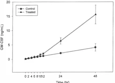
number of each sample was also determined. Our data
showed that although the number of ce lIs in treated
and control groups was the same (fig. 3) , the amount
of GM-CSF production in treated samples began in
creasing significantly from 10 hours post stimulation,
reaching the highest stimulation level at 48 hours (Fig.
TGF-� Up-Regulates GM-CSF Gene
�o
�
a;
.D
E
"
z
Qi U
800
700
600
500
400
300
200
100
Time (hr)
10 12 24 48
Fig. 3. Number of viable HTB 5637 cells in TGF-P2 stimulated and unstimulated cultures. Data was presented as the mean value ±S.E. derived from three independent experiments.
20 ,---�
15
::J"
-§,
10.s u.. C/l U
� 5 (j
o
02 4681012 24
Time (hr)
48
Fig. 4. The influence ofTGF-P2 on GM-CSF production: A time
course study. Cultures were incubated at the presence or absence of SnglmL TGF-P2. At the indicated times the supernatant of the cultures was removed and saved for ELISA. Data are shown as the mean value ±S.E. derived from three independent experiments.
4).
Biological activity of
GM-CSFTwenty-four hours exposure to TGF-�2 increased the number of CFU-GM colonies which were harvested from the supernatant of cultures. This increase (95%) was lower than that expected trom ELISA results (200% increase for 24-hrs exposure). So in another experiment, recombinant human GM-CSF (400 unit/mL of assay media) was added to control and treated cells culture media. Interestingly the number of CFU-GM colonies for control culture media in creased and reached that harvested from treated cells
cul-_25 ,---________________________ �
.!!! "ii .. u
� 20
e
III .,
'g
15 "0 u :<(!) 10
::::. u.. u '0 � ., .c E :::I Z 0
A B c o E
Fig. 5. Effects ofTGF-P2 on the CSA activity.of the culture me dia. The number ofCFU-GM colonies which formed in the pres ence of 10% control culture media (A), or 10% treated cells cul ture media (24-hrs exposure to TGF-P2) (B). The number of colo nies formed in the presence of A + 400 u/mL rhGM-CSF (C) and
B + 400 u/mL of rhGM-CSF (D). CFU-GM colonies formed in
the presence of 400u/mL rhGM-CSF (E). Data was presented as the mean value ±S.E derived from two independent experiments.
ture media, whereas the number of CFU-GM colonies har vested from treated cells culture media (in the presence and/ or absence of rhGM-CSF) was the same (Fig 5).
TGF-�2
stimulates expression of
GM-CSF mRNAin
HTB 5637-
cell line
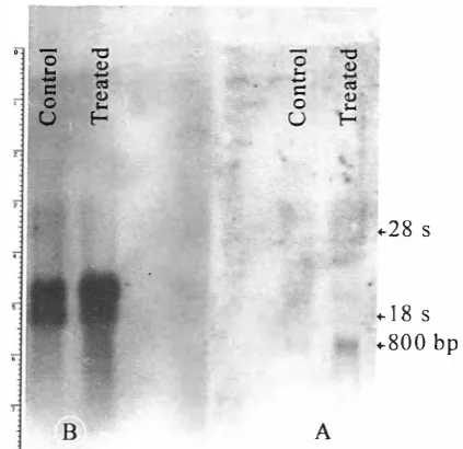
Total RNA was extracted from treated and controls samples and Slot-Blot experiment was performed. R e sults clearly showed an increase in GM-CSF mRNA content fr0111 treated cells. This effect was not seen for hy-Actin (Fig. 6). Quantitative analysis of slots was performed using a densitometer (Table I). According to slot blotting data, 24-hrs exposure to TGF-�2 in creased GM-CSF transcripts by 50%. We analyzed the effect of TGF-�l on the expression of GM-CSF mRNA by performing Northern blot analysis of cytoplasmic RNA extracted from HTB 5637 cells which had been treated with 5ng/mL of TGF-�, for 24 hrs. Results clearly showed an increase in the GM-CSF mRNA con tent from treated cells (Fig 8). There appeared to be a slight change in actin mRNA levels from the analysis of the Northern blot. However, this discrepancy may be due to differences in the amount of cytoplasmic RNA loaded on this particulate gel (Fig. 7). Experiments have confirmed that TGF-�, does not alter actin mRNA levels as expected.
-DISCUSSION
TGF-�, like many other peptide growth factors, is
Z.
Soheili and B. Goliaei
Table I. The ratio of treated: control mRNA content for y-Actin or GM-CSF slot blot analysis of total R NA.
GM-CSF mRNA 'Y-Actin mRNA Hours post stimulation Treated/Control Treated/Control
A
Treated
10
12
24
Treated
48
B
Control
10
12
24
10
12
24
48
Time (hrs.)
10
12
24
48
48
Control
Time ( hrs.)Fig. 6. Slot blot analysis of total RNA. a time-course study: I O�g of total RNA, from control and treated cultures, was loaded on slots. The membrane was hybridized with spe cific DIG labeled probe.A; GM-CSF mRNA content, D; y Actin mRNA content.
tifunctional and many of its most important activities have little to do with the transformation system in which it was first discovered. Much of the current interest in TGf-13 re search reflects its importance as a mediator of inflamma tion, repair and angiogenesis, as well as its importance as a negative regulator for many epithelial cells and for both T and B-lymphocytes.'3,'4 Because essentially all cells have
1.1
1.2 1.5 1.5
1
I 0.9 1
Fig. 7. Cultures were exposcd to T GF-f32 for 24-hrs.
cytoplasmic RNA was extracted and 25 Ilg of cytoplas
mic RNA was l o aded on each l ane of the gel. (A) The gel w as duplicated. Ethid i u m b r o m i d e stained gel. which was used for North ern analysis of hGM-CSF m R NA .
(8)
Ethi dium b r omide stained gel, which was used for Northern analysis ofy-Actin m RNA; on this par ticulate gel, a higher level of cytoplasmic R NA was loaded from the treated sample.receptors for TGF-13 15.16 TGF-13 has the potential to regu late physiological function in almost all tissues of the body. In various hemopoietic systems TGF-13 has been shown to play an inhibitory role on the production of cytokines.'7.'8 It suppresses the production of colony-stimulating activity by bone marrow stromal cells and the synthesis of GM-CSF by activated T cells, apparently at a pretranslational level.5 TGF-13 may be an active suppressor factor regulating he mopoiesis. It is supported by the reported inhibition of GM CSF mRNA transcription in a T cell line by the TGF-13.19 It is also possible that TGF-13 may induce the production of other suppressive factors.
However these inhibitory functions seem to be tissue dependent and there are several reports on the stimulating activity of TGF-13 on CSF production by tissues (or cells) other than hemopoietic types. For example in human thy roid follicular cells (TFC) and human retinal pigment
TGF-p Up-Regulates GM-CSF Gene
' '0
.b
c:: o U
B
o r-. .... c:: o U
.. 28
s.. 18
s.. 800 bp
A
Fig. 8. TGf-�, stimulates GM-CSf transcripts in the HTB 5637-cell. Northern blot analysis of cytoplasmic RNA. After blotting the mcmbrane was hybridized with GM-CSF cDNA probe (A). Arter blotting. the membrane was hybridized with y-actin eDNA
[Jl'Obc (B).
An incn:ase in the level oCy-actillmRNA in the treated sample was related to overloading.thelial cells TGF-�7 stimulates the expression of GM-CSF."·2o
Here we report an instance in which TGF-� stimulates
G
M-CSF production in a non-hemopoietic system. We re
cru ited human bladder tumor cell line HTB 5637 as a re I i
nllle and potent source for GM-CSF production. RNA
phenotyping for colony stimulating factors by reverse tran
scription and polymerase chain reaction reveals the expres
sion of mRNA for GM-CSF along with n range of other
cytokines, in
RNApreparntion of these ceIls.7
In contrast to other reports showing that TGF-� inhibits
the proliferation of normal and certain tumor derived epi
thelial cell lines.:l we didn't observe any antiproliferative
effect ofTGF-�, on the HTB 5037-cell line.
Quite illtel�estingly we
�
onfronted an increase in the
number ofCFU-GM colonies which was derived from a bio
logical assay of the treated
(24
hrs) cells med ia. However
this increase (95%) was lower than that expected fi'om ELISA
(200%). Since TGF-�2 does not have a remarknble effect
011hemopoietic progenitor cells22 it seems that some inhibitory
fnctor(s)
Illnyproduce an increase in response to TGF-�2
treatment.
The mechanism of this inhibition requires further study.
Analysis of
RNAstability has shown that the GM-CSF
IllRNAcontent of HTB 5637-cell line is very stable with a
halt�life of four hoursY The stabilization of GM-CSF mRNA
in these cells is translatiollnl dependentY
Both Slot blot and Northern analysis showed an increase
in GM-CSF mRNA in treated cells. Concluding from these
results we would like to suggest a stimulating role for TGF
�
in increasing GM-CSF production through changes
adapted at the mRNA level. These observations
mayintro
duce TGF-� as
apositive regulator for GM-CSF gene ex
pression in HTB 5637-cell line.
REFERENCES
1. Derynek R. Jarrett JA, Chen EY. Eaton 011, Bell JR. Aassoian RK. Roberts AB. Sporn MB. Goeddel DV: Human transform ing growth factor-beta: complementary DNA sequence and expression in normal and transformed cell. Nature 3 1 6 :70 I . 1 9 85.
2. Massague J:
The
TGF-J3 family of growth and differentiationHlctors. Cdl 49(4):437-438,1987.
3. Sargiacomo M, Valtieri M, Gabbanelli M. Pelosi E, Testa U.
Camagna A, Pcschle C: Pure human hematopoietic progeni
tors: direct inhibitory effect of transforming growth factors
beta I and beta2. Ann NY Acad Sci 628:84-91. 199 I.
4. Lotem J, Sachs L: Selective regulation of the activity of differ
ent hematopoietic regulatory proteins by transforming growth factor beta I in normal and leukemic myeloid cells. Blood 76(7): 1315-1322.1990.
5. Moore Sc. Theus SA, Barnett lB, Soderberg LS: Bone marrow
natural suppressor cells inhibit the growth of myeloid progeni tor cells and the synthesis of colony stimulating factors. Ex perimental hematology 20 (10): 1178-1183.1992.
6. Matsumura M, Banba N, Motohashi S, Hattori Y: Interleukin-6 and transforming growth factor-beta regulate the expression of monocyte chemoattractant protein-I and colony-stimulating fac:tors in human thyroid follicular cells. Life Sci 65:
129-135,1999.
7. Sorg R, Enczmann J, Sorg U, Heenneier K, Schneider EM,
Wernet 1': Rapid and sensitivc mRNA phenotyping for interleukins (IL-I to IL-6) and colony stimulating factors (G CSF. M-CSF, and GM-CSF) by reverse transcription and sub sequcnt polymaase chain reaction. Experimental Hematology 19:882- 887, 199 I .
8. Chomczynski P. Sacchi N: Single-step method of R NA isola
tion by acid guanidinium thiocyanate-phenol-chloroform ex
traction. Analytical Biochemistry 162: 156-I 59,1987.
9. Clemens MJ: Purification of cukaryotic messenger RNA. In:
Hames BO, [·[iggins S.L (eds.), Transcription and Translation:
A Practical Approach. Oxford: lRL Press. p. 21 I. 1984.
10. Wong GG. Witek .IS, Temple PA. Wilkens KM, L
e
aryAC, Lux enb e r g 01'. Jones SS, B rown EL, Kay RM,
O r r EC. S h o e m a ker C, G o l d e OW. Kaufman RJ,
Hewiek RM. Wan g EA, Clark SC: H uman GM-CSf.
molecular cloning of complementary D NA and puri
fication of t h e natural and r e combinant protei ns.
Science 22 8: 810-814,1985.
II. Sambrook J, Fritsch EF, Maniatis T: Molecular cloning: a
Z.
Soheili and B. Goliaei
ratory manual. New York: Cold Spring Harbor Laboratory, pp.
1.21-1.52,1989.
12. Lord Br, Marsh lCW: Enrichment of progenitor cell popula
tions from murine and human haemopoietic tissue. In: Testa NG, Molineux G, (eds.), Haemopoiesis, A Practical Approach. New York: Oxford University Press, p. 21,1993.
13. Kehrl lH, Roberts AB, Wakefield LM, lakowlew S, Sporn
MB, Fauci AS: Transforming growth factor beta is an impor tant immunomodulatory protein for human B lymphocytes. 1
Immunol 137: 3855-3860, 1986.
14. Kehrl lH. Wakefield LM, Roberts AB, lakowlew A, Alvares
mon M, Derynck R, Sporn MB, Fauci AS: Production of trans forming growth factor beta by human T lymphocytes and its potential role in the regulation of T-cell growth. 1 Exp Med
163: 1037-1050,1986.
15. Frolik CA, Wakefield LM, Smith OM, Sporn MB: Character
ization of a membrane receptor for transforming growth fac tor-beta in normal rat kidney fibroblasts. 1 BioI Chem 259: 10995-1100� 1984.
16. Kelly DL, Rizzino A: GrO\v1h regulatory factors and carcino
genesis: the roles played by transforming growth factor beta, its receptors and signaling pathways. Anticancer Res 19 (6A): 4791-4807,1999.
17. Heinrich MC, Dooley DC, Keeble W W: Transforming growth factor beta l inhibits expression of the gene prod ucts for steel factor and its receptor (c-kit). Blood 85 (7):
1769-1780, 1995.
18. Wetzler M, Talpaz M, Lowe DG, Baiocchi G, Gutterman JU,
Kurzrock R: Constitutive expression of leukemia inhibitory
factor RNA by human stromal cells and modulation by IL-1,
TNF-a, TGF-p. Experimental Hematology 19 (5): 347-3 5 1,
1991.
19. Ruegemer 11, Ho SN, Augustine lA, Schlager lW, Bell MP,
Mckean 01, Abraham RT: Regulatory effects oftransforming growth factor beta on IL-2 and IL-4 dependent T cell-CYcle
progression. lImmunol 144: 1767-1776, 1990.
20. Crane I.J, Kupper Me, McKillop-Smith S, Wallace CA,
Forrester lV: Cytokine regulation of granulocyte-macrophage colony-stimulating factor (GM-CSF) production by human reti nal pigment epithelial cells. Clin Exp Immunol 115: 288-293,1999
21. Tucker RF, Shipley GO, Moses HL, Holley RW: Growth in
hibitor from BSC-l cells closely related to platelet type
�
trans forming growth factor. Science 226: 705-707, 1984.22. Hino M, Tojo A, Miyazono K, Urabe A, Takaku F: Effects of
type p transforming growth factor on haemopoietic cells. Brit ish 10urnal of Hematology 70: 143-147, 1 9 88.
23. Ross 1-11, Sato N, Ueyama Y, Koeffier HP: Cytokine messen ger RNA stability is enhanced in tumor cells. Blood 77:
1787-1795,1991.
24. Buzby ]S, Lee SM, W inkle PV, DeMaria CT, Brewer G ,
Cairo MS: Increased granulocyte-macrophage colony stimulating factor mRN A instability in cord versus adult mononuclear cells is translation dependent and associ ated with increased level of A+U rich elements binding
factor. Blood 88: 2 889-2 897, 1996.