Medical Journal of the Islamic Republic ofIran
PURIFICATI ON AND CHARACTERIZATION OF
PRO TEIN ANTIGENS I SO LATED FROM
MYCOBACTERIUM TUBERCULOSIS (H37Rv STRA IN)
AND THEIR EFFECTS ON CELL-MEDIATED
I MMUNE RESPONSES IN GUINEA PIGS
A. ZAVARAN HOSSEINI, Ph.D.,
M.B.
ESLAMI, Ph. D*.,
AND
M.
JALALI, Ph.D. **
From the Department of Immunology, Tarbiat Modarres University, the
*Dept. of Immunology, and the **Dept. of Biochemistry, School of Public Health, Tehran University of Medical Sciences, Tehran, Islamic Republic of Iran.
ABSTRACT
Mycobacterium tuberculosis
(H37Rv strain) was used in this study. The
bacterial cells were disintegrated by sonication. The separation and characteriza
tion of the soluble molecules were attempted by various techniques including gel
filtration, ion exchange chromatographies and polyacrylamide gel electrophore
sis, using
SDS
and 2ME. Eight protein molecules with molecular weights ranging
from 6.3 up to 204
kD
were identified. Following reduction of the 204
kD
molecule
with 2ME, six smaller molecules with 12a, 12b, 21, 29,45 and 81.5
kD
molecular
weights were obtained.
All isolated protein molecules were able to induce delayed hypersensitivity
skin reaction in sensitized guinea pigs and proliferation of T-cells
in vitro.
Regarding the fact that an effective protective immunity in tuberculosis is
dependent mainly on T-cell response, it is suggested that the molecules isolated in
this study may be useful in conceiving a vaccine and/or diagnostic tests for
tuberculosis.
Keywords: M. tuberculosis, Protein Antigens, eMI Responses, Purification
MJIRI, Vol. 10, No.4, 291-297,1997.
Number 4 Winter 1375 February 1997
INTRODUCTION
Tuberculosis remains a major worldwide health prob lem, resulting in an estimated
3
million deaths and 8-10 million new cases each year}·3 Itis
estimated that 1.3 million new cases and 45,000 deaths from tuberculosis in developing countries occur annually in .children under theage of 15 years
.
2 Moreover more than3
million people are dually infected with the tubercle bacillus andmy
through out the world.2my
infectionis
the highest risk factor recently identified which increases the chance of latent infection with tubercle bacilli progressing to active tubercu losis by reducing the protection provided by cell-mediated immunity .4.36.38.40Correspondence:
A. Zavaran HO$seini, Ph.D., Tarbiat Modarres University, School of Medical Sciences, P.O. Box 141: �·4838, Tehran, Islam. Rep. Iran.
BCG vaccine has been used for a long time all over the world for the prevention of the
disease
and protection of individuals against TB. 42 However, varying rates oftion by this vaccine have been reported in different parts of the world,42,43 and the need for an effective vaccine for this very contagious disease is increasing.
It is therefore essential initially to identify molecules in the bacterial cell which effectively stimulate the immune system and induce a protective immunity. While tubercu lous patients develop high titers of specific M. tuberculosis reactive antibodies, there is no evidence indicating these have any role in protection. 10 Cell-mediated immunity is the competent protective immune response in human disease caused by intracellular pathogens.4,8,9 A number of research ers have attempted to isolate and identify the molecules from M.
tuberculosis
which could effectively activate T cells.16.18,21.29,32,41 The present research program aimed at identifying the dominant protein antigens which are directly involved in the induction of the cell-mediated immune responses (CM!) against M. tuberculosis. Considering the fact that T-cells recognize and respond mainly to protein antigens,S we focused our investigation on the proteins which are present in M. tuberculosis. For this purpose, the protein antigens from a standard strain of M. tuberculosis (H37Rv) were purified and characterized and their effects on cell-mediated immune responses were evaluated by delayed-type hypersensitivity skin tests in sensitized guinea pigs and the ability of T-cells to recognize and proliferate against these protein antigensin vitro
were also assessed.MATERIAL AND METHODS
Bacteria
M.
tuberculosis
H37Rv (ATCC 27294) and H37Ra (ATCC 25177) were provided kindly by Pasteur Institute of France.The bacteria were grown for eight weeks as a surface pellicle on modified Souton medium (sodium pyruvate 5 g, glucose 5 g, tween 80 O.5mL, L-asparagine 4.0 g, glycerl 60 g, citric acid 2g,
K2HP04
0.5 g, magnesium sulfate 0.5 g, ferric ammonium citrate 0.05 g, distilled water 950 mL, pH 7.0). This medium was sterilized at 121°C, 15 psi for 15 min.Sonic Extracts
The bacteria were separated from the medium by cen trifugation (4000x g for 30 min at 4°C). The pellet was washed three times in tris buffer and resuspended in the same buffer for sonication. The bacilli were sonicated by subjecting to ultrasonic pulses in a Virosonic Cell Disruptor model 16-850 at 4 °C, with 5 min rest periods, to alleviate the effect of heat, for a total exposure time of 20 min. The sonicate was centrifuged at 6000x g and subsequently 100,000x g at 4°C for I h. Ammonium sulphate (80% saturation) was used to precipitate and concentrate the proteins in the supernatant.
After diaIysis in 0.1 MTris-HCl pH 7.3, the sample was
FRACTIONATION
H37Rv(PPD-v)
0.6,---___________ ----,
0.5
S
0.4 � 0 0.300 N
U) 0.2
�
0.1
OLL---________ � __ �L_� '181m'
Fig.
1. Elution profile of soluble proteins of M. tuberculosis H37Rv. Gel: SephacryI200-HR flow rate 2mL/cm2 /h, fraction size 3mL/tube, height and diameter of gel: 90x2.5 cm, buffer Tris 0.1 M and sodium chloride 0.5 M pH 8,0. The sample contained 80 mg/5mL buffer.ION EXCHANGE
CHROMATOGRAPHY
H37Rv (PPD-v)0.12 0.35
.3 �
0.1
.9
... ell
S
0.08 0.25 .l:l �� a)
0 0,2 u
�
0.06 0 �U) 0.15 U
�
0.04 ell0.1
Z
0.02 0.05
0
0 0
\#e/ml
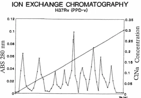
Fig. 2.
The elution profile of the soluble proteins of M. tuberculosisH37Rv. Column: 20x1.5 cm, elution buffer, 1400 mL of Tris buffer pH 8.0 with a linear concentration gradient of sodium chloride from zero up to 0.3 M, flow rate 90 mL/h, gel DEAE cellulose.
passed through a 0.22 J..I.m sterile filter. The protein content of the solution was determined by the method of Lowry et aI,IO and was stored at -70°C.
Gel Filtration Chromatography
A column (K26/100; LKB Pharmacia, Sweden) was packed with sephacryl 200-HR. Protein solution (80 mg) was then applied to the gel filtration column and eluted under pressure with O.IM Tris-HCI containing O.5M NaCI at a flow rate of 2mL/cm2/hr.
DEAE-CeIIulose Chromatography
A DEAE-cellulose column, 1.5 by 20 cm, was equili brated with 30 mM Tris buffer pH 8 containing a linear concentration gradient of sodium chloride from zero to 0.3
Fig.
3. Non-dissociated PAGE of soluble proteins of M. tubercu·losis H37Rv and H37Ra. Gel gradient 5-20%, staining coomassie brilliant blue R250.
116kD
-97kD
--.
-....
66kD
.
'� ... .
.... -_ ... .. _ _
.. .. �
tIIII ...__
29kD
,
-.=
Fig.
4. SDS-PAGE of the 2 ME-treated Fl and F4 fractions. Fraction1
has six bands and other fractions ha"\'e only one band. Gel: Acrylamidewith a concentration of 12.5%, staining coomassie brilliant blue R250.M at a rate of 90 mL/h. The fractions were collected and
store,d at 4°C until use.
Protein Analysis by SDS-PAGE
SDS-PAGE was carried out on 10, 15 and 5-20 percent gradient of acrylamide gel as described by Laemmli.13 The gels were stained with coomassie brilliant blue R -250 and it
was also silver stained according to Morrisey, 14 or by Sigma AG-25 silver stain kit (Sigma AG25).
Molecular Weight Determination
Two methods were used for molecular weight determi nation: 1) SDS-PAGE was carried out with Sigma high and low molecular weight standards (Sigma MW-SDS 70L, SDS-64, MW-SDS 175 kit), and 2) molecular sieving was conducted on sephacryl S-200HR. The column was cali brated with the protein standards with relevant molecular weights 2xW' - 13.7x10³.
Immunization of Guinea Pigs
Albino female guinea pigs weighing 400-500 g were purchased from Pasteur Institute of Iran. The guinea pigs were immunized by two injections in the hind legs and backs with 0.5 L of the chosen immunogen.
The route of injections was subcutaneously except for the BCG vaccine. The following immunogens were used: standard live M.
bovis
BCG vaccine (attenuated live bacilli, 1077 strain, Pasteur Institute, France) was injected LD., killed M.tuberculosis
H37Rv and H37Ra suspended in incomplete Freund's adjuvant (IFA) were injected S.c. A group of control guinea pigs were injected with the eluate buffer from gel filtration column in IF A. A second group of control guinea pigs were injected with Souton medium.Skin Test
The animals were skin tested seven weeks after sensiti zation. Each group of animals was given intradermal injec tions of 2 Ilg of each fraction, PPD standard or protein extract from M.
tuberculosis
H37Rv or H37Ra. The two groups of guinea pigs which served as negative controls were injected with PPD standard. Skin reaction from all groups of animals was read from 24 up to 72 h after injection .Lymphocyte Proliferation Assay
Lymphocytes were isolated in a density gradient and washed twice in culture media. The cell suspension was adjusted to 2x10⁶ viable cells per mL containing 5mM glutamine, 100 IU/mL penicillin, 100 llg/mL streptomycin and 20% inactivated fetal calf serum (GIBCO). The cell suspension was distributed in wells of U-shaped 96-well microculture plates (Nunc, Denmark). In each well 2xlOS cells in a volume of 200
IlL was added and an appropriate
amount of antigen was added according to the following scheme: either 5-10 llg/mL of one of the fractions or 101lg/ mL of PPD (Serum Institute-Denmark, batch RT 146) or 10 Ilg/mL of concanavalin-A (Sigma).Following 4 days of incubation at 37°C in a humidified 5% CO2 incubator, the cultures were pulsed with 1 IlCi of tritiated thymidine (Amersham International, U.K.). The cultures were subsequently incubated for a further 16 h at
Table I. The fractions characteristics by gel nitration chroma
tography.
Fraction·
ve*(mL)
Kav*=�
Molecular vt-vonumber
weights
(kD)
1
221
0.1 14
204
2
254
0.200
124.4
3
293
0.315
66
4
338
0.447
3 1.0
5
353
0.490
23.4
6
377
0.560
15.8
7
398
0.620
1 1.2
8
434
0.720
6.3
*ve and Kav and molecular weights of fraction obtained from sephacryl
S200-HR
column.vo= void volume, vt= total volume, ve= elution volume.
Table ll. Molecular weights of the molecules
in the fractions asdetermined by gel filtration and
SDS-PAGE.
Fradion·
. . Molecularweigbts
(kD)
njamber
Gel nitrationSDS-PAGE
1
204
8 1.5,45,29,21, * 12
2
124.4
1 18
3
66
66-67
4
3 1
30.2
5
23.4
**
6
15.8
**
7
1 1.2
12
8
6.3
**
*
Two molecules WIth very close molecular weights.**
Electrophoresis was not possible due to inadequate amount of the sample.37°C, and harvested on to glass fiber filters.The incorpo rated radioactivity was measured in a beta scintillation counter (Pharmacia, Wallack �-counter). The results were expressed as mean counts per minute ± standard error of the means.
Statistical Methods
Statistical analysis was attempted by ANOV A test using SPSS package.
RESULTS
Chromatographic Analysis
Fig.
I
illustrates the peaks obtained from gel filtration chromatography of soluble proteins from M.tuberculosis
(H37Rv). Eight fractions were obtained after elution from sephacryl 200-HR column. Fig. 2 shows the peaks and fractions obtained from DEAE-cellulose ion exchange chro matography of the same soluble proteins from M.tubercu-losis
(H37Rv). Nine fractions were collected after elution from the column. Fraction 5 was found not to be a protein. It could probably be derived from other components of the bacterium.Molecular Weight Determination
Molecular weights of fractions were determined by gel ftltration chromatography and SDS-PAGE. In order to choose a suitable gel for gel ftltration chromatography, a sample of the soluble protein was analysed by non-dissoci ated PAGE. The results indicated that the native proteins of the sample were in the approximate range of 5-250
kD
(Fig. 3).The results
of
gel ftltration chromatography regarding molecular weight determination are given in Table I. Fol lowing 2ME
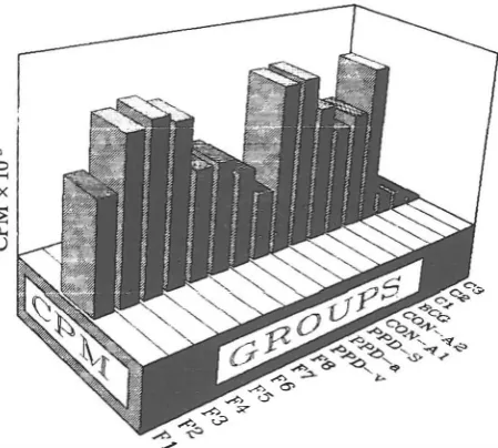
treatment of each fraction, the molecular weights of the molecules in fraction I (FI) yielded 6 molecules with various molecular weights (Fig. 4). The results of this analysis for fraction 1 and other fractions are presented in Table II.T-Lymphoc;yte Proliferation
Blood lymphocytes from different groups of guinea pigs immunized to killed M.
tuberculosis
H37Rv or BCG were exposed to either PPD, Con-A or each fraction,in vitro.
The cells were incubated at 37°C for four days. 3H-thymidine was then added in each well and the uptake of3H-thymidine by cells was measured 16 hours later. The results as assessed by uptake of 3H-thymidine indicated that the lymphocytes from all groups of i mmunized guinea pigs showed a higher proliferation responsein vitro
in comparison to the lympho cytes derived from control groups (p<O.OOOl). The prolif eration response of the lymphocytes which had been ex posedin vitro
to either whole soluble proteins of M.tuber
culosis
H37Rv, PPD standard or the fraction with 66kD
were not statistically significant (p<O.OOOl). Among the various fractions which were testedin vitro,
the lowest response was that of the fraction with 6.3kD MW.
More over the difference of proliferation response of the three fractions with 204, 23.4 and 15.8kD
molecular weights were not statistically significant (p<O.OOOl) (Fig. 5).F2, F3, F4 with molecular weights of 1 18, 66, 30.2
kD
respectively, as well as the soluble protein extracts of M.
tuberculosis
H37Rv (PPD-v) and H37Ra (PPD-a) induced the highestin vitro
response (p<O.OOOl) (Fig.'5).Regarding the dose of Con-A, there was no difference
between the
in vitro
response to 5 and 10 llg/mL of Con-A(the positive control).
Skin Reaction
All groups of guinea pigs which were skin tested showed a positive delayed type skin reaction to the intradermal injection of the antigen in comparison to that of the negative
controls (p<O:OOOl) (Table
III).
Table III. The
results of
ind
ura
ti
on due tointradermal injecti
on
of va
rious fra
ctions
, soluble extracts of M. tuberculosisH37Rv
(PPD-vl, H37Ra (PPD-al and PPD standard in each group of guinea pigs.
Antigens 204kD H8kD 66kD FI 'F2 , F3
Diameter of Indura· tion(mm)
(mean±SD) 6.2±1.3 8.8±2 8.44±2.18
* sensitized to M. tuberculosis H37Rv
** sensitized
to
BCG30.2 kD 23.4kD 15.8 kD
F4 FS F6
8.5±1.8 6±1.3 6.2±1.47
*** sensitized to culture medium plus incomplete adjuvant
**** No previous sensitization
10.0
8.0
6.0
4.0
2.0
SKIN TEST
fU
Fig.
5. The in vitro response of peripheral guinea pig lymphocytes to various fractions, PPD and Con-A. No antigen was added to the controls(CI-C3).
.-<'>6' ..-¢> """U' ..-¢> ex ..-¢>0'>
..-¢>-C=>
-?
Fig. 6. The results of induration due to intradermal injection of various fractions and PPD in different groups of guinea pigs (n= 10).
112kD 6.3kD PPD-V PPD·a PPD-S' PPD-S** PPD-S" * PPD·S" **
F7 F8 (H37Rv). H37Ra)
5.7±1.l5 5.1±O.99 8.3±1.41 9.1±1.79 7.4±O.84 7.7±1.82 2±0.63 Q.45±O
Regarding the diameter of the induration, F2, F3, F4, PPD-v, PPD-a and PPD-S induced the highest and F8 induced the weakest skin reaction (p<O.OOOl). Statistically there was no difference between the diameters of the indu ration induced b)' F1, F5, F6 and
F7
(p<O.OOOl) (Fig. 6).DISCUSSION
Purification of eight antigens from M. tuberculosis H37Rv was achieved by simple procedures using gel filtra tion and ion-exchange chromatographies. These antigens consisted of proteins with m olecular weights of204, l24.4,. 66,31,23.4,15.8, 11.2and 6.3
kD. All
isolated proteins with. the exception of the protein with the molecular weight of 204leD
appeared to be single chain polypeptides since they' migrated to the same position in the gels under reducing and non-reducing conditions. Following 2ME treatment, the 204leD
molecule was fragmented into smaller molecules with molecular weights of 81.5,29,21 and 12 kD (Fig. 4). SDS-PAGE also revealed a somewhat different molecular weight for one isolated molecule. This particular molecule' was the 124.4leD
molecule as determined by gel filtration. chromatography, which by SDS-PAGE was shown to have. a molecular weight of 118 kD. 2ME treatment of this molecule could have split a very small fragment of it, which did not seem to be a protein since no trace of a visible band was seen in polyacrylamide gel following SDS-PAGE.In recent years several antigens of M. tuberculosis have been identified by using monoclonal antibodies and other procedures.17•18,20,24,26 Regarding chemical nature and mo lecular weights, the antigens identified by these re searchers are to a great extent similar to the antigens isolated and identified in the present study. However, in the present study two relatively large protein antigens were isolated with molecular weights of 118 and 204
leD
which to our knowledge have not previously been reported. It seems by using mild procedures as well as high resolution sephacryl-200, these two molecules were isolated without fragmenta tion. It seems probable that the molecule with 204leD MW
is labile to reduction and fragmentation as the result of certain procedures and the procedures such as those men tioned in the present study are able to isolate and identify it. The immunological relevance of the purified antigens were
clearly demonstrated by positive skin test and lymphocyte transfonnation assay in guinea pigs previously sensitized to BCG or killed M. tuberculosis (H37Rv). It is therefore inferred from these results that the chains of these protein molecules comprise sequences of amino acids which struc turally make up the epitopes recognized by T-cells. Since the CM! immune responses against these antigens were quantitatively unequal, it is reasonable to suggest that these proteins bear epitopes which regarding structure and or the number of epitopes in the polypeptide chains, are different. It was found that the proteins of 118, 66 and 31
leD
which appear to be the major antigens of M. tuberc�losis H37Rv were very immunogenic. They were able to mduce a pro nounced delayed hypersensitivity reaction and promote a significant level of lymphocyte proliferation in sensitized guinea pigs. .The antigenicity of protein molecules with molecular weights in the range of those reported in this article has previously been demonstrated by skin t
�
st and l��
phoc�
te proliferation assay in guinea pigs or mIce sensItIZed WIth BCG or M. tuberculosis.10,16,24,2S,26,28,29,33,34,41In the present investigation, an attempt was made to minimize denaturation of protein molecules. Moreover, the immune response of each indi vidual guinea pig against each antigen was evaluated by skin test as well as lymphocyte proliferation assay. The results of skin tests and
�
ose of lymphocyte proliferation assays agreed closely WIth each other.The results reported by other researchers on this line 19,24,26,28,29,41 as well as the results of the present study
indi
�
ated that the majority of protein molecules in M.tuberculosis
are
able to activate and to induce CM! responses as assessed by delayed hypersensitivity skin
�
e�
tand in vitro proliferation of T-Iymphocytes. However, It IS
quite unlikely
�
at each protein anti. gen alone coul�
inducea protective immune response against tuberculosIS; these two tests may not be able to trace and identify the protective antigens in the bacterial cell. Quantitative measurement of
certain cytokines such as IL-2 and
y-
interferon produced bythe subsets44 and enumeration ofT-cell subsets including y8 T-cells which increase in mycobacterial infection4s may
also be needed to provide a vivid profile of CM! responses
for any test antigen. The protective antigen isolated from a pathogen should be able to protect the susceptible immu nized host against a challenging dose of live pathogen.
ACKNOWLEDGEMENTS
We thank Dr. R. Behin, from the WHO, Immunology Research and Training Center, Lausanne for his kind coop eration. We also thank S. Phaghi-Zadeh, Ph.D., Dept. of Statistics, School of Medical Sciences, Tarbiat Mo
�
es University, for very valuable advice in statistical analysIs.REFERENCES
1. Ehlers S, Mielke EA, Hahn H: Progress in TB research: Robert Koch's dilemma revisited. Immunol Today 15 (1): 1-4, 1994. 2. Kochi A: The global tuberculosis situation and the new control strategy of the World Health Organization. Tubercle 72: 1-6, 1991.
3. Stead WW: Pathogenesis of tuberculosis: clinical and epidemiologic perspective. Rev Infect Dis 11 (suppI2): 366-368, 1989.
4. Rieder HL, Cauthen CM, Comstock GW, Snider D Jr: Epide miology of tuberculosis in the United States. Epidemiol Rev 11: 29-30,1989.
5. Klaus GGB, Humphrey JH: The fate of antigen. In: Lachmann PJ, et al. (eds.), Clinical Aspects of Immunology. Blackwell Scientific Publications Vol. I, Chap. 7, pp. 107-126, Boston: 1993.
6. Dannenberg
AM
Jr: Immune mechanisms in the pathogenesis of pulmonary tuberculosis. Rev Infect Dis 11 (suppI2): 369-377,1989.7. Dannenberg
AM
Jr: Cellular hypersensitivity and cellular immunity in the pathogenesis of tuberculosis specificity, sys temic and local nature and associated macrophage enzymes. Bact Rev 32: 85-102, 1968.8. Youmans GP, Youmans A: Recent studies on acquired immu nity in tuberculosis. Curr Trop Microbiol Immunol 48: 129-178,1969.
9. Orme IM, Andersen P, Boom WH: T-cell response to
Mycobacterium tuberculosis. J Infect Dis 167: 1481-97, 1993.
10. Havill DV, Wallis RS, Boom
WH
, Daniel T, Chervenak K, Ellner II: Human immune response to Mycobacterium tuber culosis antigen. Infect Immun 59(2): 665-670, 1991.11. Selwyn P A, Hartel D, Lewis VA, Schoenbaum E, Vermund S, Klein R, Walker A, Frieland GH: A prospective study of the risk of tuberculosis among intravenous drug users with human immunodeficiency virus infection. N Engl J Med 320: 545-550,1989.
12. Lowry OH, Rosebrough NJ, Farr AL, Randall RJ: Protein measurement with the folin phenol reagent. J BioI Chern 193: 265-275, 1951.
13. Laemmli UK: Cleavage of structural proteins during the assembly of the head of bacteriophage T4. Nature 227: 680-685,1970.
14. Morrissey J: Silver stain for proteins in polyacrylamide gels: a modified procedure with enhanced uniform sensitivity: Anal Biochem 117: 307-310,1981.
15. Cooper TG: Gel permeation chromatography. In: Cooper TG (ed.), The Tools of Biochemistry . Chap. 5, John Wiley & Sons Inc, p. 169, 1977.
16. Andersen P, Askgaard, D, Ljungqvist L, Bennedsen J, Heron
I: Proteins released from Mycobacterium tuberculosis during growth. Infect Immun 59 (6): 1905-1910, 1991.
17. Young RA: Mycobacterial antigens, genes and vaccines:
Vaccines: New Concepts and Developments: Proceedings of the 10th International Convocation on Immunology, Buffalo, New York, p. 245,1986.
18. Young SK, et al: Results of the Third Immunology of Leprosy, Im m unology of Tuberculosis, Antimycobacterial Monoclonal Antibody Workshop. Infect Immun 60 (9): 3925-3927, 1992.
19. Dannenberg AM Jr: Controlling tuberculosis: The pathologist's point of view: Res MicrobioI141(2): 192, 1990.
20. Lyons J, et al: Expression of Mycobacterium tuberculosis and
Mycobacterium lepraeproteins by vaccinia virus. InfectImmun
58 (12): 4089-4098,1990.
21. Pal PG, Horwitz MA: Immunization with extracellular pro teins of Mycobacterium tuberculosis-induced cell-mediated immune responses and substantial protective immunity in a guinea pig model of pulmonary tuberculosis. Infec Immun 60
(11): 4781-4792, 1992.
22. Annapura PS, Prasad HK: Human immune response of frac tionated antigens of Mycobacterium tuberculosis: Abstract, 8th International Congress of Immunology, Budapest, Hungary, August 23-28, 1992.
23. Carlucci S, et al: Mycobacterial A60-specific T-cell repertoire in pulmonary tuberculosis: Abstract, 8th International Congress of Immunology, Budapest, Hungary, p. 491,1992.
24. Collins FM, Lamb J, Young, D: Biological activity of protein antigens isolated from Mycobacterium tuberculosis culture filtrate. Infec Immun 56 (5): 1260-1266, 1988.
25. Fifs T, Costopoulos C, Randford AJ, Basic A, Wood PR: Purification and characterization of major antigens from a
Mycobacterium bovis culture filtrate. Infec Immun 59 (3): 800-807, 1991.
26. Nagai S, Wilker HG, Harboe M, Kinomoto M: Isolation and partial characterization of major antigens in the culture of Mycobacterium tuberculosis. Infec Immun 59 (1): 372-382,
1991.
27. Young D, Kent L, Rees A, Lamb J, Ivanyi J: Immunological activity of 38-kilodalton protein purified from Mycobacterium tuberculosis. Infec Immun 54 (1): 177-183, 1986.
28. Andersen PA, Askgaard D, Ljunqvist L, Bentzon MW, Heron T: T-cell proliferative response to antigens secreted by Myco bacterium tuberculosis: Infect Immun 59 (4): 1558-1563, 1991.
29. Worsaae A, Ljungqvist TL, Haslov K, Heron I, Bennedsen J: Allergenic and blastogenic reactivity of three antigens from
Mycobacterium tuberculosis in sensitized guinea pigs. Infec Immun 55 (12): 2922-2927, 1987.
30. Nagai S, Matsumoto J, Nagasuga T: Specific skin reactive protein from culture filtrate of Mycobacterium bovis BCC.
Infec Immun 31 (3): 1152-1160,1981.
31. Jong AD, Hoentjen AH, Zanden VD: A rapid method for identification of mycobacterium species by polyacrylamide gel electrophoresis of soluble cell proteins. J Med Microbiol 34: 1-5, 1991.
32. Lamb JR, Lathigra R, Thbard JR, Sweetzer D, Young RA, Ivanyi J, Young DB: Identification of mycobacterial antigens recognized by T lymphocytes. Rev Infect Dis 11 (suppl 2): 443-447, 1989.
33. Harboe M, Wiker HG, Nagai S: Protein antigens of mycobac teria studied by quantitative immunologic techniques. Clin Infec Dis 14: 313-9,1992.
34. Dudani AK, Gupta RS: Immunological characterization of a human homologue of the 65-kilodalton mycobacterial antigen. Infect Immun 57 (9): 2786-2793, 1989.
35. Bruyn JD, Bosmans R, Turneer M, Weckx M, Nyabenda J, Vooren JPV, Falmagne P, Wiker HG, Harboe M: Purification, partial characterization and identification of a skin reactive protein antigen of Mycobacterium bovis BCG. Infect Immun 55 (1): 245-252, 1987.
36. Quinn TC: Interactions of the human immunodeficiency virus and tuberculosis and the implications for BCG vaccination. Rev Infec Dis 11 (suppI2): 379-384, 1989.
37. Harboe M, Nagai S, Patarroyo ME, Turres ML, Ramirez C, Cruz N: Properties of proteins MPB64, MPB70, and MPB80 of Mycobacterium bovis BCG. Infec Immun 52 (1): 293-302, 1986.
38. Collins FM: Antituberculous immunity: new solutions to an old problem. Rev Infec Dis 13: 940-950, 1991.
39. Wiegeshaus E, Balasubramanian V, Smith DW: Immunity to tuberculosis from the perspective of pathogenesis. Infectlmmun 57 (12): 3671-3676, 1989.
40. Young LS, Wormser GP: The resurgence of tuberculosis. Scan J Infect Dis (soppl) 93: 9-19, 1994.
41. Young DB, Garbe TR: Heat shock proteins and antigens of mycobacterial tuberculosis. Infect Immun 59: 3086-3093, 1991.
42. Fine PEM: The BCG story: lessons from the past and implica tions for the future. Rev Infect Dis 11 (suppI2). 353-359, 1989.
43. Stanford JL: Improving on BCG. APMIS 99: 103-113,1991.
44. Prete GD, Romagnani SL: The role ofTHI and TH2 subsets in human infectious diseases. Trend in Microbiol 2 (1): 4-6, 1994.
45. O'Brien RL, Happ MP, Dallas A, Palmer ED, Kubo R, Born WK: Stimulation of amajor subset of lymphocytes expressing
T-cell receptoryo by an antigen derived from Mycobacterium
tuberculosis: Cell 57: 667-674, 1989.