R E S E A R C H A R T I C L E
Open Access
Thyroid hormone actions are temperature-specific
and regulate thermal acclimation in zebrafish
(Danio rerio)
Alexander G Little
1*, Tatsuya Kunisue
2, Kurunthachalam Kannan
2,3and Frank Seebacher
1Abstract
Background:Thyroid hormone (TH) is best known for its role in development in animals, and for its control of metabolic heat production (thermogenesis) during cold acclimation in mammals. It is unknown whether the regulatory role of TH in thermogenesis is derived in mammals, or whether TH also mediates thermal responses in earlier vertebrates. Ectothermic vertebrates show complex responses to temperature variation, but the mechanisms mediating these are poorly understood. The molecular mechanisms underpinning TH action are very similar across vertebrates, suggesting that TH may also regulate thermal responses in ectotherms. We therefore aimed to determine whether TH regulates thermal acclimation in the zebrafish (Danio rerio). We induced hypothyroidism, followed by supplementation with 3,5-diiodothyronine (T2) or 3,5,30-triiodothyronine (T3) in zebrafish exposed to different chronic temperatures. We measured whole-animal responses (swimming performance and metabolic rates), tissue-specific regulatory enzyme activities, gene expression, and free levels of T2and T3.
Results:We found that both T3and the lesser-known T2, regulate thermal acclimation in an ectotherm. To our knowledge, this is the first such study to show this. Hypothyroid treatment impaired performance measures in cold-acclimated but not warm-acclimated individuals, whereas supplementation with both TH metabolites restored performance. TH could either induce or repress responses, depending on the actual temperature and thermal history of the animal.
Conclusions:The low sensitivity to TH at warm temperatures could mean that increasing temperatures (that is, global warming) will reduce the capacity of animals to regulate their physiologies to match demands. We suggest that the properties that underlie the role of TH in thermal acclimation (temperature sensitivity and metabolic control) may have predisposed this hormone for a regulatory role in the evolution of endothermy.
Keywords:Thyroid hormone, Zebrafish, Temperature, Cold acclimation, Hypothyroid, Ectotherm, Metabolism, Thermal plasticity, Thermal response
Background
During thermal acclimation, ectotherms can shift their re-action norms by modifying the thermal sensitivities of their metabolic and other physiological pathways. This process can be mediated by changes in enzyme concentra-tions and mitochondrial biogenesis, modification of cell
and mitochondrial membranes, and conformational
changes that optimize enzyme efficiencies at different temperatures [1-3]. Despite the importance of acclimation
for physiology, ecology, and conservation, the overarching mechanisms governing this process in ectotherms remain unknown [3]. We hypothesize that thyroid hormone (TH) regulates the thermal-acclimation response. Testing this hypothesis is important to understand how animals respond to temperature change. It is also important in an evolutionary context, because the ancestral function(s) of TH may have predisposed it for its central regulatory role in the evolution of endothermy.
TH has garnered much attention for its roles in meta-bolic heat production (thermogenesis) and energy ex-penditure [4-8] in mammals, which could be exploited for the treatment of diseases including obesity, type 2
* Correspondence:[email protected] 1
School of Biological Sciences, A08 University of Sydney, Science Road, Sydney, NSW 2006, Australia
Full list of author information is available at the end of the article
diabetes, and metabolic syndromes [9-15], but the com-plexity of the TH system is far from resolved. Several TH metabolites can stimulate physiological responses through a wide range of signaling pathways that are sub-ject to many levels of biological regulation [16-21]. In vertebrates, TH is produced in the thyroid gland primar-ily as thyroxine (T4), and is metabolized to 3,5,30 -triiodothyronine (T3) and 3,5-diiodothyronine (T2) by deiodinase enzymes (D1, D2 and D3) in peripheral tissues [22]. Other TH isomers exist, but are either physiologically inactive or have very low activity [23]. T3 was originally believed to be the only physiologically active TH because of its unique affinity for TH receptors, which regulate the expression of target genes transcriptionally by binding to thyroid response elements (TREs) in their promoters [23]. Recently, however, T2was also found to stimulate metabolism, but through non-genomic (post-transcriptional) pathways, which are as yet poorly under-stood [23]. T2 acts at different cellular levels (including plasma membrane, cytosol, and mitochondria) and elicits much quicker responses than T3[18,23]. Although T2has been shown to stimulate metabolism, its physiological relevance is still in question [23].
TH is ubiquitous across all vertebrates [4,24], and even stimulates growth and development in many invertebrate groups [25-32]. Interestingly, TH can produce drastically different responses in different animal groups [24,33]. Gen-erally, TH regulates growth and development in vertebrates and invertebrates, but additionally regulates metabolism and thermogenesis during cold exposure in mammals. However, these functionally distinct roles are underpinned by overlapping physiological and biochemical pathways [33]. Energy metabolism and its control are highly con-served in vertebrates [34], but it is unknown whether the role of TH in mediating thermal responses is independently derived in mammals, or whether it is also present in earlier vertebrates. In all animals, biochemical pathways are sensi-tive to acute changes in temperature. However, endotherms and many ectotherms regulate, or acclimate, their metabol-ism to compensate for longer-term (days to weeks) thermal variation in their environments. We hypothesize that, as in endotherms, TH regulates these physiological re-sponses of ectotherms to chronic changes in their thermal environment.
Specifically, we assessed the metabolic role of TH du-ring thermal acclimation in the zebrafish (Danio rerio). The zebrafish was chosen as a model because fish occupy an early position in vertebrate evolution, and the zebrafish in particular has become an important biomedical model for thyroid-related disease, including obesity, cardiovascu-lar disease, and diabetes [35,36]. We used a multi-factorial experimental design in which we induced hypothyroidism, followed by supplementation with T2and T3(plus normal thyroid controls) in zebrafish exposed to different chronic
and acute temperature combinations. We measured whole-animal responses (swim performance, metabolic rates, and metabolic scope), and determined tissue-specific protein function (activities of regulatory enzymes), metabolic gene expression (liver and muscle), and levels of free T2and T3to determine whether TH is the mechan-ism that drives thermal acclimation in ectotherms. We hypothesized that hypothyroidism would impair acclima-tion responses, and that supplementaacclima-tion with T3and T2 would restore acclimation in hypothyroid fish.
Results
Thyroid hormone levels
Levels of both T3 and T2 were lower in muscle tissue from cold-acclimated fish (Table 1). The TH levels in cold-acclimated hypothyroid fish supplemented with T3 and T2 verified that our hypothyroid and supplementa-tion treatments were effective (Table 2).
Effects of cold acclimation
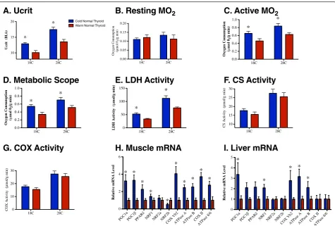
Cold acclimation significantly increased sustained swim-ming performance (critical sustained swim speed; Ucrit) at acute test temperatures of both 18°C and 28°C (Figure 1A; Table 3; see Additional file 1: Table S1). Cold-acclimated fish (kept for 3 weeks at 18°C) compensated for the limit-ing effect of low temperature, and swam as well at the 18°C acute test temperature as warm-acclimated (3 weeks at 28°C) fish did at a test temperature of 28°C.
There was no effect of acclimation temperature on resting metabolic rate, but cold acclimation significantly increased active metabolic rate and, hence, metabolic scope (Figure 1B–D; Table 3). There was a significant in-crease in maximal lactate dehydrogenase (LDH) activity in the cold-acclimated fish, but no effect of acclimation treatment on maximal citrate synthase (CS) or cyto-chrome c oxidase (COX) activities (Figure 1E–G; Table 3; see Additional file 1: Table S1). Cold-acclimation signifi-cantly increased transcript levels of the transcriptional
Table 1 Muscle-specific levels of T2and T3in
cold-acclimated and warm-acclimated normal thyroid zebrafish
Cold-acclimated Warm-acclimated
Sample T2, ng/g T3, ng/g T2, ng/g T3, ng/g
1 2.45 3.35 3.11 6.23
2 <1.00 <1.00 1.25 9.57
3 1.91 2.83 1.56 7.05
4 4.27 7.00 2.96 28.70
5 1.40 1.95 1.70 39.10
6 <1.00 1.54 5.25 531.00 Mean 1.71 to 2.00 2.80 to 2.95 2.64 103.61
coactivators PGC1αand PGC1β, their target transcription factors PPARδ, NRF1, and NRF2b, subunits of the mito-chondrial enzymes COX (COX VB2 and COX II), and FoF1-ATPase (ATPase A, ATPase B, and ATPase 8/6) in muscle, and PGC1α, NRF1, NRF2b, COX VB2, ATPase A, and ATPase B in liver (Figure 1H,I; Table 4; Table 5; see Additional file 1: Table S1).
The effects of hypothyroid treatment on responses that acclimated
Hypothyroid treatment significantly decreased the sus-tained swim speed in the cold-acclimated fish, but it had no effect on swimming performance in the warm-acclimated fish (Figure 2A; Table 3; see Additional file 1: Table S1). Hypothyroid treatment significantly decreased active metabolic rate and metabolic scope at both accli-mation temperatures (Figure 2B,C; Table 3). Hypothy-roid treatment significantly decreased LDH activity in muscle of cold-acclimated fish but it had no effect on warm-acclimated fish (Figure 2D; Table 3; see Additional file 1: Table S1). There was also a significant interaction Table 2 Muscle-specific levels of T2and T3in
cold-acclimated hypothyroid zebrafish supplemented with T2or T3
T2-supplemented T3-supplemented
Sample T2, ng/g T3, ng/g T2, ng/g T3, ng/g
1 2.66 0.00 0.00 107.00
2 <5.00 0.00 0.00 40.80
3 1.73 0.00 0.00 52.90
4 <5.00 0.00 0.00 30.60 5 <5.00 0.00 0.00 40.00 6 <5.00 0.00 0.00 63.10 7 56.50 <1.00 0.00 147.00
8 52.10 1.97 0.00 77.00
9 44.00 1.75 NA NA
Mean 17.50 to 19.70 0.42 to 0.52 0.00 69.80
SEM 2.80 0.09 0.00 4.95
Abbreviations: NA, not applicable.
18C 28C 10 15 20 U cri
t (
B
L
/s
)
*
* Warm Normal Thyroid Cold Normal Thyroid
18C 28C 0.0 0.2 0.4 0.6 0.8 1.0 O x y g en C o ns um pt io n ( µ mo l 0 2 /g m in ) * * 18C 28C 0 10 20 30 C OX Ac ti v it y ( µ mo l/g min ) 18C 28C 0.00 0.05 0.10 0.15 0.20 O x yge n C o ns um pt io n (µ mo l 0 2 /g min ) 18C 28C 0 50 100 150 LD H A ct iv it y ( µ mo l/ g mi n ) * * Re la ti v e m R NA L ev el
PGC1 PGC 1
PPAR NRF1 NRF2a NRF2b
COX Vb2ATPase AATPase BCOX IIATPase 8/6
0 2 4 6 * * * * * * * * * * 18C 28C 0.0 0.2 0.4 0.6 0.8 1.0 O xyge n C o n su m p ti o n ( µ mo l 0 2 /g m in ) * * 18C 28C 10 15 20 25 30 C S A ctiv ity ( µ mo l/g min ) Re la ti v e m R NA L ev el
PGC1PGC1PPAR NRF1 NRF2 a
NRF2b COX Vb
2
ATPase AATPase BCOX IIATPase 8/6
0 1 2 3 4 5 * * * * *
A. Ucrit
B. Resting MO
2C. Active MO
2D. Metabolic Scope
E. LDH Activity
F. CS Activity
G. COX Activity
H. Muscle mRNA
I. Liver mRNA
Figure 1Thermal-acclimation responses in normal thyroid fish.(A) Ucrit, (B) resting metabolic rate, (C) active metabolic rate, (D) metabolic
between acclimation treatment and test temperature on the activity of LDH.
Paralleling responses of swimming performance and LDH activity, there were significant interactions be-tween hypothyroid treatment and acclimation tempe-rature for transcript levels of PGC1α, COX VB2, COX II, ATPase A, ATPase B, and ATPase 8/6 in muscle, whereby hypothyroid treatment significantly decreased transcript levels in cold-acclimated fish but had no effect on warm-acclimated fish (Figure 2E; Table 4; see Additional file 1: Table S1). There were
significant effects of hypothyroid treatment on COX II and ATPase 8/6 in muscle and COXVB2 in li-ver (Figure 2E,F; Table 4; Table 5). Hypothyroid treat-ment had no significant effect on transcript levels of PGC1β, PPARδ, NRF1, and NRF2b in muscle, or on PGC1α, ATPase A, and ATPase B in liver (Figure 2E,F; Table 4; Table 5). For those genes that responded to acclimation treatments, there was a tendency for hypothyroid treatment to decrease transcript levels in cold-acclimated fish and increase transcript levels in warm-acclimated fish.
Table 3 Three-way permutational multivariate analysis of variance (PERMANOVA) results comparing parameters between cold-acclimated and warm-acclimated normal thyroid and hypothyroid fish
ATa Hb TTc ATa× Hb ATa× TTc Hb× TTc ATa× Hb× TTc
Ucritd
F 5.319 12.900 14.768 5.711 0.0088 2.079 0.838
d.f 1, 69 1, 69 1, 69 1, 69 1, 69 1, 69 1, 69
P 0.014* <0.001** <0.001** 0.017* 0.883 0.121 0.351 RMR
F 3.767 1.216 3.069 1.153 0.427 4.479 2.411
d.f 1, 59 1, 59 1, 59 1, 59 1, 59 1, 59 1, 59
P 0.031* 0.273 0.048 0.297 0.653 0.032* 0.103
AMR
F 12.790 8.354 12.430 0.567 1.064 0.112 0.200
d.f 1, 59 1, 59 1, 59 1, 59 1, 59 1, 59 1, 59
P <0.001** 0.004** <0.001** 0.471 0.305 0.829 0.717 MS
F 8.488 9.140 8.251 1.280 0.772 0.352 1.031
d.f 1, 59 1, 59 1, 59 1, 59 1, 59 1, 59 1, 59
P 0.004** 0.004** 0.007** 0.247 0.371 0.637 0.293 LDHe
F 11.547 0.642 145.460 5.611 3.886 0.291 2.346
d.f 1, 72 1, 72 1, 72 1, 72 1, 72 1, 72 1, 72
P <0.001** 0.445 <0.001** 0.018* 0.045* 0.645 0.107 CSe
F 0.505 0.010 45.700 2.410 0.040 0.006 0.246
d.f 1, 72 1, 72 1, 72 1, 72 1, 72 1, 72 1, 72
P 0.497 0.991 <0.001** 0.122 0.935 0.992 0.674 COXe
F 1.920 2.122 16.849 3.872 1.027 0.361 0.406
d.f 1, 56 1, 56 1, 56 1, 56 1, 56 1, 56 1, 56
P 0.159 0.148 <0.001** 0.044* 0.314 0.551 0.548 Abbreviations: AMR, active metabolic rate, CS, citrate synthase; COX, cytochrome oxidase; MS, metabolic scope; LDH, lactate dehydrogenase; RMR, resting metabolic rate.
a
Acclimation temperature.
b
Thyroid treatment.
c
Test temperature.
d
Swimming performance.
e
The effects of hypothyroid treatment on response measures that did not acclimate
Hypothyroidism decreased resting metabolic rate at the 18°C test temperature, but increased resting metabolic rate at the 28°C test temperature (Figure 3A; Table 3). Hypothyroid treatment had no significant effect on CS ac-tivity or muscle transcript levels for NRF2a (Figure 3B,D; Table 3; Table 4). There was a significant interaction
between hypothyroid treatment and maximal COX ac-tivity, whereby hypothyroidism significantly increased COX activity in muscle of the warm-acclimated fish but had no effect on the cold-acclimated fish (Figure 3c; see Table 3, Additional file 1: Table S1).
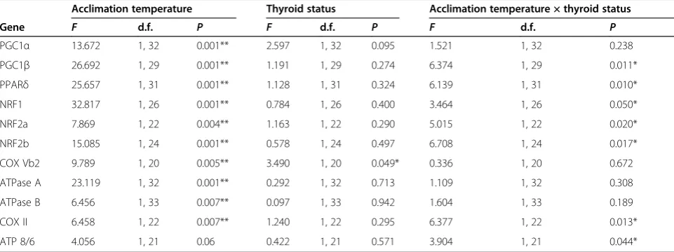
There were significant interactions between hypothyroid treatment and acclimation temperature for liver transcript levels of PGC1β, PPARδ, NRF1, NRF2a, NRF2b, COX Table 4 Two-way permutational multivariate analysis of variance (PERMANOVA) results comparing muscle mRNA levels between cold-acclimated and warm-acclimated normal thyroid and hypothyroid fish
Acclimation temperature Thyroid status Acclimation temperature × Thyroid Status
Gene F d.f. P F d.f. P F d.f. P
PGC1α 3.330 1, 34 0.041* 6.454 1, 34 0.001** 4.357 1, 34 0.026* PGC1β 12.690 1, 34 0.001** 2.677 1, 34 0.09 2.068 1, 34 0.108 PPARδ 5.708 1, 31 0.010* 0.172 1, 31 0.827 2.511 1, 31 0.117 NRF1 5.069 1, 28 0.021* 0.921 1, 28 0.343 0.268 1, 28 0.714 NRF2a 1.434 1, 28 0.239 0.145 1, 28 0.885 1.086 1, 28 0.302 NRF2b 4.108 1, 17 0.045* 0.557 1, 17 0.537 0.060 1, 17 0.948 COX Vb2 1.996 1, 33 0.124 3.786 1, 33 0.029* 7.293 1, 33 0.002** ATPase A 3.612 1, 27 0.048* 2.260 1, 27 0.099 3.136 1, 27 0.043* ATPase B 1.948 1, 27 0.148 1.216 1, 27 0.269 5.903 1, 27 0.012* COX II 12.410 1, 25 0.001** 3.850 1, 25 0.026* 2.478 1, 25 0.079 ATP 8/6 7.221 1, 24 0.004** 8.229 1, 24 0.002** 2.631 1, 24 0.068 Abbreviations: ATPase A, F0F1-ATPase subunit A; ATPase B, F0F1-ATPase subunit B; ATPase 8/6, F0F1-ATPase subunits 8 and 6; COX II, Cytochrome c oxidase subunit
2; COX Vb2, cytochrome c oxidase subunit 5b2; d.f. degrees of freedom; NRF1, Nuclear respiratory factor 1; NRF2a, Nuclear respiratory factor 2a; NRF2b, Nuclear respiratory factor 2b; PGC1α, Peroxisome proliferator-activated receptorγcoactivator 1-α; PGC1β, Peroxisome proliferator-activated receptorγcoactivator 1-β; PPARδ, Peroxisome proliferator-activated receptorδa andδb.
*P<0.05. **P<0.01.
Table 5 Two-way permutational multivariate analysis of variance (PERMANOVA) results comparing liver mRNA levels between cold-acclimated and warm-acclimated normal thyroid and hypothyroid fish
Acclimation temperature Thyroid status Acclimation temperature × thyroid status
Gene F d.f. P F d.f. P F d.f. P
PGC1α 13.672 1, 32 0.001** 2.597 1, 32 0.095 1.521 1, 32 0.238 PGC1β 26.692 1, 29 0.001** 1.191 1, 29 0.274 6.374 1, 29 0.011* PPARδ 25.657 1, 31 0.001** 1.128 1, 31 0.324 6.139 1, 31 0.010* NRF1 32.817 1, 26 0.001** 0.784 1, 26 0.400 3.464 1, 26 0.050* NRF2a 7.869 1, 22 0.004** 1.163 1, 22 0.290 5.015 1, 22 0.020* NRF2b 15.085 1, 24 0.001** 0.578 1, 24 0.497 6.708 1, 24 0.017* COX Vb2 9.789 1, 20 0.005** 3.490 1, 20 0.049* 0.336 1, 20 0.672 ATPase A 23.119 1, 32 0.001** 0.292 1, 32 0.713 1.109 1, 32 0.308 ATPase B 6.456 1, 33 0.007** 0.097 1, 33 0.942 1.604 1, 33 0.189 COX II 6.458 1, 22 0.007** 1.240 1, 22 0.295 6.377 1, 22 0.013* ATP 8/6 4.056 1, 21 0.06 0.422 1, 21 0.571 3.904 1, 21 0.044* Abbreviations: ATPase A, F0F1-ATPase subunit A; ATPase B, F0F1-ATPase subunit B; ATPase 8/6, F0F1-ATPase subunits 8 and 6; COX II, Cytochrome c oxidase subunit
2; COX Vb2, Cytochrome c oxidase subunit 5b2; d.f. degrees of freedom; NRF1, Nuclear respiratory factor 1; NRF2a, Nuclear respiratory factor 2a; NRF2b, Nuclear respiratory factor 2b; PGC1α, Peroxisome proliferator-activated receptorγcoactivator 1-α; PGC1β, Peroxisome proliferator-activated receptorγcoactivator 1-β; PPARδ, Peroxisome proliferator-activated receptorδa andδb.
VB2, COX II, and ATPase 8/6, whereby hypothyroidism significantly increased the transcript levels of PGC1β, PPARδ, NRF1, NRF2a, and NRF2b in cold-acclimated fish and significantly reduced transcript levels of PGC1β and COX II in warm-acclimated fish (Figure 3D; Table 5; see Additional file 1: Table S1). Overall, for those genes that
did not respond to acclimation treatments, there was a tendency for hypothyroidism to increase transcript levels in cold-acclimated fish and decrease transcript levels in warm-acclimated fish. This trend was the reverse of the pattern seen in the genes that did respond to thermal ac-climation in muscle.
18C 28C
-40 -20 0
C
h
an
g
e in
U
cr
it (
%
)
H
H ATxH
Cold Hypothyroid Warm Hypothyroid
Change in M
e
tabolic
S
c
ope
(%
)
18C 28C
-40 -30 -20 -10 0
H H
H
H
PGC1 PGC1 PPAR NRF1 NRF2b
COX Vb2ATPase AATPase B
COX II ATPase 8/6
-100 -50 0 50 100
Change in m
R
NA
lev
e
l (
%
)
H H
H H
ATxH ATxH ATxH ATxH
H H
H H
18C 28C
-30 -20 -10 0
Change in M
O2
(%
)
H H
H
H
18C 28C
-40 -20 0 20 40
Change in LDH A
c
ti
v
it
iy
(%
)
H
H ATxH
PGC1
COX Vb2 ATP ase A
ATPas e B 0
50 100
Change in m
R
NA
lev
e
l (
%
)
H
H
A. Ucrit
B. Active MO
2C. Metabolic Scope
D. LDH Activity
E. Muscle mRNA
F. Liver mRNA
Figure 2Effects of hypothyroid treatment on cold-acclimation responses.Percentage difference in (A) Ucrit, (B) active metabolic rate,
The effects of T2and T3supplementation for responses sensitive to hypothyroid treatment
Supplementation of hypothyroid fish with either T2 or T3resulted in a significant recovery of sustained swim-ming performance in cold-acclimated fish at both the 18°C and 28°C test temperatures (Figure 4A; Table 6). There was no significant effect of T2or T3 supplementa-tion on resting metabolic rate, but T2supplementation significantly restored active metabolic rate and metabolic scope at both test temperatures (Figure 4B,C; Table 4). There was a significant effect of T2and T3 supplementa-tion on muscle LDH activity, with T2 tending to de-crease activity levels, and T3 increasing activity levels (Figure 4D; Table 6). In muscle, T2 supplementation resulted in a significant recovery of transcript levels for
PGC1α and COX VB2, whereas T3 supplementation
resulted in a significant recovery of transcript levels for PGC1α, COX VB2, ATPase B, COX II and ATPase 8/6 (Figure 4F; Table 7). In liver, T2 or T3supplementation had no significant effect on the transcript levels of any of the genes previously shown to be sensitive to hypothyroid treatment (Figure 4G; Table 7).
Discussion
We have shown that TH regulates thermal acclima-tion of metabolism in an ectothermic vertebrate. Our
principal novel findings were that 1) the actions of TH are temperature-specific, and 2) TH regulates thermal acclimation in ectotherms. We also showed that 3) T2 has a functional role in this thermal response, which, to our knowledge has not been shown in any other system. Thus, to our knowledge, this is the first time that an en-vironmental factor as pervasive as temperature has been shown to determine not just the magnitude of a hormone-mediated response, but also the direction.
The traditional model for hormonal regulation is based on homeostatic control [24], by which the bioavailability of a hormone is adjusted to regulate its action. Opposing responses are typically mediated by antagonistic hor-mone pairs [37], but single horhor-mones can also drive dif-ferent responses depending upon the physiological context [24]. In the current study, we identified a novel signaling response, by which TH elicits a positive or negative response depending on the actual temperature and thermal history of the animal. TH has long been known to act in a tissue-specific manner [38,39], and it is possible that the same mechanisms that underlie its tissue specificity also underlie its temperature specificity. Phenotypic differences between tissues are primarily driven by differential patterns in gene expression defined during ontogeny [40,41], but gene-expression patterns, and therefore tissue phenotypes, are plastic, and can be
18C 28C
-40 -20 0 20 40
Change in M
O2
(%
) Cold Hypothyroid
Warm Hypothyroid
18C 28C
0 10 20 30
Change in CO
X
A
c
ti
v
it
y
ATxH H
H
18C 28C
0 5 10 15
Change in CS
A
c
ti
v
it
y
(
%
)
NRF2a PGC1 PPAR NRF1 NRF2a NRF2b COX II ATPase 8/6 -100
-50 0 50 100
Change in m
R
NA
lev
e
l (
%
)
Muscle Liver
ATxH ATxH ATxH ATxH ATxH ATxH ATxH
H
H
H H
H H
H
A. Resting MO
2B. CS Activity
C. COX Activity
D. Muscle and Liver mRNA
adjusted in response to environmental factors such as temperature [1-3]. Thus, the thermal-acclimation re-sponse may change the tissue phenotype temporally to alter sensitivity to TH in a way that may parallel how different tissue types respond to TH.
The temperature specificity of TH action is evident at multiple levels of physiological organization, and mediates
performance functions that determine fitness. We have shown that TH regulates energy metabolism and loco-motor performance in response to chronic exposure to cold. This is the first time, to our knowledge, that a central regulator of thermal acclimation has been identified in an ectotherm, and provides a model that could explain vertebrate radiation during thermal-niche expansion. TH
18C 28C
0 50 100 150
Ucrit Recovery (%)
R
R R
R
Cold Hypothyroid + T2
Cold Hypothyroid + T3
18C 28C
-50 0 50 100
MO
2
Max Recovery (%)
R
R
LDH Activity Recovery (%) 18C 28C -200
-150 -100 -50 0
50 R p<0.05
Liver RNA Recovery (%)
PGC1 PPAR NRF1 NRF2a NRF2b COX Vb2
0 50 100 150 200
Resting MO
2
Recovery (%)
18C 28C
-100 -50 0 50
Scope Recovery (%)
18C 28C
-50 0 50 100
R
R
PGC1
COX Vb2ATPase AATPase BCOX IIATPase 8/6
0 50 100 150 200 250
Muscle RNA Recovery (%)
R R
R R
R
R
R
A. Ucrit
B. Resting MO2
C. Active MO
2D. Metabolic Scope
E. LDH Activity
G. Liver mRNA
F. Muscle mRNA
Figure 4Effects of T2and T3supplementation on thyroid hormone (TH)-sensitive measures in cold-acclimated fish.Percentage recovery in (A) Ucrit, (B) resting metabolic rate, (C) active metabolic rate, (D) metabolic scope, (E) muscle LDH activity, (F) muscle mRNA transcript levels
and(G)liver mRNA transcript levels in cold-acclimated hypothyroid fish supplemented with either T2(teal) or T3(purple); R, significant recovery
appears to have evolved as an environmental signaling molecule prior to vertebrate evolution [26-30,42,43]. In many invertebrates, TH suppresses larval structures, and promotes the growth and development of the juvenile ru-diment [28]. Although many of these animals require ex-ogenous THs ingested from food, others can synthesize
THs or TH-like compounds endogenously [25,32,44-46]. It is interesting to note that in echinoderms, endogenously synthesized TH has been suggested to be a derived trait [25]. Growth and developmental rates are intrinsically linked to energy metabolism, and it is therefore likely that TH has always regulated these processes, at least in part by regulating metabolism. It is unknown whether the temperature specificity of TH is con-served in invertebrates, but it is conceivable that TH pathways evolved their sensitivity to temperature be-cause both play such major roles in development [47-49]. If TH regulated metabolism to promote de-velopment at thermally challenging temperatures, then selection could favor this additional role. With an en-dogenous store of TH in the form of the thyroid gland, vertebrates could regulate these responses au-tonomously, and exploit novel thermal environments while maintaining important performance parameters such as locomotor capacity.
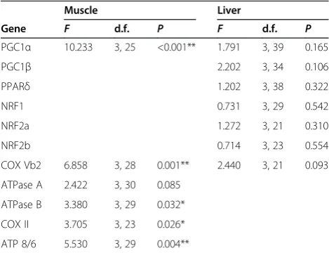
The properties that underlie the role of TH in thermal acclimation, temperature sensitivity, and metabolic con-trol may have predisposed this hormone for a regulatory role in the evolution of endothermy. Of the response variables related to cold acclimation that we measured, most were highly sensitive to TH. The genes that were upregulated by TH during cold acclimation in zebrafish are homologous to those that control mammalian ther-mogenesis. In mammals, TH modulates the transcrip-tional regulation of metabolism by controlling expression levels of PGC1α[50], which plays a master role in coord-inating the cross-genome expression of transcription fac-tors involved in mitochondrial biogenesis and proteins that drive oxidative metabolism, including COX and F0F1-ATPase [51]. Our findings show that this same pathway underlies cold acclimation in an ectotherm. The most parsimonious explanation is that these pathways were conserved in early vertebrate ancestors; however, without Table 6 Two-way ANOVA results comparing parameters between cold-acclimated normal thryoid, hypothyroid, and T2- or T3-supplemented fisha
Thyroid status Test temperature Thyroid status × test temperature
F d.f. P F d.f. P F d.f. P
Ucrit b
12.689 3, 65 0.001** 31.085 1, 65 0.001** 0.959 3, 65 0.417 RMR 1.466 3, 55 0.234 4.253 1, 55 0.044* 0.209 3, 55 0.890 AMR 3.535 3, 55 0.020* 11.032 1, 55 0.002** 0.050 3, 55 0.985 Scope 3.170 3, 55 0.006** 50.558 1, 55 0.005** 0.132 3, 55 0.899 LDHc 11.025 3, 64 0.001** 130.539 1, 64 0.001** 0.516 3, 64 0.673 Abbreviations: AMR, active metabolic rate; d.f., degrees of freedom; MS, metabolic scope; LDH, lactate dehydrogenase; RMR, resting metabolic rate.
a
Citrate synthase was not measured because there was no significant effect of thyroid hormone in the cold-acclimated and warm-acclimated normal thyroid and hypothyroid fish.
b
Swimming performance.
c
Maximal activity. *P<0.05. **P<0.01.
Table 7 One-way ANOVA results comparing muscle and liver mRNA levels between cold-acclimated normal thyroid, hypothyroid, and T2-supplemented or
T3-supplemented fisha
Muscle Liver
Gene F d.f. P F d.f. P
PGC1α 10.233 3, 25 <0.001** 1.791 3, 39 0.165
PGC1β 2.202 3, 34 0.106
PPARδ 1.202 3, 38 0.322
NRF1 0.731 3, 29 0.542
NRF2a 1.272 3, 21 0.310
NRF2b 0.714 3, 23 0.554
COX Vb2 6.858 3, 28 0.001** 2.440 3, 21 0.093 ATPase A 2.422 3, 30 0.085
ATPase B 3.380 3, 29 0.032* COX II 3.705 3, 23 0.026* ATP 8/6 5.530 3, 29 0.004**
Abbreviations: ATPase A, F0F1-ATPase subunit A; ATPase B, F0F1-ATPase
subunit B; ATPase 8/6, F0F1-ATPase subunits 8 and 6; COX II, Cytochrome c
oxidase subunit 2; COX Vb2, Cytochrome c oxidase subunit 5b2; d.f. degrees of freedom; NRF1, Nuclear respiratory factor 1; NRF2a, Nuclear respiratory factor 2a; NRF2b, Nuclear respiratory factor 2b; PGC1α, Peroxisome proliferator-activated receptorγcoactivator 1-α; PGC1β, Peroxisome proliferator-activated receptorγcoactivator 1-β; PPARδ, Peroxisome proliferator-activated receptor
δa andδb.
a
Missing values represent genes that were not sensitive to TH in the cold-acclimated and warm-cold-acclimated normal thyroid and hypothyroid fish, and therefore were not analyzed in cold-acclimated normal thyroid, hypothyroid, and T2-supplemented or T3-supplemented fish.
similar analyses of other ectotherms, we cannot preclude the possibility that they were independently derived in both fish and mammals. PGC1αhas also been shown to adjust skeletal muscle phenotype in ways that affect loco-motion [52]. As is the case in mammals, PGC1αappears to be conserved as a target of TH in zebrafish, and prob-ably also in other ectotherms. With the same pathways underlying both processes, the evolution of thermogenesis in mammals and birds [53-57] may have already been pprogrammed as a component of the cold-acclimation re-sponse in ectotherms.
T3 is often considered to be the only TH capable of genomic action because of its unique affinity for thyroid receptors [23]. However, T2has recently been shown to stimulate metabolism in mammals and fish [23]. Our work supports the notion that T2 is also an important transcriptional regulator [58,59]. In many cases, T2was just as effective as, if not more effective than, T3at regu-lating the transcription of metabolic genes. Although T2 has poor affinity for thyroid receptors, it could exert its transcriptional control through cell surface receptors that are also known to respond to TH [5,18,60], or through reversible epigenetic modifications to histone complexes [61]. Importantly, the current study shows that T2modulates performance parameters in the whole animal, which means that it is of ecological relevance and probably also of medical relevance. TH is associated with many modern lifestyle-induced conditions, and zebrafish have emerged as an important model for hu-man endocrine diseases including obesity, diabetes, and metabolic syndromes [35,36]. The fact that T2regulates metabolism and locomotor performance in a manner that is potentially very different from that of T3 means that the mechanistic basis of TH action is far broader than realized to date. A corollary is that thyroid-related diseases may be more complex, but also that there may be novel avenues for treatments that specifically target T2.
Several studies have measured changes in T3 and T4 plasma levels during thermal response in fish, but the combined results are ambiguous. In the current study we found a high level of variation in T3levels in warm-acclimated euthyroid fish. There was also much variation in the TH levels of the fish receiving T2and T3 supple-ment treatsupple-ments, although all individuals measured had supplementation levels greater than or equal to their euthyroid counterparts. Overall, the muscle-specific con-centrations of T3and T2in our study decreased drastic-ally with cold acclimation, when their effects were most pronounced. However, TH concentrations alone do not indicate the bioavailability and/or bioactivity of the hor-mone within the target tissue. Instead, the action of TH is modulated by downstream regulators, which include plasma distributor proteins, TH transporters, deiodinase enzymes, intracellular reservoir proteins, target proteins
(thyroid receptors, cell surface receptors, and specific enzymes), and transcriptional regulators [16-21,62]. The actions of TH may be more sensitive to changes in these downstream regulatory elements than to absolute free TH levels.
In many cases, the warm-acclimated zebrafish were far less sensitive to TH than cold-acclimated fish. This was especially evident in measures of locomotor perform-ance. Decreased responsiveness to TH at warmer tem-peratures suggests that chronic increases in temperature, such as those brought about by global warming, will re-duce the capacity of animals to adjust to environmental variation. Chronic warming could conceivably alter tis-sue phenotypes in ways that dampen, or reverse, TH-mediated response. This would interfere with TH as an environmental signaling molecule, and would compromise its crucial role in ectotherm growth and development. Importantly, many aquatic pollutants found worldwide, such as dioxins, bisphenol A, and phthalates, are thyroid-disrupting chemicals (TDCs) and bioaccumulate higher up the food chain [63]. Our findings indicate that the toxic effects of these chemicals may be temperature-specific, which is of crucial importance to the ecological influence of these pollutants. Together, climate change and rising levels of global pollution may amplify these risks. Warming temperatures are likely to result in higher con-centrations, longer durations, and increased distributions of TDCs throughout the water column [64].
Conclusions
Methods
Ethics statement
All experiments were carried out with the approval of the University of Sydney Animal Experimentation Ethics Committee (approval number L04/6-2010/2/5325).
Animals and treatments
Zebrafish were purchased from commercial suppliers (Kim’s Aquatic World, Sydney, NSW, Australia, and Livefish, Bundaberg, QLD, Australia) and maintained at 23°C in 10-liter tanks of dechlorinated water at densities between 1.5 and 2 fish/l for at least 1 week before the start of the treatment regimens. Fish were fed ad
libitum and maintained in a 14-hour light/10-hour
dark photoperiod.
Fish were split into two temperature treatments, a cold acclimation group kept at 18°C, and a warm acclimation group at 28°C, and held at these temperatures (±0.5°C) for 3 weeks. Within these acclimation groups, fish were separated into a normal thyroid group and a hypothyroid treatment group. Within the cold-acclimated hypothyroid group, fish were further divided into three treatment groups to be given daily supplements of T3(3,5,30 -triiodo-thyronine; Sigma), T2(3,5-diiodothyronine; Sigma), or the ethanol vehicle. There were five replicate tanks per treatment.
We induced hypothyroidism by maintaining the tank water with 0.3 mmol/l propylthiouracil (PTU; Sigma, Australia), which blocks the production of T4 at the thyroid gland [23], dissolved in a DMSO ve-hicle. Every 4 days, 80% of the tank water was changed, and the PTU was added again to return the concentration to 0.3 mmol/l. The hypothyroid groups
were also treated with 5 μmol/l iopanoic acid
(Thermofisher Scientific Inc., Australia) daily to in-hibit deiodinase activity, thereby preventing the per-ipheral metabolism of TH. Tanks holding the normal thyroid groups were maintained with proportionate amounts of the respective vehicles (0.05% DMSO and 0.025% ethanol).
Sustained swim performance
Sustained swim performance (critical sustained swim speed, Ucrit [65]) was measured in a flume consisting of a clear Perspex tube 150 mm in length and 26 mm in diameter, tightly fitted into the single exit of a Y-shaped rubber connector (total length 0.15 meters). Two 12 V inline submersible pumps (iL500; Rule, Miami, FL, USA) were fitted into the other openings of the Y connector. A plastic grid separated the flume from the two pumps, and bundles of hollow straws were positioned at each end of the flume to promote laminar flow. The flume and pumps were submerged in a plastic tank (645 × 423 × 276 mm). We used a variable DC power source (MP3090;
Powertech, Osborne Park, WA, Australia) to adjust the flow speed, which was calibrated using a flow meter (FP101; Global Water, Gold River, CA, USA). Ucritwas de-termined in accordance with published protocols [65,66], with a time interval between speed increments of 600 sec-onds, a speed increment of 0.06 meters and an initial flow rate of 0.2 m/s. Fish were allowed 15 minutes to equili-brate to the flume conditions before swim trials were begun. Animals were swum until fatigued, which was de-fined as the time when fish could no longer hold their position in the water column [65]. Each fish was swum at 18°C and 28°C in random order, with at least 24 hours be-tween swim trials. Ucritis reported as body length (BL) per second (s).
Metabolic scope
C2–C1
ð Þ V
ð Þ=ðMTÞ;
where C1 is the initial concentration of O2, C2 is the final concentration of O2, V is the volume of the cham-ber, M is the mass of the fish and T is the trial time. Metabolic scope was calculated as the difference bet-ween active and resting metabolic rates.
Thyroid hormone quantification
Unfortunately, the limits of detection did not allow us to analyze the fish liver tissue because of its small size. Levels of T3and T2(Table 3, Table 4) were quantified in muscle tissue as described previously [67,68], and ex-pressed as means, and as ranges of the mean in cases where the TH metabolite was detected in amounts below the limits of quantification.
Enzyme assays
We measured the activities of LDH, CS and COX to as-sess how acclimation temperature and thyroid status affect maximal rates of metabolic enzymes. We chose these en-zymes because they are important components of glyco-lytic and oxidative metabolism [69,70]. Tail muscle was collected, transferred into liquid nitrogen, and stored at −80°C for later analysis. Enzyme activities were de-termined in accordance with published protocols [71].
mRNA concentrations
Upon completion of treatments, the zebrafish were eu-thanized by immersion in a buffered MS222 (tricaine methane sulfonate) solution (0.4 g/l MS222 + 0.8 g/l Na2HCO3). Liver and muscle were dissected and stored
in RNAlater (Ambion, USA) at −20°C. RNA was
extracted from samples (TRIreagent; Molecular Research Center, Cincinnati, OH, USA), in accordance with the manufacturer’s instructions. RNA concentration and qual-ity were verified using a spectrophotometer (NanoDrop Technologies, Australia) and a microfluidics-based platform (Bioanalyzer 2100; Agilent Tecnologies, Australia) when necessary. An aliquot (2 μg) of total RNA from each sample was treated with DNAse I (Sigma-Aldrich) and reverse-transcribed using RNAse HMMLV reverse transcriptase (Bioscript; Bioline, Australia) and random hexamer primers (Bioline).
Quantitative reverse transcriptase (qRT)-PCR was performed on a qRT-PCR machine ( 7500; Applied Biosystems, Foster City, CA, USA) in accordance with published protocols [66]. Primers for important tran-scriptional regulators of metabolic enzymes, peroxisome proliferator-activated receptorγ coactivators 1-α and 1-β (PGC1α and PGC1β), peroxisome proliferator-activated receptors δa andδb (PPARδ), nuclear respiratory factors 1, 2a and 2b (NRF1, NRF2a and NRF2b), and subunits of metabolic enzymes, cytochrome c oxidase subunits VB2
and II (COX VB2 and COX II), and F0F1-ATPase subunits A, B, 8 and 6 (ATPase A, ATPase B and ATPase 8/6) were adopted from published works, or designed from their respective Genbank sequences (see Additional file 2: Table S2). The gene transcripts were chosen because they encode enzymes, enzyme subunits, transcription factors, and coactivators that are important regulators of oxidative phosphorylation [3,70,72]. All qPCRs were run in 96-well optical plates (BIOplastics, Landgraaf, the Netherlands). Real-time PCR reactions contained 1× SensiMix SYBR (Bioline, Australia), 4.5 mmol/l MgCl2, 50 to 900 nmol/l of each primer and approximately 100 ng cDNA. The cycle consisted of 95°C for 7 minutes, followed by 40 cycles of 95°C for 20 seconds and 58°C for 1 minute. Dissociation-curve analysis was performed after the ampli-fication step to verify the presence of only a single PCR product. Transcript expression levels of the 11 target genes in each treatment group were normalized to elong-ation factor 1 (EF1)-α in accordance with recent recom-mendations for zebrafish housekeeping genes [73], and were expressed relative to the warm-acclimated normal thyroid treatment for the warm/cold hypothyroid experi-ment, and to the cold-acclimated normal thyroid treat-ment for the T3/T2supplementation experiment.
Statistical analyses
Data are presented as means ± standard error of the mean (SEM) Trifactorial datasets (acclimation tempera-ture × thyroid status × test temperatempera-ture) and bifactorial datasets for mRNA analysis (acclimation temperature × thyroid status) were analyzed by permutational multi-variate analysis of variance (PERMANOVA) using the Primer 6 and PERMANOVA+ packages (PRIMER-E Ltd, Plymouth, Cornwall UK). In the case of interactions involving acclimation temperature and test tempe-rature, a priori planned contrasts were conducted in PERMANOVA to determine the main effects of accli-mation temperature in the normal thyroid treatments, and of thyroid status in the cold and warm accli-mation treatments. The remaining bifactorial datasets (thyroid status × test temperature) and single-factor datasets (thyroid status) were analyzed by ANOVA followed by Tukey post hoc tests using PASW Statistics 18. Where ANOVA was used, all data were tested for nor-mality and homogeneity of variance using Levene’s test. If a dataset tested significant for Levene’s test, it was transformed (log10, sqrt,x2). Significance was considered asP<0.05.
We used the truncated product method [74] to assess the effect of multiple comparisons on the validity of
type 1 errors. Multiple hypothesis testing did not bias the statistical results presented here (P<0.0001).
Data presentation
The effects of acclimation treatment are presented as ab-solute values for Ucrit, metabolic rate, metabolic scope, and maximal enzyme activity, and as relative values for mRNA transcript levels. For hypothyroid treatments, the data are presented as percentage change from the nor-mal thyroid treatments:
ðnormal thyroid treatment value–hypothyroid treatment valueÞ 100:
For T2 and T3 supplementation treatments, the data are presented as percentage recovery from hypothyroid treatment
ððsupplemented treatment value–hypothyroid treatment valueÞ=
ðnormal thyroid treatment value–hypothyroid treatment valueÞÞ 100:
Additional files
Additional file 1: Table S1.Pairwise planned comparison results comparing the effects of acclimation temperature between cold control (CC) and warm control (WC) groups, and of hypothyroidism between CC and cold hypothyroid (CH) treatments and WC and warm hypothyroid (WH) treatments.PMC, Monte CarloP-value; t,t-value; d.f., degrees of
freedom; m-, muscle RNA; l-, liver RNA.
Additional file 2: Table S2.List of primers used in this study with original source (published article or Genbank accession number for which primers were designed).
Abbreviations
ANOVA:analysis of variance; ATPase A: F0F1-ATPase subunit A; ATPase B:
F0F1-ATPase subunit B; ATPase 8/6: F0F1-ATPase subunits 8 and 6;
COX: Cytochrome c oxidase; COX II: Cytochrome c oxidase subunit 2; COX Vb2: Cytochrome c oxidase subunit 5b2; CS: Citrate synthase; d.f.: degrees of freedom; DMSO: dimethyl sulfoxide; LDH: Lactate dehydrogenase; NRF1: Nuclear respiratory factor 1; NRF2a: Nuclear respiratory factor 2a; NRF2b: Nuclear respiratory factor 2b; nTRE: Negative thyroid response element; PERMANOVA: permutational multivariate analysis of variance; PGC1α: Peroxisome proliferator-activated receptorγcoactivator 1-α; PGC1β: Peroxisome proliferator-activated receptorγcoactivator 1-β; PPARδ: Peroxisome proliferator-activated receptorδa andδb; T2:
3,5-diiodothyronine; T3: 3,5,30-triiodothyronine; TH: Thyroid hormone;
TDC: Thyroid-disrupting chemical; TRE: Thyroid response element.
Competing interest
The authors declare no competing interest.
Authors’contributions
AGL and FS conceived the project and designed the experiments; TS and KK developed the analytical protocols; AGL, TS and KK performed the experiments; AGL analyzed the data; and AGL and FS wrote the manuscript. All authors read and approved the final manuscript.
Acknowledgements
Funding
This work was funded by an Australian Research Council Discovery Grant to FS.
Financial disclosure
The funders had no role in study design, data collection and analysis, decision to publish, or preparation of the manuscript.
Author details
1
School of Biological Sciences, A08 University of Sydney, Science Road, Sydney, NSW 2006, Australia.2School of Public Health, Wadsworth Center,
New York State Department of Health, Albany, NY 12201-0509, USA.3State Key Laboratory of Urban Water Resources and Environment, IJRC PTS, Harbin Institute of Technology, Harbin 150090, China.
Received: 8 February 2013 Accepted: 19 March 2013 Published: 26 March 2013
References
1. Guderley H:Metabolic responses to low temperature in fish muscle.
Biol Rev Camb Philos Soc2004,79:409–427.
2. Seebacher F:Responses to temperature variation: integration of thermoregulation and metabolism in vertebrates.J Exp Biol2009, 212:2885–2891.
3. O’Brien KM:Mitochondrial biogenesis in cold-bodied fishes.J Exp Biol
2011,214:275–285.
4. Hulbert AJ, Else PL:Comparison of the“mammal machine”and the “reptile machine”: energy use and thyroid activity.Am J Physiol1981, 241:R350–R356.
5. Moeller LC, Broecker-Preuss M:Transcriptional regulation by nonclassical action of thyroid hormone.Thyroid Res2011,4:S6.
6. Silva JE:Thyroid hormone control of thermogenesis and energy balance.
Thyroid1995,5:481–492.
7. Cannon B:Brown adipose tissue: function and physiological significance.
Physiol Rev2004,84:277–359.
8. López M, Varela L, Vázquez MJ, Rodríguez-Cuenca S, González CR, Velagapudi VR, Morgan DA, Schoenmakers E, Agassandian K, Lage R, de Morentin PBM, Tovar S, Nogueiras R, Carling D, Lelliott C, Gallego R, Orešič M, Chatterjee K, Saha AK, Rahmouni K, Diéguez C, Vidal-Puig A: Hypothalamic AMPK and fatty acid metabolism mediate thyroid regulation of energy balance.Nat Med2010,16:1001–1008.
9. Erion MD, Cable EE, Ito BR, Jiang H, Fujitaki JM, Finn PD, Zhang BH, Hou J, Boyer SH, Van Poelje PD, Linemeyer DL:Targeting thyroid hormone receptor-βagonists to the liver reduces cholesterol and triglycerides and improves the therapeutic index.Proc Natl Acad Sci USA2007,104:15490. 10. Hollenberg AN, Forrest D:The thyroid and metabolism: the action
continues.Cell Metab2008,8:10–12.
11. Baxter JD, Webb P:Thyroid hormone mimetics: potential applications in atherosclerosis, obesity and type 2 diabetes.Nat Rev Drug Discov2009, 8:308–320.
12. Dayan CM, Panicker V:Novel insights into thyroid hormones from the study of common genetic variation.Nat Rev Endocrinol2009, 5:211–218.
13. Skarulis MC, Celi FS, Mueller E, Zemskova M, Malek R, Hugendubler L, Cochran C, Solomon J, Chen C, Gorden P:Thyroid hormone induced brown adipose tissue and amelioration of diabetes in a patient with extreme insulin resistance.J Clin Endocr Met2010,95:256–262. 14. Tseng Y-H, Cypess AM, Kahn CR:Cellular bioenergetics as a target for
obesity therapy.Nat Rev Drug Discov2010,9:465–482.
15. Pei L, Leblanc M, Barish G, Atkins A, Nofsinger R, Whyte J, Gold D, He M, Kawamura K, Li H-R, Downes M, Yu RT, Powell HC, Lingrel JB, Evans RM: Thyroid hormone receptor repression is linked to type I pneumocyte-associated respiratory distress syndrome.Nat Med2011,17:1466–1472. 16. Hulbert AJ:Thyroid hormones and their effects: a new perspective.Biol
Rev Camb Philos Soc2000,75:519–631.
17. Flamant F, Gauthier K, Samarut J:Thyroid hormones signaling is getting more complex: storms are coming.Mol Endocrinol2006,21:321–333. 18. Moreno M, de Lange P, Lombardi A, Silvestri E, Goglia F:Metabolic effects
of thyroid hormone derivatives.Thyroid2008,18:239–253.
19. Nelson ER, Habibi HR:Thyroid receptor subtypes: structure and function in fish.Gen Comp Endocr2009,161:90–96.
20. Bianco AC:Minireview: cracking the metabolic code for thyroid hormone signaling.Endocrinology2011,152:3306–3311.
21. Visser W, Friesema E, Visser TJ:Minireview: thyroid hormone transporters: the knowns and the unknowns.Mol Endocrinol2011,25:1–14. 22. Köhrle J:The deiodinase family: selenoenzymes regulating
23. Goglia F:Biological effects of 3,5-diiodothyronine (T2).Biochem Mosc
2005,70:203–213.
24. Norris DO:Vertebrate Endocrinology.London: Elsevier Academic Press; 2007:1–573.
25. Heyland A, Price D:Thyroid hormone metabolism and peroxidase function in two non-chordate animals.J Exp Zool Mol Dev Evol2006, 306:551–556.
26. Heyland A, Moroz L:Cross-kingdom hormonal signaling: an insight from thyroid hormone functions in marine larvae.J Exp Biol2005, 208:4355–4361.
27. Miller AEM, Heyland A:Endocrine interactions between plants and animals: Implications of exogenous hormone sources for the evolution of hormone signaling.Gen Comp Endocr2010,166:455–461. 28. Heyland A, Hodin J:Heterochronic developmental shift caused by thyroid
hormone in larval sand dollars and its implications for phenotypic plasticity and the evolution of nonfeeding development.Evolution2004, 58:524–538.
29. Flatt T, Moroz LL, Tatar M, Heyland A:Comparing thyroid and insect hormone signaling.Integr Comp Biol2006,46:777–794.
30. Crockford S:Evolutionary roots of iodine and thyroid hormones in cell-cell signaling.Integr Comp Biol2009,49:155–166.
31. Paris M, Escriva H, Schubert M, Brunet F, Brtko J, Ciesielski F, Roecklin D, Vivat-Hannah V, Jamin EL, Cravedi JP, Scanlan TS, Renaud JP, Holland ND, Laudet V:Amphioxuspostembryonic development reveals the homology of chordate metamorphosis.Curr Biol2008,18:825–830.
32. Eales J:Iodine metabolism and thyroid-related functions in organisms lacking thyroid follicles: are thyroid hormones also vitamins?Exp Biol Med1997,214:302.
33. Tata JR:Looking for the mechanism of action of thyroid hormone.
J Thyroid Res2011,2011:730630.
34. Koch LG, Britton SL:Aerobic metabolism underlies complexity and capacity.J Physiol (Lond)2007,586:83–95.
35. Craig PM, Moon TW:Fasted zebrafish mimic genetic and physiological responses in mammals: a model for obesity and diabetes?Zebrafish2011, 8:109–117.
36. Löhr H, Hammerschmidt M:Zebrafish in endocrine systems: recent advances and implications for human disease.Annu Rev Physiol2011, 73:183–211.
37. Koeslag JH, Saunders PT, Wessels JA:The chromogranins and the counter-regulatory hormones: do they make homeostatic sense?J Physiol1999, 517:643–649.
38. Izumo S, Nadal-Ginard B, Mahdavi V:All members of the MHC multigene family respond to thyroid hormone in a highly tissue-specific manner.
Science1986,237:597–600.
39. Brent GA:Tissue-specific actions of thyroid hormone: insights from animal models.Rev Endocr Metab2000,1:27–33.
40. Weissman IL:Stem cells: units of development, review units of regeneration, and units in evolution.Cell2000,100:157–168. 41. Maniatis T, Goodbourn S, Fischer J:Regulation of inducible and
tissue-specific gene expression.Science1987,236:1237–1245.
42. Davey K:From insect ovaries to sheep red blood cells: a tale of two hormones.J Insect Physiol2007,53:1–10.
43. Paris M, Brunet F, Markov GV, Schubert M, Laudet V:The amphioxus genome enlightens the evolution of the thyroid hormone signaling pathway.Dev Genes Evol2008,218:667–680.
44. Laudet V:The origins and evolution of vertebrate metamorphosis.
Curr Biol2011,21:R726–R737.
45. Paris M, Hillenweck A, Bertrand S, Delous G, Escriva H, Zalko D, Cravedi JP, Laudet V:Active metabolism of thyroid hormone during metamorphosis of amphioxus.Integr Comp Biol2010,50:63–74.
46. Saito M, Seki M, Amemiya S, Yamasu K, Suyemitsu T, Ishihara K:Induction of metamorphosis in the sand dollarPeronella japonicaby thyroid hormones.Dev Growth Differ1998,40:307–312.
47. Byrne M, Ho M, Selvakumaraswamy P, Nguyen HD, Dworjanyn SA, Davis AR: Temperature, but not pH, compromises sea urchin fertilization and early development under near-future climate change scenarios.Proc R Soc B: Biol Sci2009,276:1883–1888.
48. Sewell MA, Young CM:Temperature limits to fertilization and early development in the tropical sea urchinEchinometra lucunter.J Exp Mar Biol Ecol1999,236:291–305.
49. Hoegh-Guldberg O:Temperature, food availability, and the development of marine invertebrate larvae.Integr Comp Biol1995, 35:415–425.
50. Weitzel JM, Iwen KAH, Seitz HJ:Regulation of mitochondrial biogenesis by thyroid hormone.Exp Physiol2003,88:121–128.
51. Lin J, Handschin C, Spiegelman BM:Metabolic control through the PGC-1 family of transcription coactivators.Cell Metab2005, 1:361–370.
52. Li J, Kinoshita T, Pandey S, Ng CKY, Gygi SP, Shimazaki K-I, Assmann SM: Modulation of an RNA-binding protein by abscisic-acid-activated protein kinase.Nature2002,418:793–797.
53. Ruben J:The evolution of endothermy in mammals and birds: from physiology to fossils.Annu Rev Physiol1995,57:69–95.
54. McNab BK:The evolution of endothermy in the phylogeny of mammals.
Am Nat1978,112:1–21.
55. Walter I, Seebacher F:Endothermy in birds: underlying molecular mechanisms.J Exp Biol2009,212:2328–2336.
56. Walter I, Seebacher F:Molecular mechanisms underlying the
development of endothermy in birds (Gallus gallus): a new role of PGC-1 alpha?Am J Physiol-Reg I2007,293:R2315–R2322.
57. Nespolo RF, Bacigalupe LD, Figueroa CC, Koteja P, Opazo JC:Using new tools to solve an old problem: the evolution of endothermy in vertebrates.Trends Ecol Evol (Amst)2011,26:414–423.
58. Mangiullo R, Gnoni A, Damiano F, Siculella L, Zanotti F, Papa S, Gnoni GV: 3,5-diiodo-L-thyronine upregulates rat-liver mitochondrial F0F1-ATP synthase by GA-binding protein/nuclear respiratory factor-2. BBA-Bioenergetics2010,1797:233–240.
59. Garcia-G C, Lopez-Bojorquez L, Nunez J, Valverde-R C, Orozco A: 3,5-Diiodothyronine in vivo maintains euthyroidal expression of type 2 iodothyronine deiodinase, growth hormone, and thyroid hormone receptor beta 1 in the killifish.Am J Physiol-Reg I2007, 293:R877–R883.
60. Cheng S-Y, Leonard JL, Davis PJ:Molecular aspects of thyroid hormone actions.Endocr Rev2010,31:139–170.
61. Pandya K, Pulli B, Bultman S, Smithies O:Reversible epigenetic modifications of the two cardiac myosin heavy chain genes during changes in expression.Gene Expr2010,15:51–59.
62. Suzuki S, Suzuki N, Mori J-I, Oshima A, Usami S, Hashizume K: Micro-Crystallin as an intracellular 3,5,30-triiodothyronine holderin vivo.
Mol Endocrinol2007,21:885–894.
63. Casals-Casas C, Desvergne B:Endocrine disruptors: from endocrine to metabolic disruption.Annu Rev Physiol2011,73:135–162.
64. Lamon L, Dalla Valle M, Critto A, Marcomini A:Introducing an integrated climate change perspective in POPs modelling, monitoring and regulation.Environ Pollut2009,157:1971–1980.
65. Brett JR:The relation of size to rate of oxygen consumption and sustained swimming speed of sockeye salmon (Oncorhynchus nerka).
J Fish Board Can1965,22:1491–1501.
66. Seebacher F, Walter I:Differences in locomotor performance between individuals: importance of parvalbumin, calcium handling and metabolism.J Exp Biol2012,215:663–670.
67. Kunisue T, Fisher JW, Fatuyi B, Kannan K:A method for the analysis of six thyroid hormones in thyroid gland by liquid chromatography-tandem mass spectrometry.J Chromatogr B Analyt Technol Biomed Life Sci2010, 878:1725–1730.
68. Kunisue T, Fisher JW, Kannan K:Determination of six thyroid hormones in the brain and thyroid gland using isotope-dilution liquid chromatography/tandem mass spectrometry.Anal Chem2011, 83:417–424.
69. Gladden LB:Lactate metabolism: a new paradigm for the third millennium.J Physiol (Lond)2004,558:5–30.
70. Moyes CD, Hood DA:Origins and consequences of mitochondrial variation in vertebrate muscle.Annu Rev Physiol2003,
65:177–201.
71. Seebacher F, Guderley H, Elsey RM, Trosclair PL:Seasonal acclimatisation of muscle metabolic enzymes in a reptile (Alligator mississippiensis).J Exp Biol2003,206:1193–1200.
73. McCurley A, Callard G:Characterization of housekeeping genes in zebrafish: male–female differences and effects of tissue type, developmental stage and chemical treatment.BMC Mol Biol2008, 9:102–114.
74. Zaykin DV, Zhivotovsky LA, Westfall PH:Truncated product method for combiningP-values.Genet Epidemiol2002,22:170–185.
doi:10.1186/1741-7007-11-26
Cite this article as:Littleet al.:Thyroid hormone actions are temperature-specific and regulate thermal acclimation in zebrafish
(Danio rerio).BMC Biology201311:26.
Submit your next manuscript to BioMed Central and take full advantage of:
• Convenient online submission
• Thorough peer review
• No space constraints or color figure charges
• Immediate publication on acceptance
• Inclusion in PubMed, CAS, Scopus and Google Scholar
• Research which is freely available for redistribution