214
Formulation
and
Evaluation
of
Mucoadhesive
Microspheres
of
Ciprofloxacin
Shiv Shankar Hardenia1*, Ankit Jain1, Ritesh Patel1, Anu Kaushal2
1. College of Pharmacy, Indore Professional Studies (IPS) Academy, Knowledge Village, Agra-Bombay Road, Rajendra Nagar, Indore (Madhya Pradesh), India.
2. ASBASJSM College of Pharmacy, Bela, Ropar, Punjab, India.
*Corresponding author: [email protected]
ABSTRACT:
In the present research work, ethylcellulose microspheres containing ciprofloxacin were prepared and evaluated for in-vitro performance of ciprofloxacin. Ciprofloxacin microspheres containing ethylcellulose were prepared by emulsion solvent diffusion evaporation method. The surface morphological characteristics of ethylcellulose microspheres were investigated using scanning electron microscopy. The polymer ratio, stirring speed and the temperature affected the particle size, shape and surface morphology of the microspheres. The in-vitro drug release was carried out using USP paddle type dissolution rate test apparatus in 0.1N Hcl dissolution medium at 291nm. It was found that drug release from the formulations was different at different concentrations of polymers and different RPM and temperature. The best cumulative release was achieved after 24 hrs i.e. 91.6%. The Mucoadhesive property of the ethylcellulose microspheres was evaluated by in-vitro wash off test. The microspheres exhibited 75% mucoadhesion and showed good drug entrapment efficiency. By, above results it was concluded that ethylcellulose microspheres showed reproducible results, with good Mucoadhesive properties and good surface morphology.
Keywords: ciprofloxacin, mucoadhesive microsphere, ethyl cellulose, oral delivery
INTRODUCTION:
215
time of dug formulation. One of the approaches the formulation of Gastro retentive dosage forms in the form of Mucoadhesive microspheres. Microsphere carrier systems, made from natural polymers are attracting considerable attentions for several years, for sustained drug delivery. Today, those dosage forms which can control the release rates and which are target specific have a great impact in development of novel drug delivery systems. Microspheres are part of such novel delivery systems [1-3].
The success of these microspheres is limited because due to short residence time at the site of absorption. Therefore, it would be advantageous to provide an intimate contact of the drug delivery systems with the absorbing membranes. This can be achieved by coupling bioadhesion characteristics to microspheres and formulating bioadhesive microspheres. These microspheres provide advantages such as efficient absorption and increased bioavailability of drugs owing to high surface-to-volume ratio, a much more intimate contact with the mucus layer and specific targeting of drugs to the absporption site [4-7].
Ethycellulose was used as matrix polymer in which drug was dispersed because of its hydrophobic characteristic. Hydroxypropylmethylcellulose (K 100M) and Carbomer (934P) were used as Mucoadhesive polymers [8]. These polymers are selected because of their good Mucoadhesive properties. Ciprofloxacin is a first generation fluoroquinolones, it has a biological life of 4hrs [9]. Thus, development of controlled release dosage forms would clearly be advantageous. So, here an attempt is made to prepare microspheres by using HPMC (K100M) and Carbomer (934P) as Mucoadhesive polymers.
MATERIALS AND METHODS: Materials:
Ciprofloxacin was obtained as a gift sample from Cipla (Baddi), ethylcellulose, HPMC (100M) and carbomer (934P) were purchased from sigma Aldrich. All other chemicals were of analytical grade.
Methods:
216
constant 15ºC all through the process and microspheres were washed with petroleum ether and dried at room temperature. Total 37 batches were prepared with different drug: polymer ratios using combinations of HPMC/CARBOPOL with EC. For all batches RPM was maintained at 100 and temperature was maintained at 15ºC. Total seven formulations were prepared with different drug polymer ratio. These seven formulations were included in the optimization study and evaluated [10-14].
RESULTS OF TRIAL BATCHES:
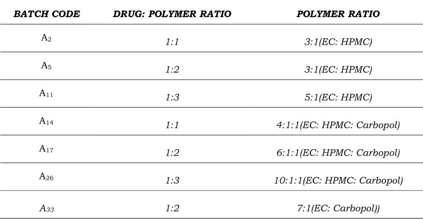
Table 1. Batches that gave smooth discrete microspheres
BATCH CODE DRUG: POLYMER RATIO POLYMER RATIO
A2 1:1 3:1(EC: HPMC)
A5 1:2 3:1(EC: HPMC)
A11 1:3 5:1(EC: HPMC)
A14 1:1 4:1:1(EC: HPMC: Carbopol)
A17 1:2 6:1:1(EC: HPMC: Carbopol)
A26 1:3 10:1:1(EC: HPMC: Carbopol)
A33 1:2 7:1(EC: Carbopol))
Particle size analysis
The particle size of the microspheres was determined by using optical microscopy method. Approximately 100 microspheres were counted for particle size analysis by using calibrated optical microscope [15].
Shape and surface morphology
The surface and inner part of the microspheres were observed via scanning electron microscopy (SEM, Hitachi S 502, Tokyo Japan). The sample was mounted on to an aluminium stub and sputter coated for 120s with platinum in an argon atmosphere [16].
Drug entrapment efficiency
217
absorbance at 291nm in 0.1N HCL. The volumetric flask was stirred continuously for 24 hr on a magnetic stirrer. Dilutions were properly made and measured for the drug entrapment [17].
% Entrapment efficiency= (Wa/Wt) × 100
Where, Wa. is the actual ciprofloxacin content and Wt. is the theoretical ciprofloxacin content.
In-vitro washes off test
The Mucoadhesive property of the microspheres was evaluated by in-vitro adhesion testing method known as wash off method. Piece of rat stomach mucosa 1×1 cm was tied on to a glass slide using a thread. Approximately 100 microspheres were spread on to the wet rinsed tissue specimen and the prepared slide was hung on to one of the grooves of a USP tablet disintegrating test apparatus. The disintegrating test apparatus was operated whereby the tissue specimen was given up and down movements regularly in the beaker of the disintegrating apparatus, which contained the gastric fluid (pH 1.2). At the end of 30 min, 1 hr and at hourly intervals up to 4 hrs, the number of microspheres still adhering to the tissue was counted [18].
Percent mucoadhesion = (weight of adhered microspheres/weight of applied microspheres) × 100
In-vitro release studies
The drug release study was carried out using USP paddle type apparatus at 37±0.5ºC and at 100 RPM using 900ml of 0.1N HCL medium as a dissolution medium. 5ml of aliquot was withdrawn at a predetermined time intervals, up to 24 hrs. The medium was replenished with 5 ml of fresh buffer each time. The absorbance is measured via U.V spectrophotometry at the wavelength of 291nm and then calculates the % cumulative release of formulation [19].
Stability studies
The stability study of the drug was determined through thin layer chromatography, % drug content and in vitro drug release study. The batch A5P2 was packed in an
218
RESULTS AND DISCUSSION:Ethylcellulose microspheres were prepared by the emulsion solvent diffusion evaporation method. The particle size of the microspheres was determined by optical microscopy. The average particle size was found to be in the range of 61.4 to 199.9µm. Batch A5P2 showed the least particle size of 86.7µm which is due to spherical nature of
microspheres as showed by the SEM. It was investigated that on increasing the concentration of polymer the particle size increases. The mean particle size of microspheres increased from 71.1µm to 190.9 with increase in concentration of polymer from 1 to 3%. The particle size of microspheres increased with the increase in the concentration of polymer, since at higher concentrations the polymer solution dispersed into larger droplets, at concentrations lower than the optimum level the solution became less viscous and dispersed into various fine droplets that easily coalesced, resulting in larger microspheres.
The mean particle size of microspheres decreased from 199.9µm to 87.6µm with the increase in the rotational speed that was 900RPM, 1200 RPM and 1500RPM. It was revealed that particle size of microspheres prepared at 1500 RPM was smaller than that of 900 and 1200 RPM. This was due to the effect of stirring speed on the size of globules. The preparation of ethylcellulose microspheres involved the maintenance at elevated temperature. The mean particle diameter of the microspheres varied from 90.8µm to 87.6µm, when temperature was increased from 10ºC to 15ºC, whereas further increase in the temperature to 20ºC lead to increase in the particle size to 152.4µm. This suggested that the temperature of system determines the size of the microspheres.
Optical microscopy of batch A5P2
219
Figure 1. Scanning electron microscopy of A5P2
After the determination we found that the batch A5P2 gave the highest % drug
entrapment efficiency than the other batches with RPM 1500 and temperature 15ºC. % drug entrapment was determined by using U.V spectrophotometer at 291nm.
The Mucoadhesive property of the microspheres was evaluated by in vitro adhesion testing methods called in-vitro wash off test. This test was done with the help of USP disintegration apparatus in which beaker contained 1.2 pH buffer solution. The numbers of microspheres adhering to the tissue were calculated after 30 min, 1 hr and hourly at 4 hr. After determination it was found that batch A5P2 showed highest percent
75% mucoadhesion than other batches.
Table 2. Result of In-Vitro wash off test to affects mucoadhesive properties of the microspheres
% MUCOADHESION TO STOMACH MUCOSA
BATCH CODE
AFTER
1 HR
AFTER
2 HR
AFTER
3 HR
AFTER
4 HR
A2P1 (1:1) 64 43 32 24
A5P2 (1:2) 90 85 75 75
A11P3 (1:3) 80 75 69 65
220
A17P5 (1:2) 82 74 70 70
A26P6 (1:3) 85 80 75 72
A33P7 (1:2) 85 65 60 50
% Mucoadhesion of A2P1, A5P2 and A11P3 batch.
IN VITRO WASH OFF TEST
0
10
20
30
40
50
60
70
80
90
100
0
1
2
Time(hr)
3
4
5
%
M
u
c
o
a
d
h
e
si
o
n
A2P1
A5P2
A11P3
Figure 2. In-vitro wash off test between % mucoadhesion and time in hr. of A2P1, A5P2 and A11P3 batch
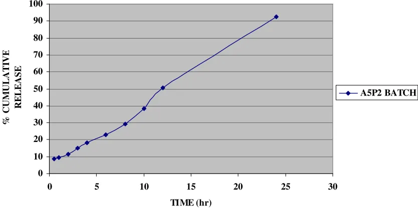
In vitro release studies were performed, in 0.1N HCL at 291nm. The drug release from the formulations was different at different concentrations of polymers, at different RPM and temperature. After 24 hrs the release was found to be 83.6, 92.5, 75.5, 82.3, 85, 72, 85, 75.5, 84, 91.6 and 84.6% of the batches A2P1 TO A5T6. A5P2 batch containing EC
221
0 10 20 30 40 50 60 70 80 90 100
0 5 10 15 20 25 30
TIME (hr)
%
C
U
M
U
L
A
T
IV
E
R
E
L
E
A
S
E
A5P2 BATCH
Figure 3. % Cumulative release of batch A5P2 at 291 nm
Stability studies of batch A5P2 was performed by TLC method, % drug entrapment
efficiency and in-vitro release studies. The batch A5P2 showed reproducible results,
there was no any change in the Rf value, % drug entrapment efficiency and in vitro release. Rf value was 0.56 determined with the help of TLC method. % drug entrapment was 70.3% determined with the help of UV method. In vitro release was 92.5% determined by UV method.
Thin layer chromatography
Table 3. Rf value of batch A5P2
BATCH A5P2
RH/TEMP. Rf VALUE
75/400C 0.50
60/250C 0.54
Room Temp. 0.56
% Drug content
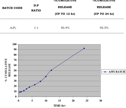
Table 4. % Drug content of batch A5P2
BATCH CODE D:P RATIO %DRUG CONTENT
222
Invitro drug release studyTable 5. % Cumulative release content of batch A5P2
BATCH CODE D:P RATIO
%CUMULATIVE
RELEASE
(UP TO 12 hr)
%CUMULATIVE
RELEASE
(UP TO 24 hr)
A5P2 1:1 50.4% 92.5%
0 10 20 30 40 50 60 70 80 90 100
0 5 10 15 20 25 30
TIME (hr)
%
C
U
M
U
L
A
T
IV
E
R
E
L
E
A
S
E
A5P2 BATCH
Figure 4. % Cumulative release at 291 nm release of A5P2 batch after 3 months
After 3 months, it was found that there was no degradation of ciprofloxacin drug and was maintained for the period.
REFERENCES:
223
2. Capan Y, Jiang G,Giovagnoli S, De Luca PP. Prepararion and characterization of poly (D,L-lactide-co-glyclide) microspheres for controlled release of human growth hormone. AAPS Pharm Sci Tech. 2003; 4:E28.
3. Gogel MC,Amin AF. Formulation optimization of controlled release diclofenac sodium microspheres using factorial design. J Control Relase.1998;51:115-122 4. Ikeda K, Murata K, Kobayashi M, Noda K. Enhancement of bioavailability of
dopamine via nasal route in beagle dogs. Chem Pharm Bull (Tokyo). 1992; 40:2155-2158.
5. Nagai T, Nishimoto Y, Nambu N, Suzuki Y, Sekine K. Powder dosage form of insulin for nasal administration. J Control Release. 1984;1:15-22.
6. Ilium L, Farraj NF, Critchley H, Davis SS. Nasal administration of gentamicin using a novel microsphere delivery system. Int J Pharm. 1988; 46:261-265.
7. Schaefer MJ, Singh J. Effect of isopropyl myristic acid ester on the physical characteristics and in vitro release of etoposide from PLGA microspheres. AAPS PharmSciTech. 2000; 1:E32.
8. Rowe C Raymond, Sheskey J. Handbook of pharmaceutical Excipients. American pharmaceutical association IV and V edition (2003):235, 297, 89.
9. Benita S. Microencapsulation methods and industrial applications. Drug and the pharmaceutical sciences. (2006) 73:99-118.
10.Ranga Rao KV, Devi KP. Swelling controlled release systems: recent developments and applications. Int J Pharm.1988; 48:1-16.
11.Chaurasia MK, Jin SK. Potential of guar gum microspheres for target specific drug release to colon. J Drug Target. 2004; 12:435-442.
12.Wong TW, Chan LW, Lee HY, Heng PW. Release characteristics of pectin microspheres prepared by an emulsification technique. J Microencapsul. 2002; 19:511-522.
13.Vasir JK, Tambwekar K, Garg S. Bioadhesive microspheres as a controlled drug delivery system. Int J Pharm. 2003; 255:13-32.
14.Lehr CM, Bouwstra JA, Schacht EH, Junginer HE. In vitro evaluation of Mucoadhesive properties of chitosan and some other natural polymers. Int J Pharm. 1992; 78:43-48.
15.Thanoo BC, Sunny MC, Jayakrishnan A. Cross-linked chitosan microspheres: preparation and evaluation as a matrix for the controlled release of pharmaceuticals. J Pharm Pharmacol. 1992; 44:283-286.
224
17.Patel JK, Bodar MS, Amin AF, Patel MM. Formulation and optimization of Mucoadhesive microspheres of metoclopramide. Ind J Pharm Sci. 2004; 66:300-305.
18.Dubey RR, Parikh RH. Two-stage optimization process for formulation of chitosan microspheres. AAPS Pharm. Sci. Tech. 2004; 5: E5.
19.Deasy PB .Microencapsulation and related drug processes. New York, NY: Marcel Dekker, 1984.