Ultrasonography examination is the main diag-nostic tool for the detection and determination of thyroid nodule presence and for describing its clinical characteristics. It is also used for nee-dle guidance during fine-neenee-dle aspiration biopsy (FNAB) [3–5].
Nowadays it is advised that the initial preop-erative diagnostic evaluation of thyroid nodules The incidence of thyroid lesions which are
un-detectable in physical examination, and only de-tectable using ultrasonographic imaging, accounts for up to 50%. Numerous thyroid nodules can be detected only by high resolution ultrasonography examination [1]. Most of these lesions are benign, however the detection of such tumors involves some concern about possible malignancy [2].
Krzysztof Kaliszewski
1, A–F, Agnieszka Zubkiewicz-Kucharska
2, C, D,
Beata Wojtczak
1, E, Marta Strutyńska-Karpińska
1, E,
Urszula Zaleska-Dorobisz
3, E, Elżbieta Leśków
4, BUltrasound Guided Fine-Needle Aspiration Biopsy
of Thyroid Nodules: Does Radiologist Assistance
Decrease the Rate of Unsatisfactory Biopsies?
1 1st Department and Clinic of General, Gastroenterological and Endocrine Surgery, Wroclaw Medical University,
Poland
2 Department of Endocrinology and Diabetology for Children and Adolescents, Wroclaw Medical University,
Poland
3 Department of General and Pediatric Radiology, Wroclaw Medical University, Poland 4 Department of Pathomorphology, Wroclaw Medical University, Poland
A – research concept and design; B – collection and/or assembly of data; C – data analysis and interpretation; D – writing the article; E – critical revision of the article; F – final approval of article
Abstract
Background. Ultrasound guided fine-needle aspiration biopsy (UG-FNAB) is the main presurgical, minimally invasive, accurate and generally safe procedure for the diagnosis of thyroid pathology. At present it is recom-mended as a valuable diagnostic tool for the management of thyroid nodules.
Objectives. This study aimed to evaluate if a radiologist’s assistance in the UG-FNAB procedure decreased the rate of unsatisfactory biopsies.
Material and Methods. Over a 3-year period, 385 (100%) patients were enrolled to the study. All individuals had UG-FNAB performed for the first time due to multiple nodules of the thyroid gland. Patients with a family history of thyroid cancer, receiving radioactive iodine and other predispositions for thyroid malignancy were excluded. 184 (47.79%) patients were examined using UG-FNAB with a radiologist’s assistance (group 1) and 201 (52.21%) without such support (group 2). All biopsies were performed by the same surgeon. All specimens obtained were examined by two cytologists experienced in thyroid pathology.
Results. The specimens from the UG-FNAB were more frequently diagnostic when obtained from procedures performed with a radiologist’s assistance (77.8% vs. 56.8%, p < 0.0001). The cellularity of the specimens obtained from the UG-FNAB performed with a radiologist’s assistance was higher than those obtained without such support (66.7% vs. 56.9%, p < 0.0001).
Conclusions. UG-FNAB of the thyroid nodules performed with a radiologist’s assistance makes it possible to obtain more valuable specimens, which may improve diagnostic accuracy in the preoperative management of thy-roid pathology (Adv Clin Exp Med 2016, 25, 1, 93–100).
Key words: thyroid nodules, UG-FNAB, radiologist assistance.
ORIGINAL PAPERS
Adv Clin Exp Med 2016, 25, 1, 93–100
should be based on ultrasonography and ultra-sound-guided fine-needle aspiration biopsy (UG--FNAB), which in the opinion of some authors, are the most accurate diagnostic procedures leading to correct presurgical diagnosis [6]. Others add, how-ever, that both of these examinations need to be improved [7]. They have proposed an addition-al molecular evaddition-aluation of the materiaddition-al obtained from UG-FNAB, with regard that the thyroid cells gained in this procedure need to be not only in a very good state but also in a sufficient amount for such testing. Nevertheless, UG-FNAB is consider to be one of the most valuable and accurate diag-nostic procedures used in almost every case of thy-roid nodule [8, 9], to distinguish benign and ma-lignant lesions [10].
UG-FNAB procedures may be performed by physicians of numerous specializations, e.g. endo-crinologists, pathologists, cytologists, oncologists and surgeons, but the assistance of a radiologist is not clearly established. The diagnostic accuracy of UG-FNAB and the adequacy of obtained speci-mens differ from each other due to various factors. One of the most important is the state of the nod-ule’s area which is undergoing biopsy [11]. There-fore, some authors emphasize the role of a radiolo-gist in the UG-FNAB procedure [6]. They add that the assisting radiologist should have a basic knowl-edge of thyroid disease, be familiar with specimen processing and recognize the cytology and radio-logic appearances of thyroid nodules. Some radi-ologists have proposed algorithms for the manage-ment of a solitary thyroid nodule, multinodular goiter and casually-discovered nodule [6]. In their opinion, this should increase the value of subse-quently performed UG-FNAB. Very often this procedure is performed to make a decision for fur-ther treatment of thyroid nodules. It is also used in a follow-up of patients who were qualified for sur-gery due to thyroid nodules but who refused the treatment.
Numerous studies have been conducted to as-sess the ultrasonographic parameters of nodules which, when used during UG-FNAB, should in-crease its predictive value, but their results are not certain [12]. The authors indicate some ultraso-nography-specific areas of thyroid nodules that should be biopsied. Some investigators have pro-posed ultrasonography feature-oriented criteria to characterize benign thyroid nodules that do not require UG-FNAB [13]. In their opinion, the com-bination of four ultrasonography (US) features (calcification, no increase in vascularity, well-de-fined margin and no lymphadenopathy) is high-ly predictive for a biopsied thyroid tumor to in-crease its results. A high number of fluid lesions in a multinodular goitre or high vascularization of
biopsied nodules decreases the diagnostic value of UG-FNAB, which sometimes leads to a false neg-ative diagnosis. This then leads to detection of the malignancy only in post-operative histopatholog-ical examination. Yoon et al. constructed US no-mograms of thyroid nodule areas which should be biopsied during UG-FNAB to increase the ac-curacy of cytological diagnosis. It may be useful in selecting patients who are at high risk for the malignancy [14]. Notwithstanding, the only ultra-sonography area selected for the UG-FNAB pro-cedure, proven to increase the diagnostic value of specimens in the detection of the malignancy, is microcalcification, which was presented in Ian-nuccilli’s study [15]. Urgurlu et al. also showed that a biopsy of an area with microcalcifications increased the prognostic value of specimens and subsequently the detection of malignancy by up to 39 times [16].
As contradictory information is given regard-ing which of the ultrasonography features should be elaborated on during the UG-FNAB procedure to obtain the most accurate, cellular rich, colloid-poor or blood-ingredient-colloid-poor specimen in order to decrease the number of non-diagnostic results of UG-FNAB, it seems appropriate to consider ra-diologist support to increase the predictive value of this procedure [16].
To the best of our knowledge, no reports on the efficacy of radiologist assistance during UG-FNAB have been published to date. Therefore, we undertook this study to assess the potential bene-fits of a radiologist’s assistance during UG-FNAB. The aim of the study was to evaluate if a radi-ologist’s assistance in the UG-FNAB procedure de-creased the rate of unsatisfactory biopsies.
Material and Methods
The agreement for our study was prepared and approved by the Bioethics Committee of Wroclaw Medical University (signature no.: KB-419/2015).
Three hundred eighty five patients (352 fe-males 91.4% and 33 fe-males 8.6%) with thyroid nod-ules, referred to the General and Endocrine Sur-gery Outpatients Clinic by the 1st Department and
ultrasonography probe set was used. The UG--FNAB was performed using 0.5 mm gauge nee-dles and 10 cc syringes for each procedure. When accurate placement of the needle in the targeted ar-ea of the nodule was confirmed on the ultrasonog-raphy monitor screen by the assisting radiologist in the first group and in the second group by the surgeon performing the procedure, an ultrasound photograph was taken. The ultrasound character-istics of each nodule, such as echogenicity, inter-nal fluid or solid structure, the presence of micro-calcifications, irregular margins and intranodular vascularity, were analyzed to indicate the area of the nodule for a biopsy. A sample was taken and the specimen was prepared on glass slides, fixed in 95% ethanol and sent for cytology examination to cooperating cytologists. All specimens obtained in both groups were examined by the two idem cytol-ogists experienced in thyroid pathology. Patients with a family history of thyroid cancer, who had received radioactive iodine or had any other pre-disposition to thyroid malignancy, such as a histo-ry of head or neck irradiation in childhood or the presence of the multiple endocrine neoplasia syn-drome in first degree relatives, were excluded from this study.
Statistical Analysis
The statistical analysis was conducted with the use of the STATISTICA data analysis software sys-tem, v. 12 (StatSoft, Inc. 2014), based on the license owned by the Wroclaw Medical University.
In this trial, the following statistical measures were used: arithmetical mean (x), median and standard deviation (SD), and ranges of the deter-mined parameters in the study groups.
The Shapiro-Wilk test was used to confirm the consistency of the analyzed samples’ age distribu-tion within the normal distribudistribu-tion. As the dis-tribution of the analyzed samples was not signif-icantly different from the normal distribution of statistically identical variance, to assess the even-tual differences, the t-test was used. An intergroup frequency assessment was performed with the use of a χ2 test. Yate’s correction was applied when
the expected frequency was less than 5 or the to-tal count was less than 50. Any difference with a p-value < 0.05 was statistically significant, while a p-value from 0.05 to < 0.10 was considered on the borderline of statistical significance.
Results
Altogether 453 biopsies in 385 patients were performed, 212 (in 184 patients) in group 1 (with a radiologist’s assistance) and 241 (in 201 patients)
in group 2 (without a radiologist’s assistance). The patients’ characteristics are presented in Table 1. The cytological diagnosis was significantly more frequently possible to be obtained in 77.8% of the UG-FNAB with a radiologist’s assistance and in 56.8% of UG-FNAB without a radiologist’s assis-tance (p < 0.0001). Specimen cellularity obtained during the UG-FNAB performed with a radiolo-gist’s assistance was of a more sufficient amount (Fig. 1., 1a., 1b.) compared to those obtained with-out a radiologist’s assistance (Fig. 2., 2a., 2b., 66.7% vs. 56.9%, p < 0.0001). There was no statistical sig-nificance between gender, age, vascularization, type of biopsied nodule (dominant/not dominant) and the type/number of complications after the UG-FNAB procedures.
Discussion
Many articles, reports and reviews about the techniques of performing FNAB have been writ-ten so far. The first description of the use of nee-dle aspiration biopsy as a diagnostic procedure for the evaluation of thyroid nodules was made by Martin and Ellis in 1930 [17]. They used an 18 gauge aspiration technique but in 1960 these investigators presented a new diagnostic tool and introduced fine-needle biopsy as more profitable and friendlier for patients. Some other authors have described a slightly different technique to obtain thyroid cells, which has been called fine-needle non-aspiration biopsy (FNNAB) [18]. In this technique we do not use a syringe or suc-tion, which causes less blood contamination of the specimens. Instead of suction, the physician performing FNNAB moves the needle forward and backward inside the nodule for 5–10 s. Gen-erally, fine-needle aspiration biopsy of the thyroid gland is a safe method and no serious complica-tions like hemorrhage or hematoma have been observed [19]. The implantation of thyroid cell carcinoma in the needle track is extremely rare, poorly documented and is not taken under con-sideration as essential problem [20].
repeated UG-FNAB procedures were not includ-ed in our analysis.
To our knowledge, there are no recommen-dations on the team performing UG-FNAB: how many physicians and with what specialization. The recommendations of numerous authors focus on the quality of the sample. Some authors suggest taking at least 6 aspirations in order to increase the quality of thyroid samples [21]. They also recom-mend avoiding a central puncture of a bigger nod-ule because of the high probability of fluid or de-generated tissue presence. Other investigators also agree about the great importance of the biopsied area which should be carefully selected for UG-FNAB. To emphasize the role of US in this pro-cedure, they concluded that UG-FNAB provides a better representative sample and has a higher di-agnostic value than free-hand FNAB in the eval-uation of thyroid lesions [22]. Therefore, in our
opinion, the assistance of a radiologist may be helpful while performing UG-FNAB, to localize the most accurate area of the thyroid nodule to ob-tain diagnostic samples rich in thyroid cells.
Papini et al. say that ultrasonography guidance in the hands of an experienced radiologist makes possible the correct and safe sampling of even small and deeply-located thyroid nodules [23]. What is more, some cases may benefit from ultrasonogra-phy-guided minimally-invasive procedures as an alternative to surgery. The cost-effectiveness and relatively high safety of the fine-needle aspiration biopsy of the thyroid nodule has made this proce-dure the standard, initial and main test in the eval-uation of this pathology [24].
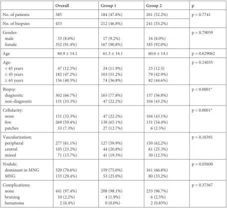
Inadequate specimens are treated as non-di-agnostic or unsatisfactory and account for 2–20% of all specimens obtained during UG-FNAB pro-cedures [8, 25]. The authors do not say if there Table 1. Characteristics of patients with UG-FNAB performed with (Group 1) and without (Group 2) radiologist’s assis-tance. Descriptive data are presented as numbers (n) and percent (%) or the mean + standard deviation (+ SD)
Overall Group 1 Group 2 p
No. of patients 385 184 (47.8%) 201 (52.2%) p = 0.7741
No. of biopsies 453 212 (46.8%) 241 (53.2%)
Gender: male
female 33 (8.6%)352 (91.4%) 17 (9.2%)167 (90.8%) 16 (8.0%)185 (92.0%)
p = 0.79059
Age 60.9 ± 14.1 61.3 ± 14.1 60.6 ± 14.1 p = 0.629062
Age: < 45 years ≥ 45 years ≥ 65 years
47 (12.2%) 182 (47.2%) 156 (40.5%)
24 (11.9%) 103 (51.2%) 74 (36.8%)
23 (12.5) 79 (42.9%) 82 (44.6%)
p = 0.24035
Biopsy: diagnostic
non-diagnostic 302 (66.7%)151 (33.3%) 165 (77.8%) 47 (22.2%) 137 (56.8%)104 (43.2%)
p < 0.0001*
Cellularity: none few patches
151 (33.3%) 269 (59.4%) 33 (7.3%)
47 (22.2%) 138 (65.1%) 27 (12.7%)
104 (43.1%) 131 (54.4%) 6 (2.5%)
p < 0.0001*
Vascularization: peripheral central mixed
277 (61.1%) 105 (23.2%) 71 (15.7%)
127 (59.9%) 44 (20.8%) 41 (19.3%)
150 (62.2%) 61 (25.3%) 30 (12.5%)
p = 0.10391
Nodule:
dominant in MNG
MNG 320 (70.6%)133 (29.4%) 159 (75.0%) 53 (25.0%) 161 (66.8%) 80 (33.2%)
p = 0.05600
Complications: none bruising hematoma
441 (97.4%) 10 (2.2%) 2 (0.4%)
208 (98.1%) 4 (1.9%) 0 (0.0%)
233 (96.7%) 6 (2.5%) 2 (0.83%)
p = 0.37367
is any correlation between inadequate speci-mens and the type of specialist performing it or the number of physicians doing this procedure. In our study we observed 22.2% of non-diagnos-tic specimens in the group where UG-FNAB pro-cedures were performed with a radiologist’s assis-tance (group 1) and approximately two times more (44.6%) in the group without a radiologist’s assis-tance (group 2). This difference was statistically significant (p < 0.0001).
Some authors have tried to explain the rel-atively high amount of non-diagnostic results. They have noticed some especially important fac-tors that influence the non-diagnostic number of FNAB results. They pointed to the skill of the phy-sician performing the procedure, thyroid nodule vascularity, criteria for qualification of the speci-men as suitable for cytologic examination and the
fluid component of the nodule [26]. In our study, the types of vascularization in both groups did not differ from each other and did not have any in-fluence on the quality of specimens (p = 0.10391).
It is estimated that the diagnostic smear should include at least 6 clusters of well-preserved cells with each group containing at least 10–15 cells. In our study the assistance of a radiologist during the UG-FNAB procedures (group 1) significant-ly increased the cellularity in this group (66.7% vs. 56.9%, p < 0.0001). The percentage of specimens which did not contain any thyroid cells was two times higher in the group of patients in whom UG- -FNAB procedures were performed without a radi-ologist’s assistance. It is worth noticing that non-di-agnostic smears may be a significant problem in the
aspect of thyroid carcinoma prevalence, as a malig-nancy rate up to 7% was reported in patients with initial unsatisfactory or non-diagnostic smears [27].
In 2006, the American Association of Clinical Endocrinologist (AACE) and Associazione Medici Endocrinologi (AME) published the guidelines for performing FNAB to improve the adequacy and ac-curacy of cytologic specimens [8]. They suggest that FNAB should be performed by a physician trained in both thyroid gland examination and biopsy. The material should be obtained from various parts of the nodule and especially peripheral areas of low vascularity should be biopsied. The histopathol-ogist who interprets the specimens should be ex-perienced in thyroid gland cytology. A number of studies have suggested some following steps to re-duce the number of non-diagnostic results of UG- -FNAB [8, 19, 21]. For example, they recommend
Fig. 1a. UG-FNAB in a 54-year-old woman. Procedure performed with radiologist’s assistance. Transverse sonogram of the right lobe of the thyroid demonstrates a solid nodule with a hypoechoic, colloid foci
rebiopsy if the cytology is non-diagnostic and have 2–4 aspirates going from one nodule.
UG-FNAB cannot be treated as a screening test for all thyroid nodules in the population, but should be used as an initial diagnostic tool for all patients with palpable nodule or with suspicion of malignancy. Several ultrasonographic charac-teristics should be taken into consideration while choosing the area of the nodule for biopsy, to re-ceive a higher diagnostic value of UG-FNAB, in-cluding: microcalcifications, hypoechogenicity, ir-regular margins and low intranodule vascularity. It is confirmed that ultrasonography used as guid-ance for FNAB significantly increases the sensitiv-ity, positive predictive value and negative predic-tive value [15], however, no information regarding if a radiologist’s assistance in the UG-FNAB pro-cedure increases the value of the examination was given. In our opinion, this information is especially
interesting considering the fact that UG-FNAB re-sults are operator-dependent [8, 26].
As sufficient experience is recommended to carry out the procedure, some authors suggest that at least 1 to 5 aspirations per month should be do-ne by a UG-FNAB-performing, “dedicated”, physi-cian [29]. However, FNAB does not have to be con-fined only to large medical centers. Good results of this procedure might also be obtained in commu-nity and rural hospitals, if the number of aspirators and cytopathologists in one center is not large [30].
The other argument for the presence of the ra-diologist during the UG-FNAB procedure may be safety. Although FNAB is a minimally invasive pro-cedure, some complications may occur,
fortunate-ly rarefortunate-ly. Bleeding or hematoma are almost always self-limiting, when gentle but firm pressure is ap-plied to the aspiration site. During our study, hema-toma developed in only two patients, and both were in the group where the UG-FNAB procedure was performed without a radiologist’s assistance. Those hematomas did not lead to tracheal compression. Those unfortunate incidences happened in the very beginning of our study, pointing out once again the importance of the UG-FNAB team’s experience. We cannot be sure, however, if the presence of a radiol-ogist would have prevented those complications.
It appears that radiologists can point to some areas for FNAB which in their opinion are more suitable for obtaining diagnostic material and in some cases are more suspected for malignancy. In our study, the radiologist’s assistance during UG- -FNAB performed by a surgeon led to the iden-tification of unique and ultrasonography-specific
Fig. 2a. UG-FNAB in a 55-year-old woman. Procedure performed without radiologist’s assistance. Transverse sonogram of the left lobe of the thyroid demonstrates a partially cystic tumor with solid internal projections
areas of the thyroid nodules which caused a bet-ter quality of the specimens for further cytologic examination.
UG-FNAB of thyroid nodules performed with a radiologist’s assistance makes it possible to
obtain more valuable specimens, which may im-prove the diagnostic accuracy in the preoperative management of thyroid pathology. UG-FNAB per-formed with a radiologist’s assistance may increase the safety of this procedure.
References
[1] Pfestroff A, Luster M: How are thyroid nodules diagnosed? Dtsch Med Wochenschr 2015, 140, 565–572. [2] Baier ND, Hahn PF, Gervais DA, Samir A, Halpern EF, Mueller PR, Harisinghani MG: Fine-needle aspiration
biopsy of thyroid nodules: experience in a cohort of 944 patients. AJR Am J Roentgenol 2009, 193, 1175–1179. [3] Yan Z, Yu-Kun L, Jie T, Min LI, Zhi-Li W, Quan W: Clinical value of ultrasonography in diagnosing diffuse
thy-roid diseases accompanied with suspicious nodules. Zhongguo Yi Xue Ke Xue Yuan Xue Bao 2015, 37, 290–293. [4] Zhou L, Xu D, Chen L: Uni- and multi-variate analyses for ultrasonic signs of smaller than 5mm nodules of
mul-tifocal papillary thyroid carcinoma. Zhonghua Yi Xue Za Zhi 2015, 95, 917–919.
[5] Kim SK, Lee JH, Woo JW, Park I, Choe JH, Kim JH, Kim JS: Prediction table and nomogram as tools for diagno-sis of papillary thyroid carcinoma: combined analydiagno-sis of ultrasonography, fine-needle aspiration biopsy, and BRAF V600E mutation. Medicine 2015, 94, 760.
[6] Cortázar García R, Quirós López R, Acebal Blanco MM: The role of radiologists in the management of thyroid nodules. Radiologia 2008, 50, 471–480.
[7] Syrenicz A, Koziołek M, Ciechanowicz A, Sieradzka A, Bińczak-Kuleta A, Parczewski M: New insights into the diagnosis of nodular goiter. Thyroid Res 2014, 7, 6.
[8] AACE/AME Task Force on Thyroid Nodules. American Association of Clinical Endocrinologists and Associazione Medici Endocrinologi medical guidelines for clinical practice for the diagnosis and management of thyroid nod-ules. Endocr Pract 2006, 12, 63–102.
[9] Hegedus L: Clinical practice: the thyroid nodule. N Engl J Med 2004, 351, 1764–1771.
[10] Frates MC, Benson CB, Charboneau JW, Cibas ES, Clark OH, Coleman BG, Cronan JJ, Doubilet PM, Evans DB, Goellner JR, Hay ID, Hertzberg BS, Intenzo CM, Jeffrey RB, Langer JE, Larsen PR, Mandel SJ, Middleton WD, Reading CC, Sherman SI, Tessler FN: Management of thyroid nodules detected at US: Society of Radiologists in Ultrasound consensus conference statement. Radiology 2005, 237, 794–800.
[11] Nachiappan AC, Metwalli ZA, Hailey BS, Patel RA, Ostrowski ML, Wynne DM: The thyroid: review of imaging features and biopsy techniques with radiologic-pathologic correlation. Radiographics 2014, 34, 276–293.
[12] Kovacevic DO, Skurla MS: Sonographic diagnosis of thyroid nodules: correlation with the results of sonographi-cally guided fine-needle aspiration biopsy. J Clin Ultrasound 2007, 35, 63–67.
[13] Tay SY, Chen CY, Chan WP: Sonographic criteria predictive of benign thyroid nodules useful in avoiding unnec-essary ultrasound-guided fine needle aspiration. J Formos Med Assoc 2015, 114, 590–597.
[14] Yoon JH, Lee HS, Kim EK, Moon HJ, Kwak JY: A nomogram for predicting malignancy in thyroid nodules diagnosed as atypia of undetermined significance/follicular lesions of undetermined significance on fine needle aspiration. Surgery 2014, 155, 1006–1013.
[15] Iannuccilli JD, Cronan JJ, Monchik JM: Risk for malignancy of thyroid nodules as assessed by sonographic crite-ria: the need for biopsy. J Ultrasound Med 2004, 23, 1455–1464.
[16] Ugurlu S, Caglar E, Yesim TE, Tanrikulu E, Can G, Kadioglu P: Evaluation of thyroid nodules in Turkish popu-lation. Intern Med 2008, 47, 205–209.
[17] Martin HE, Ellis EB: Biopsy by needle puncture and aspiration. Ann Surg 1930, 92, 169–181.
[18] Santos JE, Leiman G: Nonaspiration fine needle cytology: application of a new technique to nodular thyroid dis-ease. Acta Cytol 1988, 32, 353–356.
[19] Belfiore A, La Rosa GL: Fine-needle aspiration biopsy of the thyroid. Endocrinol Metab Clin North Am 2001, 30, 361–400.
[20] Hales MS, Hsu FS: Needle tract implantation of papillary carcinoma of the thyroid following aspiration biopsy. Acta Cytol 1990, 34, 801–804.
[21] Hamburger JI: Diagnosis of thyroid nodules by fine needle biopsy: use and abuse. J Clin Endocrinol Metab 1994, 79, 335–339.
[22] Krishnappa P, Ramakrishnappa S, Kulkarni MH: Comparison of free hand versus ultrasound-guided fine needle aspiration of thyroid with histopathological correlation. J Environ Pathol Toxicol Oncol 2013, 32, 149–155. [23] Papini E, Pacella CM, Hegedus L: Diagnosis of endocrine disease: thyroid ultrasound (US) and US-assisted
pro-cedures: from the shadows into an array of applications. Eur J Endocrinol 2014, 170, 133–146.
[24] Baloch ZW, LiVolsi VA: Fine-needle aspiration of thyroid nodules: past, present, and future. Endocr Pract 2004, 10, 234–241.
[25] Gharib H, Goellner JR: Fine-needle aspiration biopsy of the thyroid: an appraisal. Ann Intern Med 1993, 118, 282–289.
[26] Schmidt T, Riggs MW, Speights VO: Significance of nondiagnostic fine-needle aspiration of the thyroid. South Med J 1997, 90, 1183–1186.
[28] Caleo A, Vigliar E, Vitale M: Cytological diagnosis of thyroid nodules in Hashimoto thyroiditis in elderly patients. BMC Surg 2013, 13, 41.
[29] Suen KC: Fine-needle aspiration biopsy of the thyroid. CMAJ 2002, 167, 491–495.
[30] Pepper GM, Zwickler D, Rosen Y: Fine-needle aspiration biopsy of the thyroid nodule. Arch Intern Med 1989, 149, 594–596.
Address for correspondence:
Krzysztof Kaliszewski
1st Department and Clinic of General,
Gastroenterological and Endocrine Surgery Wroclaw Medical University
Skłodowskiej-Curie 66 50-369 Wrocław Poland
Tel: +48 71 784 21 62
E-mail address: [email protected]
Conflict of interest: None declared