NEUROTROPHIN-CHEMOKINE RECEPTOR INTERACTIONS ON MACROPHAGES REGULATE HIV NEUROPATHOGENESIS
Kimberly Williams
A dissertation submitted to the faculty of the University of North Carolina at Chapel Hill in partial fulfillment of the requirements for the degree of Doctor of Philosophy in the
Curriculum of Neurobiology in the School of Medicine.
Chapel Hill 2015
Approved by: Rick Meeker Joseph Eron
Mohanish Deshmukh Kenneth McCarthy
ABSTRACT
Kimberly Williams: Neurotrophin-chemokine receptor interactions on macrophages regulate HIV neuropathogenesis
(Under the direction of Rick Meeker)
Invading perivascular macrophages and microglial cells play a pivotal role in the neuropathogenesis of Human Immunodeficiency Virus (HIV) associated neurological disorders. As with many neurodegenerative diseases, the activation of macrophages and microglia cells in the central nervous system (CNS) leads to the secretion of unknown neurotoxins and is thought to contribute heavily to neuronal damage. However, these cells are very dynamic with a wide range of functions including neuroprotection and repair. It has become increasingly clear that strategies to modulate specific phenotypes of these cells have substantial therapeutic potential. This thesis has identified a novel neurotoxic phenotype in human macrophages that extends beyond the classical M1 inflammatory or M2
the neurotoxic phenotype while mature NGF created a more neuroprotective phenotype which was dependent on TrkA. The neurotoxic phenotype was also driven by HIV in a p75NTR and CXCR4 dependent manner. Neurotrophin signaling was able to modulate the HIV neurotoxic phenotype by exacerbating neurotoxin production (proNGF) or partially restoring the non-toxic phenotype (NGF). This thesis for the first time identified an interaction between the neurotrophin receptors p75NTR and TrkA and the HIV co-receptor CXCR4 through the use of co-immunoprecipitation techniques, which may be involved in the regulation of neurotoxic phenotypes. NGF co-stimulation with HIV increased CXCR4 phosphorylation and association with G-protein receptor kinase 2 (GRK2). This contrasted with the decrease in GRK2/CXCR4 complexes seen with HIV stimulation alone.
Phosphorylated CXCR4 (pCXCR4) was found in overlapping domains with p75NTR and TrkA, which was downregulated by HIV. NGF was able to restore the p75NTR/pCXCR4 co-localization and partially restored pCXCR4/TrkA co-co-localization. This data indicated that NGF suppressed the HIV neurotoxic phenotype by facilitating phosphorylation of the HIV co-receptor CXCR4 while proNGF enhanced HIV effects. Overall, these studies have identified novel effects of the neurotrophins on macrophages that may provide new
Dedicated to:
ACKNOWLEDGEMENTS
I would first like to thank my mentor Dr. Rick Meeker. During the past 6 years, Rick has been a constant supporter of all of my endeavors. I have truly been fortunate to have hands on training from a great scientist while also being encouraged to be involved in other activities outside of the lab. Rick selfless personality shined through during his willingness to stay late to help me with grants and preparation for conferences. He has truly been a great collaborator on this dissertation and has also taught me many things outside of lab from baking to exposing me to new cultures. I cannot begin to thank Rick for everything he has done for me over the years.
PREFACE
To go through life and get through grad school, a strong community is necessary. Though these people did not have a direct impact on the studies in this dissertation they have helped shape me in to the woman I am today , for this I thank them.
I would like to that Dr. Pat Phelps. Without Pat, my life would be completely different today. I remember our first one on one conversation where Pat was honest and straight forward with me regarding my options for graduate school and what it would take to reach my career goals. Because of this conversation, I have vowed to be honest, available and straight forward with students that I come in contact with who share similar experiences and backgrounds as myself. One of the major lessons learned from Pat is to find opportunities that coincide with my personality and that will truly make me happy. I admire Pat’s free spirit and her ability to try new endeavors (And master all of them). I aspire to be as fearless and resilient as Pat one day and am so glad to have her in my life.
Benjamin Philpot, my first mentor at UNC has taught me so much over the year. Because of him I now project and command a room much better than the girl that began in his lab in 2008. Throughout the years I have been able to really count on Ben for
encouragement and support in all of my endeavors. He has always been a floor and email away when I needed him and I truly appreciate everything he has done to support me over the years.
Arriving at UNC, I had no friends or close family in the area. The people in the Philpot lab became my first friends. Rylan Larsen, Thorfinn Riday and Maile Henson truly made the transition much more enjoyable for myself. I knew throughout the years that I could always count on them for a good laugh, all of the neurobiology gossip and any late night help (Thorfinn). Maile in particular became a cool mother figure for me that taught me not only how to be a rock star western blotting queen but also how to stay grounded and true to my values. Throughout my scientific career I will never forget her words of advice, keep your friends that are outside of science close because they will help keep life into perspective and although many around you may not believe hold true to your own belief system because though we are researchers, God is only allowing us to see small pieces of the puzzle through our research.
I would also like to thank the many other friends I have made during my graduate career. Kenton Woodard and Reginald Cannady have been true backbones for me during my graduate career. Kenton and I took every class that I had at UNC. We studied together, we encouraged each other and have become lifelong friends. I know great thing will come from him in the future and I just cant wait to see it. This is also true for Reggie. If it was not for Reggie to prep and encourage me through the long check list that is graduate school, I would have been forced to figure it out on my own. His support and friendship has truly been a blessing during this time. Also my grad school experience was balanced thanks to a group of girls that I call my friends. Grace Silva, Samira Brooks and Tikvah Hayes have been a joy to get to know. We have been there to be a listening ear and the turn up crew when needed. I have enjoyed getting to know these girls and look forward to many more turn ups!
God assigns you some family members at birth and then allows you to think you are picking some throughout life. In my almost 29 years I have gained a family that are often times more present and supportive than my blood relatives and for that I am thankful. To my two childhood best friends, Breauna Moore and Ashley Phillips, you were the sisters that I didn’t have from the age of 5. Even though we haven’t been in constant communication over these last years, you two will always hold a special place in my heart. Thank you for
everything from teaching me how to ride a bike to how to be a true friend so early in life. My high school best friends, Jennifer Lowe and Debrienne Robinson, words can not how much your love and support has meant to me over the years. Thank you for calling, encouraging and supporting me all these years. Debee thank you for joining me on conferences so that I wouldn’t have to travel alone and always having my best interest at heart during scholastic endeavors and life events.
Courtney Taylor, thank you for always being just a drive away when I need you. I don’t know what I would have done during those times where I needed to get away if it had not been for you. Our countless weekend trips were really necessary to balance the stresses of graduate school.
Shannan Tonette, we went through grad school together and I could not think of a better person to have gone through it with. Form our daily morning conversations to the many conferences that we attended together having you along for the ride has made this all the much better. I think one day you will forgive me but if it wasn’t for grad school we would have never found our favorite city and acquired this love for beignets. I cant wait to meet my newest baby, Ms. Aadyn Payne!!
Curtis Cain, how far we have come and this is only just the beginning. You have been the one that I can always count on and I cannot begin to explain how appreciative I am to have you in my life. I knew at a very young age that you were something special and that as a team we are unstoppable. I can not wait to see what we are about to do next. I love you forever and always.
To my family, thank you for all of your love support and prayers.
To my grandmother, I know that you are proud of me and have been rooting me on and probably helping out along the way. I miss you and love you everyday.I hope that I continue to make you proud.
To my brother, Brandon, the sky is the limit and I can wait to see you reach it. You have so much potential and I hope that I have been your encouragement to take on this world and be the best Brandon that you can be. I love you more than you may ever realize and I am glad God gave me you!
Mommy.
TABLE OF CONTENTS
ABSTRACT ... III
ACKNOWLEDGEMENTS ... VI
PREFACE ... VIII
LIST OF TABLES ... XV
LIST OF FIGURES ... XVI
LIST OF ABBREVIATIONS ... XIX
CHAPTER 1: INTRODUCTION ... 1
Inflammation in the Central Nervous System ... 1
Normal composition of Immune cells in the Central Nervous System ... 2
Immune surveillance in the CSF ... 3
Macrophage/Microglia activation and its role in Neuroinflammation ... 4
General overview of HIV infection ... 7
History of HIV: a public health problem ... 7
HIV Infection ... 8
Structure of the HIV Virion ... 9
HIV binding and entry into host cell ... 10
Integration into the genome and viral replication ... 11
Replication and mutations ... 12
HIV in the CNS ... 13
HIV in the CNS before the era of HAART ... 13
After the era of HAART ... 14
Target cells in the central nervous system ... 14
HIV entry into the central nervous system ... 15
Mechanisms of Neuropathogenesis ... 16
Neuroprotection ... 19
Mechanisms of neuroprotection and animal models ... 19
Neurotrophin Signaling ... 24
Functional studies of neurotrophin receptors on macrophages ... 30
CHAPTER 2: DIFFERENTIAL REGULATION OF MACROPHAGE PHENOTYPE BY MATURE AND PRO-NERVE GROWTH FACTOR ... 32
Introduction ... 32
Materials and Methods ... 34
Results ... 44
Neurotoxic activity of the hMDM ... 81
Discussion ... 83
Conclusions ... 92
CHAPTER 3: NEUROTROPHINS MODULATE MACROPHAGE RESPONSES TO HIV ... 93
Introduction ... 93
Materials and Methods ... 95
Results ... 105
Discussion ... 140
CHAPTER 4: NEUROTROPHIN-CXCR4 INTERACTIONS REGULATE THE TOXIC ACTIVATION OF MACROPHAGES BY HIV ... 148
Introduction ... 148
Materials and Methods ... 150
Results ... 158
Discussion ... 172
DISCUSSION ... 175
Summary of findings ... 175
Discussion ... 178
LIST OF TABLES
Table 1.1. Soluble protein changes in macrophage conditioned medium in
response to NGF or proNGF. ... 74
Table 2.1 Soluble protein changes in macrophage conditioned medium in
LIST OF FIGURES
Figure 1.1. Varied appearance of human monocyte-derived macrophages
(hMDM) in culture. ... 45
Figure 1.2. Neurotrophin receptors p75NTR and TrkA were concentrated in
discrete domains on human macrophages... 48
Figure 1.3. Neurotrophins promote cell survival in hMDMs. ... 52
Figure 1.4. Neurotrophin receptor expression on hMDMs and monocytes
quantified by flow cytometry. ... 54
Figure 1.5. Monocytes expressed high and low levels of p75NTR and TrkA. ... 55
Figure 1.6. Neurotrophin Receptors p75NTR and TrkA are co-localized to the
same microdomains on hMDMs ... 57
Figure 1.7. Sortilin Receptors are expressed and co-localize with p75NTR on
hMDMs. ... 59
Figure 1.8. Morphological changes in hMDMs are regulated by
neurotrophins. ... 62
Figure 1.9. Functional changes by NGF and proNGF. ... 65
Figure 1.10. Mature NGF and proNGF have different effects on macrophage
calcium activity. ... 68
Figure 1.11. Changes in TGF-β family proteins in macrophage conditioned
medium induced by NGF or proNGF. ... 76
Figure 1.12. Changes in angiogenesis family proteins in macrophage
conditioned medium induced by NGF or proNGF. ... 77
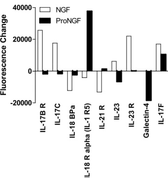
Figure 1.13. Changes in IL-17 family proteins in macrophage conditioned
Figure 1.14. Changes in MMP family proteins in macrophage conditioned
medium induced by NGF or proNGF. ... 79
Figure 1.15. Release profile of proteins typically associated with M1 (inflammatory) or M2 (anti-inflammatory) macrophage polarization in response to NGF or proNGF. ... 80
Figure 1.16. Mature and proNGF have opposing effects on the secretion of neurotoxic factors by hMDM... 82
Figure 2.1 .TrkA and p75NTR interactions are increased by HIV in hMDMs ... 107
Figure 2.2. HIV increases Sortilin/p75NTR interaction without altering sortilin expression . ... 109
Figure 2.3. HIV does not cause cell death in hMDMs... 111
Figure 2.4. HIV and neurotrophins altered the pattern of calcium responses in human monocyte-derived macrophages (hMDM). ... 114
Figure 2.5. Podosome expression and calcium activity in hMDM. ... 118
Figure 2.6. HIV increases migration in hMDMs but not phagocytosis ... 120
Figure 2.7 HIV increased neurotoxicity is increased by proNGF and partially suppressed by NGF ... 123
Figure 2.8. TrkA and Membrane Ruffling blocks Neurotoxin formation. ... 124
Figure 2.9. Changes in secretory profiles induced by HIV... 134
Figure 2.10. Proteins that were increased by HIV and correlated with toxicity. ... 135
Figure 2.11. Proteins which were decreased by HIV and also correlated with toxicity. ... 136
Figure 2.12. Effects of NGF in the absence of significant effects of HIV. ... 137
Figure 2.13. HIV decreases the M2 surface marker CD206. ... 139
Figure 3.2 CXCR4 but not CCR5 suppressed ruffles in hMDMs ... 161
Figure 3.3 CXCR4 signaling in necessary for calcium spiking. ... 163
Figure 3.4 NGF induced phosphorylation of CXCR4 in response to HIV. ... 165
Fig 3.5 CXCR4 interacts with neurotrophin receptors. ... 167
Figure 3.6 CXCR4 forms complexes with neurotrophin Receptors, ... 169
LIST OF ABBREVIATIONS
ABI 7500 Applied biosystems (PCR system)
aCSF artificial cerebrospinal fluid
ADC AIDS Dementia Complex
AIDS Acquired Immune Deficiency Syndrome
ANI asymptomatic Neurocognitive Impairment
ANOVA Analysis of variance
ART Antiretroviral Therapy
B27 Gibco B-27 supplement
BCA Bicinchoninic acid
BDNF Brain derived neurotrophic factor
BSA Bovine serum albumin
CART Combined Antiretroviral therapy
CCR5 C-C Chemokine Receptor type 5
CD Cluster of Differentiation
CD4 Cluster of Differentiation 4
CNS Central Nervous System
CRAC Calcium release activated channel
CSF Cerebrospinal fluid
CXCR4 C-X-C Chemokine Receptor type 4
DAVID
Database for Annotation, Visualization and Integrated
DMEM Dulbecco’s modified Eagle medium
FBS Fetal Bovine Serum
FGF Fibroblast growth factor
GADPH glyceraldehyde 3-phosphate dehydrogenase
GDF Growth differentiation factor
GM-CSF Granulocyte macrophage colony-stimulating factor
Gp120 Glycoprotein 120 (HIV envelope protein, 120 kDa)
GPCR G-protein coupled receptor
GRK2 G-protein coupled receptor kinase 2
HAART Highly active antiretroviral therapy
HAD HIV Associated Dementia
HAND HIV Associated Neurocognitive Disorder
HBSS Hank’s balanced salt solution
HEPES 4-(2-Hydroxyethyl)piperazine-1-ethanesulfonic acid
HIV Human Immunodeficiency Virus
hMDM Human monocyte-derived macrophage
HRP Horseradish peroxidase
IL Interleukin
IP (10) Interferon gamma-induced protein
LPS Lipopolysaccharide
MCM Macrophage conditioned medium
MEM Modified Eagle medium
MMP Matrix metalloproteinase
MND Minor neurocognitive Disorder
NGF Nerve growth factor
NT-3 Neurotrophin 3
NT-4 Neurotrophin 4
NTR Neurotrophin Receptor
p75NTR p75 neurotrophin receptor
PBMCs Peripheral Blood Mononuclear Cells
PBS Phosphate buffered saline
pCXCR4 phosphorylated CXCR4
PMSF Phenylmethylsulfonyl fluoride
proBDNF pro Brain derived neurotrophic factor
proNGF proNerve growth factor
RIPA Radio-immunoprecipitation assay
SDF-1 Stromal Derived Factor-1
SDS Sodium dodecyl sulfate
TGF Transforming growth factor
TIMP-1 Tissue inhibitor of metalloprotease 1
TrkA Tropomyosin Receptor Kinase A
TrkB Tropomyosin Receptor Kinase B
CHAPTER 1: INTRODUCTION
Inflammation in the Central Nervous System
The central nervous system (CNS), consisting of the brain and spinal cord, was once thought to be an immune privileged environment protected from debris and systemic immune cell infiltration by the blood brain and blood cerebrospinal fluid barriers. However, we now know that there is constant communication between the immune system and the central nervous system to ensure proper homeostasis and signaling in the brain. The introduction of external stressors such as injury and infection can lead to inflammation in the brain known as neuroinflammation [1]. Short periods of neuroinflammation can be beneficial in the central nervous system, however, prolonged and excessive activation of these immune responses can cause chronic inflammation that leads to neuronal damage and loss[2]. Excessive
inflammation can take many forms and is often seen in neurodegenerative diseases[3]. This thesis will focus on inflammation resulting from HIV infection that underlies the
development of HIV Associated Neurocognitive Disorders (HAND). Understanding how to control inflammation in the central nervous system is important in creating new therapeutic avenues for HIV neuropathogenesis, and a better understanding of these processes is a major goal of this dissertation. The following section reviews the normal composition and functions of immune cells in the central nervous system, the major immune cells in the brain,
Normal composition of Immune cells in the Central Nervous System
Under normal conditions, immune cell infiltration into the brain is highly restricted by the blood brain barrier resulting in a vastly different composition of immune cells inside the brain compared to other organs. For example, the population of T cells in the brain parenchyma is one tenth of its appearance in other organs. Immune surveillance in the brain is mediated primarily by microglial cells, also known as the resident macrophage of the brain. Microglia/macrophages are a part of the mononuclear phagocyte group and are among the first cells to respond to injury and infection during innate immunity [2,4]. Microglia represent 0.5%-16.6% of cells present in the brain and are found in higher quantities in white matter regions compared to grey matter regions [5]. Microglia cells are normally tightly controlled quiescent cells in the central nervous system with distinct roles in maintaining a healthy environment. These roles include surveillance of the microdomain, removal of debris and noxious agents through phagocytosis, modulation of synaptic connections and plasticity, communication with other brain cells and responding to infectious and toxic agents[6].
Astrocytes, the most abundant glial cells in the CNS, also contribute to innate
immunity, though to a much lesser extent. Astrocytes are found throughout the brain in both white and grey matter regions. Under normal conditions astrocytes support neuronal
function, secrete neurotrophic factors and aid in neural activity and homeostasis. Physically, astrocytes influence the formation and maintenance of the blood brain barrier by contributing to its structural and functional integrity[7]. While they are not classical immune cells,
astrocytes respond to insult and injury by changing cell morphology resulting in cell
hypertrophy and scar formation referred to as astrogliosis. Astrocytes are also able to secrete pro-inflammatory cytokines and complement proteins. Though astrogliosis has been
HIV associated cognitive disorders, it will not be a focus of this dissertation.
Immune surveillance in the CSF
Though immune cell populations are low within the parenchyma of the brain, constant surveillance and communication of the central nervous system is always occurring by systemic immune cells located in cerebrospinal fluid (CSF) and specialized structures just outside the CNS proper. Studies have shown that approximately 450,000 white blood cells are normally found within the CSF of healthy adults [8,9]. The composition of these cells is quite different from blood indicating that these cells are highly selected for entry into the brain compartment. Though there are reduced numbers of T cells in the brain, T cells can traffic into the CSF to survey and elicit signals to the central nervous system through interactions with ependymal and epithelial cells that line the ventricles and choroid plexus respectively[10]. Flow cytometry studies have shown that the T cells found in human and mouse CSF were CD4+, activated central memory (CD45RA-/CD27/28+) T cells, suggesting that they were recruited into the central nervous system, possibly through the choroid plexus or meninges[11]. T cells are thought to make the greatest contribution to HIV in the CSF, although the interactions of virus with cells in the parenchyma and the contributions to associated cognitive disorders are unclear.
during aging and in response to adverse conditions like infection. Varvel et al. has shown that after experimental depletion of microglia from mice, the microglia were replenished by engrafted peripheral blood derived monocytes entering the brain from small blood vessels and capillaries[13]. Similarly in a Simian Immunodeficiency Virus (SIV) animal model for neuroAIDS, labeled monocytes in bone marrow were able to travel into brain tissues more readily than in uninfected macaques [14]. Other studies, however, have tried to determine if the expansion of microglia in the parenchyma of the brain during chronic and
neurodegenerative disease were from infiltrating perivascular macrophages resembling microglia or proliferating microglial cells [15,16] and determined that the populations represented a microglia expansion.
Macrophage/Microglia activation and its role in Neuroinflammation
Neuroinflammation has been widely implicated as a pivotal event contributing to neurodegeneration. Though many neurodegenerative diseases have different causative etiologies, there are common pathologies seen among these diseases. Of importance, macrophage activation, permeability of the blood brain barrier, infiltration of systemic macrophages and secretion of neurotoxins have been noted in neurodegenerative diseases such as Alzheimer disease and HIV associated dementia (HAD)[17-20]. Though the
mechanisms for these common pathologies are not fully understood, it is well accepted that macrophage activation is a key step.
mediate functional changes in macrophages including morphology, migration, phagocytosis, cytokine secretion and antigen presentation. Though it was thought that macrophages were only able to produce pro-inflammatory factors, being one of the most secretory cells in the body, macrophages are capable of releasing factors needed in homeostasis, inflammation and host defense responses under the control of multiple mediators. Macrophage activation phenotypes are transient and reversible depending on cues received from the
microenvironment and range from strong inflammatory responses designed for elimination of invading pathogens to anti-inflammatory, protective and wound healing activities essential for tissue repair. This versatility in functions has led to the characterization of two polarized activation states, classical activation or M1 and alternative activation or M2 [21-23]. Reports have suggested that macrophages may require at least two signals to achieve polarization [24].
Classically activated inflammatory macrophages have been shown to arise from type 1 cytokines such as interferon gamma (IFN-γ), tumor necrosis factor alpha (TNF-α), and
lipopolysaccharide (LPS) stimulation. M1 induction can also occur after recognition of pathogen-associated molecular patterns (PAMPS) and Damage- associated molecular
patterns (DAMPS) on foreign or damaged agents such as viruses, bacteria and dead cells. M1 macrophages also express increased MHC class I and II receptors and secrete complement factors necessary for complement-mediated phagocytosis[25]. Activation of M1
macrophages leads to secretion of pro-inflammatory cytokines and chemokines such as IL-12, IL-23, TNF-α, IL-1, and IL-6 and production of reactive oxygen species which damage proteins.
activation state of macrophages leading to the secretion of anti-inflammatory cytokines, chemokines, growth factors and other reparative factors[26]. M2 has been sub-divided by some into 3 groups M2a, M2b and M2c [23]. M2a is induced by IL-4 and IL-13, while M2b is induced by immune complexes in combination with IL-1β or LPS and M2c is induced by IL-10, TGF-β or glucocorticoids. These activation categories however are overly simplistic and often a hurdle to understanding the wide range of phenotypes of macrophages, as many studies fail to go beyond a characterization of the classical cytokines. Hyperresponsiveness of classically activated macrophages can lead to tissue damage while exaggerated wound healing properties have been shown to increase infections in severe burn patients indicating that a balance of activities must be maintained. Understanding how these activation states may intertwine to create the proper balance in macrophage function is important for the development of precisely targeted macrophage therapies in the future.
M1 phenotypes such as increased secretion of proinflammatory factors and upregulation of the cell surface marker, CD16[8,17,29-31]. This inflammatory activation is associated with neuronal damage and a gradual progressive degeneration of the CNS without appreciable cell death. This suggests that damage caused by microglial/macrophage activation may be
reversible and interventions that restore the balance of microglia activation states may be fruitful therapeutic options for HIV associated disorders. This thesis aims to identify novel signaling interactions capable of establishing favorable macrophage phenotypes that prevent HIV neuropathogenesis.
General overview of HIV infection
In order to understand how to control the HIV-associated inflammatory cascade responsible for HIV associated cognitive disorders, it is important to understand the general nature of the virus and how it infects and interacts with its host cells.
History of HIV: a public health problem
Human immunodeficiency virus (HIV) is an epidemic affecting an estimated 1.2 million Americans in the United States and is predicted to have 50,000 new cases each year (http://www.cdc.gov/hiv/statistics/basics/ataglance.html ). HIV is transmitted through bodily fluids such as mucosal barriers and blood. The most common form of transmission is through sexual transmission, and HIV is the number one deadliest sexually transmitted disease. The first cases of HIV described in the 1980’s were 5 homosexual men presenting with
began to surface, negating these theories [32,33]. HIV quickly became an epidemic because we now know the early stages of disease present with mild symptoms, resulting in the transmission of virus from patients unaware that they were infected. To this day, 1 in 7 people infected with HIV are unaware of their status, which contributes to the continuous rise in infection. Though patients presented with various opportunistic infections, they all shared one common hallmark, depletion in CD4+ T cell counts that led to a decline in the patient’s immune system. In 1983 HIV was discovered as the virus that caused Acquired Immune Deficiency Syndrome or AIDS [34,35].
HIV Infection
symptomatic infection in 10-20% of patients. With severe HIV infection this increases to 60% of patients with clinically relevant CNS manifestation. Approximately 30-40% showed signs of dementia. Once CD4+ T cell counts fall below 200 cells per milliliter, patients are diagnosed with AIDS. During the AIDS stage, patients are more likely to suffer from opportunistic infections and severe dementia, which leads to their demise.
Currently there is no cure for HIV and the current treatments include several classes of antiviral drugs used to block the main steps in replication: entry, fusion, reverse
transcription, proteolytic processing and integration. The first antiretroviral treatment was approved by the FDA in 1987, a nucleoside reverse transcriptase inhibitor, azidothymidine (AZT, Zidovudine). This drug was ineffective in suppressing long term viral replication. Since then it has been discovered that a combination of at least three antiretroviral therapies used to block different phases of HIV replication is needed to manage infection. This
strategy became known as highly active antiretroviral therapy (HAART), currently known as combination antiretroviral therapy (CART)[36]. The use of these drugs has led to longer life expectancy as patients have suppressed viral loads systemically and increased CD4+ T cell counts. However these drugs cannot effectively cross into the brain, where virus can persist. This poses a need for neuroprotective therapies in conjunction with standard CARTs. In order to achieve this we must first understand the nature of the virus and its interaction with the central nervous system.
Structure of the HIV Virion
HIV, a member of the lentivirus subgroup of retroviruses, is a round virion (100nm in diameter) containing a double stranded RNA genome in its core. HIV has an outer
necessary for fusion and entry into the host cell. The envelope protein which mediates infection is a 160 kDa glycoprotein known as gp160. The gp160 protein is cleaved and further glycosylation occurs in the Golgi to produce a 41 kDa transmembrane protein, gp41, and a 120 kDa surface envelope protein, gp120. Gp41 serves as the stalk for gp120. The gp41/gp120 complex forms oligomers of three molecules in the membrane and is necessary for virus entry into the host cell[37].
The viral core contains the double stranded RNA and proteins necessary for
replication and integration into the host genome. The HIV genome consists of 9 genes: the structural genes that encode cell surface proteins: Gag, Pol, and Env; the regulatory genes that control replication: Tat and Rev; and the accessory genes involved in virus replication and continued infection: Vpu, Vpr, Vif, and Nef. The core also contains reverse transcriptase, integrases, and proteases which are encoded by the pol gene and are necessary for viral integration into the host genome[38].
HIV binding and entry into host cell
Viral fusion and entry into the host cell is dependent on the interaction of the viral surface glycoproteins with two classes of surface receptors on the host cell, the primary receptor CD4 and the co-receptors, CXCR4 and CCR5. CD4 is a transmembrane protein consisting of 4 glycoprotein domains (D1-D4) that are in high abundance on T cells. Macrophages also express CD4 though in much lower quantities. CD4 expression is
tropism preferences for the co-receptors, which explain the ability of some viral strains to infect T cells (CXCR4 preferring) whereas other strains infect cells of monocyte lineage (CCR5 preferring). Although the strong infection preferences under some circumstances suggested that the preferences might be mutually exclusive, it is now recognized that both chemokine receptors are expressed on each cell type and viral infection by T cell tropic or macrophage tropic strains is not exclusive to their preferred receptors. In addition, there are also dual tropic viruses, CXCR4/CCR5 that can infect through both receptors. The
efficiency of entry is determined to a large degree by the abundance of CD4 in conjunction with either co-receptor[39].
HIV entry is achieved when the HIV surface protein, gp120 binds to CD4 causing a conformational change in the gp120 trimer allowing interactions with co-receptors CCR5 or CXCR4. Once gp120 binds to the co-receptor, a second conformational change occurs in gp41, exposing a fusion domain that embeds itself into the cell membrane resulting in fusion and the formation of a pore for viral entry into the host cell. Since most infection involves transmission at mucosal and cellular barriers, initial infection is typically mediated via the infection of CCR5 expressing macrophages that are abundant at these sites. The importance of CCR5 for entry and infection is evident in a select group of individuals who are resistant to infection (approximately 10% of Caucasians) due to a deletion mutation in the CCR5 gene on allele 32 (CCR5∆32) that restricts the binding of CCR5 preferring HIV virions[40,41].
Integration into the genome and viral replication
cell and begin with reverse transcription of the viral double stranded RNA into double stranded DNA and formation of the replication complex. The replication complex also known as the pre-integration complex consists of the viral DNA/RNA, reverse transcriptase, integrase, viral matrix protein p17, viral proteases Vpr, histones and non-histone proteins. This complex is transported to the nucleus to begin integration. Once into the nucleus, the viral and host DNA is prepped for viral DNA insertion, catalyzed by integrases followed by ligation. Once integrated into the host genome the provirus begins to multiply, making viral RNA and mRNA that are later translated into viral proteins. After transcription of the viral copies, the RNA is transported out of the nucleus and the viral proteins and genomic RNA are then packaged in the cytoplasm of the host cell. Budding of the viral RNA and protein package is the final stage of virus production and occurs through exocytosis[42].
Replication and mutations
During heterosexual transmission, 80% of infection is initiated with a single
variant[43]. On average, an infected individual will produce 1010 virions in a day[44]. Unlike mammalian cells, viral DNA repair is inefficient. This leads to a high mutation rate of 3 X 10⁵ nucleotide bases per cycle of replication leading to the generation of many
variants[45,46]. Over time the viral populations in a patient becomes increasingly more diverse contributing to the genetic variability of virions. This evolution of the virus aids in its long-term survival within the host and leads to resistance of HIV drug treatments causing the need for constant changes in a patient’s therapy[47].
However, these analyses have also identified small groups of unique variants suggesting an evolution of viral sequences unique to the CNS[49]. This has led to the idea that strains of HIV compartmentalized in the CNS may have unique properties that encourage
neurodegeneration[50,51] although this is still controversial. Understanding the interactions of HIV with cells in the CNS is essential for the development of strategies that control replication and disease progression.
HIV in the CNS
HIV infection in the CNS is a persistent problem that has evolved with the
introduction and evolution of antiretroviral therapy. In this section we will review how HIV infection is thought to cause neuropathogenesis.
HIV in the CNS before the era of HAART
Before the introduction of HAART, a large proportion of patients presented with opportunistic central nervous system infections and neurological deficits ranging from mild cognitive-motor impairment to severe dementia. In 1987, patients with severe sub-acute encephalitis were identified as having AIDS dementia complex (ADC), currently termed HIV associated dementia or HAD[52-54]. HAD was characterized as subcortical damage causing severe cognitive impairment, altered psychomotor movements and severe disturbance in day to day activities [55]. Before the HAART era, 20-30% of patients were diagnosed with HAD [56,57] and it is unclear why only a subset of patients have clinically significant CNS
After the era of HAART
After the initiation of HAART treatment, the prevalence of HAD decreased
drastically[58-60]. However 18-50% of HIV patients still present with milder neurocognitive disorders, now designated HIV associated neurocognitive disorders (HAND)[61]. HAND encompasses HAD, asymptomatic neurocognitive impairment (ANI) and minor
neurocognitive disorder (MND). ANI affects 24% of HIV patients with abnormal
neuropsychological performance but no change in life activities while MND affects 52% of patients and also results in abnormal neuropsychological testing with mild interference in daily activities[62]. This reduction in clinical neurological symptoms suggested that neuronal damage created by HIV infection may be fully reversible and highly amenable to therapeutic intervention.
Target cells in the central nervous system
proteins, nef and gp120, that lead to disruptions in the blood brain barrier increasing its permeability[67,68].
The main cellular targets that carry the HIV burden in the central nervous system are the perivascular macrophages and microglial cells. These long lived cells are able to harbor virus and produce a slow steady viral replication once virus enters the brain[69].
HIV entry into the central nervous system
HIV enters and infects the central nervous system shortly after the initiation of primary infection. Once infection is established the brain acts as a protective reservoir for HIV [70]. In human studies, HIV RNA has been measured in the CSF prior to treatment and during HAART treatment even when systemic infection is suppressed[71]. This phenomenon has been termed CNS escape. Though HIV entry into the CNS has been accepted since the 1980’s, the mode of entry is still controversial today. The most accepted theory of HIV entry is the Trojan Horse hypothesis: Activated monocytes expressing CD14+/CD16+ cell surface markers, some of which are infected with HIV, are able to cross the blood brain barrier[29]. Once in the brain, monocytes are differentiated into perivascular macrophages. These activated macrophages are then able to produce more HIV, which then infects resident microglial cells.
that line the blood brain barrier and modify proteins that are essential for maintaining tight junctions in the blood brain barrier[68,73].
Another mode of entry involves HIV entry via the choroid plexus. The choroid plexus is a unique organ with specialized functions including the production of cerebrospinal fluid (CSF). This intricate structure extends from the ependymal of the lateral, third and fourth ventricles of the brain and is the home of many immune cells including a large population of macrophages that survey the central nervous system, providing immune
surveillance within the cerebral ventricles. HIV titers within the CSF can reach levels of 106 -107virions /ml yet no study has shown that HIV or infected macrophages can enter into the parenchyma of the brain via the choroid plexus and ventricular system.
Mechanisms of Neuropathogenesis
To date there has been no good indication of HIV infection of neurons although they do express co-receptors CXCR4 and CCR5 [80,81]. Some early studies showed that the neuronal damage was caused by the HIV surface protein gp120 which is cleaved from macrophages after engagement with the CD4 receptor [82,83]. Others have linked damage to cytokines such as TNF, arachidonic acid, quinolinic acid and other HIV proteins including Tat and Nef [84,85]. Of all of these factors the greatest support for a pathogenic role has been for gp120 and Tat. Various direct and indirect neurotoxic mechanisms have been
hypothesized but a common theme has been the ability of the proteins to induce an
inflammatory response. This, in turn, has led to many studies showing that soluble proteins secreted by stimulated macrophages produced neural damage. Lynn Pulliam and Dana Giulian were the first to show that HIV infected macrophages secreted neurotoxic factors leading to neuronal damage [17,18]. Lipton showed subsequently that the secretion of
neurotoxins from microglia does not require infection and that the gp120 surface protein was sufficient to induce the secretion of these factors[86]. In a feline immunodeficiency virus (FIV) model, both infectious FIV and its envelope protein FIV-PPR produce comparable levels of neurotoxicity [87]. Similar to human pathology, gp120 transgenic mouse brains have increased appearance of vacuolizations in dendrites, decrease in synapto-dendritic complexity and increased activation of macrophages. With this appreciation that macrophage secretion of neurotoxins is the culprit for neuronal damage, the search for the neurotoxic factors has been an ongoing struggle[88] yet to be resolved.
These initial studies and observations have led to the ability to study neuropathogenesis in culture. Cultured neurons in the presence of challenged
damage as it evolves during early stages of pathogenesis. Hallmarks of early neuronal damage include swelling of the soma, calcium dysregulation, and beading along dendritic processes [89,90]. Although neuronal death and apoptosis have been documented and has been highly investigated as a mechanism of neuronal death, it represents a very small number of cells seen in the late stages of infection. Indeed the cognitive symptoms in HAD can be dramatically reversed by antiretroviral therapy[60]. Thus, there is little evidence that
neuronal death plays a significant role in the development of dementia except perhaps at very late stages of disease.
Alteration in calcium homeostasis has been implicated in neurodegenerative diseases such as Alzheimer, Parkinson disease and HAD and is known as the “calcium hypothesis for neuronal disease”[91]. Although many different definitions of neuronal calcium
dysregulation have been used in different studies, studies in this thesis focus on the influx of calcium that occurs in two phases leading to neuronal damage and in extreme cases death. This bi-phasic calcium deregulation was originally worked out in cultured embryonic spinal neurons challenged with glutamate during excitotoxicity [92]. Neurons responding to
returned suggesting the potential role to reverse neurological dysfunction during HIV infection with proper therapeutic avenues.
During the delayed calcium rise, another hallmark of neuronal damage appears: the formation of beading along the dendritic processes. This beading can also be seen in post mortem tissue from HIV patients with HAD. Beading is dependent on calcium, [93] and in an FIV model it has been shown to follow the delayed calcium influx in neurons [89].
Neuroprotection
Mechanisms of neuroprotection and animal models
Though the initiation of HAART has reduced the prevalence of HIV associated dementia in patients, there is still a need for neuroprotection as many patients still present with mild to moderate cases of neurocognitive impairment[94] . It is also predicted that as patients age, HIV cognitive disorders will continue to increase in prevalence and severity. When assessing how to therapeutically target HIV in the central nervous system three general strategies have been followed: 1) complete eradication of the virus from the central nervous system, 2) blockade of neuronal damage and 3) suppression of macrophage inflammatory activation.
Eradication:
genome, eradication strategies would have to reactivate viral replication in order to identify and kill infected cells. It is unclear if there is latent virus in the CNS or a low persistent level of replication. In either case difficulties in identifying and eradicating latent virus or
destroying the resident immune cells hosting virus would be technically very challenging and potentially more damaging to the CNS than the infection. A better understanding of HIV in the central nervous system and how virus is harbored will be necessary in order to make complete eradication of virus from the brain a possibility[96].
Inhibiting Neuronal Damage:
Though the mechanisms of neuronal damage are not fully understood, calcium dysregulation via suppressed clearance processes in conjunction with activity of NMDA glutamate receptors or voltage gated calcium channels, oxidative stress and structural damage have been implicated as primary underlying causes. Neuroprotective therapies that cross the blood brain barrier and target these processes have been explored. Some have gone to clinical trials but none have yet proven to be effective.
One potential therapy that has been tested in animal models of HIV infection and in clinical trials was a non-competitive NDMA antagonist, memantine. A severe combined immunodeficiency (SCID) mouse injected with HIV infected human monocyte-derived macrophages into the caudate and putamen was used to produce HIV encephalitis (HIVE) and resulted in impaired synaptic transmission and long term potentiation in the CA1 region of hippocampus slices. Treatment with memantine significantly improved synaptic
impairments and allowing researchers the ability to study progression of CNS degeneration over a few months. Greater than 90% of macaques develop SIV encephalitis when infected with two different viruses, a biological viral strain and a neurovirulent recombinant
clone[97]. Memantine treatment administered to SIV infected rhesus macaques 2 weeks post infection, at peak viremia, prevented dopamine deficits often seen in this animal model. Memantine also upregulated mRNA and protein expression of brain derived neurotrophic factor (BDNF), suggesting a potential role in neurotrophic support [98]. Based on the beneficial effects seen in animal studies, memantine was tried in a phase 2 clinical trial to treat HIV neurocognitive disorders; however, subjects showed no significant improvement in neuropsychological performance[99]. One promising neuroprotective modulator that is currently in clinical trials to treat Alzheimer disease, LM11A-31, has been a focus in our lab for the treatment of HIV associated dementia. In these studies, a naturally occurring feline immunodeficiency virus (FIV) animal model recapitulates disease progression in HIV infected patients. FIV rapidly penetrates the CNS and leads to neuropathogenesis. Neuronal damage in this model is similar to what is seen in HIV infected patients albeit less severe. In mixed neural cultures (neurons, astrocytes and microglia), FIV induced calcium
Suppression of macrophage activation:
Much attention has begun to surface around the regulation of macrophage and microglial activation during disease. Though there is much that still needs to be understood about HIV induced macrophage activation, it has been shown that much of the toxic activity can be attributed to non-infectious interactions. HIV surface glycoprotein, gp120, can activate macrophages through the G-protein coupled co-receptors CXCR4 and CCR5, independently of binding to the primary HIV receptor CD4[86,100]. The relative role of CCR5 or CXCR4 tropic viruses in the generation of neural damage has been actively debated in attempts to determine if particular HIV strains have greater neurovirulence. HIV virions in the central nervous system are typically CCR5 tropic, however, macrophages also contain CXCR4 receptors. Studies have shown that both CCR5 and CXCR4 tropic virions result in the secretion of toxins by macrophages however the exact mechanism is not clear [101]. HIV engagement with these receptors activates downstream MAPK signaling (Erk1/2, JNK, p38) and phosphorylation of pyk2 and FAK which have been implicated in morphological changes of the cell leading to migration and secretion of cytokines and other neurotoxic
factors[102,103].
Studies have reported that HIV gp120 increases macrophage secretion of M1 markers such as TNF-α [104-106] whereas others have shown that HIV infection increases the M2 marker IL-10[107-109]. These seeming discrepancies in the polarization of macrophage activation are difficult to reconcile unless one assumes that macrophage activation states reflect much more complex phenotypes.
have failed to provide significant long-term neural protection in clinical trials for many neurodegenerative diseases. One in particular that was assessed for the treatment of HAND is minocycline. Minocycline, a tetracycline antibody, has been shown to suppress the
expression of M1 markers with no transient effect on M2 markers [110] through mechanisms that are unclear. In culture minocycline inhibits microglial activation and reduces excessive matrix metalloproteinase activity and nitric oxide synthesis in neuron/microglial co-cultures from human fetal brain. Also, minocycline treated SIV infected macaques showed a
reduction in perivascular macrophage infiltration and an overall reduction of CD68 and MHC class II expressing cells in the brain compared to untreated SIV infected animals.
Minocycline decreased SIV encephalitis by reducing HIV viral loads in brain tissue and CSF, the inflammatory chemokine MCP-1 in CSF and axonal degeneration in this animal model. [111-113]. Infected macrophages in culture treated with minocycline showed reduced viral production and prevented reactivation of virus in latent infected cells[113]. A Phase II clinical trial was conducted for treatment of HIV associated cognitive impairments. Though hopeful, no improvement in neuropsychological test was seen at the end of this
study[114,115].
Macrophages respond to many external cues depending on which tissues they are located in and the health of their environment. In the central nervous system neurotrophic support is necessary for neuronal health and stability. In the FIV model, increased microglial expansion and activation was seen in co-cultures with neurons. LM11A-31, the
neuroprotective modulator that blocked neuronal damage induced by macrophage
raised the possibility that the neuroprotective effects of LM11A-31 could be linked to actions of the drug on macrophages as well as neurons[89]. However, very limited information on expression and function was available to determine if neurotrophin receptors were likely targets. Understanding how neurotrophic support in the central nervous system may alter macrophage activation during normal and pathologic conditions such as HAND is important for future therapeutic development, and is a primary goal of this dissertation.
Neurotrophin Signaling
As early as 1996 Levi-Montalcini [116] introduced the idea that neurotrophins may
regulate the immune response. Since this early observation, a handful of studies have
explored neurotrophin receptor gene transcript expression and immunoreactivity in immune
cells in blood and various pathologic tissues[117]. These studies have shown that
neurotrophin receptors are expressed on many types of immune cells. However few details
were available to indicate the level of expression, localization and functions of the receptors.
The presence of neurotrophin receptors on macrophages and microglia would provide
considerable opportunity for neurotrophin regulation of immune cell function, particularly in
the nervous system where neurotrophins are abundant. However, this aspect of neuroimmune
interactions has received little attention and much of what we know regarding neurotrophin
signaling comes from neuronal studies.
tissue specific providing a highly versatile system for the development and maintenance of proper brain function[119-122]. The neurotrophin family is comprised of nerve growth factor (NGF), brain-derived neurotrophic factor (BDNF), neurotrophin-3 (NT-3) and neurotrophin-4 (NT-4). Each neurotrophin exerts unique actions via interactions with specific high affinity tropomyosin regulated kinase (Trk) receptors. NGF interacts with TrkA, BDNF with TrkB, NT-3 with TrkC and to a lesser extent with TrkA and TrkB and NT-4 with TrkB. In addition to the Trk receptors, both the mature neurotrophins and their pro-neurotrophin precursors can bind to the p75 neurotrophin receptor (p75NTR) with low and high affinity, respectively[123]. Trk receptors and their ligands are involved in neuronal survival, differentiation, proliferation, migration and synapse formation. Binding of neurotrophins causes dimerization of Trk receptors and activation through auto
phosphorylation. The p75NTR is a member of the tumor necrosis factor (TNF) receptor family and contains a death domain. Unlike the Trk receptors, p75NTR lacks a catalytic domain and exerts its functions by interacting with other receptors[118].
Neurotrophins are synthesized by glial cells and neurons [124,125{Huang, 2001 #11152]} as a pro-neurotrophin which is then proteolytically processed to the mature neurotrophin[126]. Initially, the pro-neurotrophins were thought to be inactive precursors of the active mature peptides. With the later recognition that the pro-neurotrophins are
physiologically active came numerous studies showing that the pro-neurotrophins are ligands for the p75NTR. It is now well recognized that neurotrophin signaling is dependent on the
recent recognition of upregulated p75NTR expression in response to injury and disease[117] . The availability of high affinity targets for pro versus mature neurotrophins is highly
regulated by the interactions of p75NTR with its receptor partner. Heteromeric association of p75NTR with Trks enhances mature neurotrophin signaling by increasing the affinity of the neurotrophin for the receptor by as much as 100-fold to a KD of approximately 10 pM,
[127-129] thereby increasing cell survival, differentiation and growth. In contrast, the p75NTR -sortilin complex binds pro-neurotrophins with high affinity and activates pathways that lead to apoptotic cell death. The best studied of the pro-neurotrophins is proNGF. ProNGF is synthesized and secreted by a variety of different cell types providing a rich source of cell-cell interactions[130-133]. Cleavage of proNGF can take place intracell-cellularly by convertases or extracellularly by the enzymes furin, plasmin and matrix metalloprotease-7 (MMP-7) [134,135]. ProNGF and proBDNF have both been shown to trigger a p75NTR-dependent apoptotic cascade[136-140]. Confirmation of the role of each pro-neurotrophin was provided by demonstrating protection using specific antibodies to proNGF[137,141] or proBDNF [142]. The apoptotic cascade induced by proNGF and proBDNF was dependent on the interaction of p75NTR with the co-receptor sortilin since addition of a fusion molecule containing the extracellular domain of sortilin prevented the apoptotic effects of proBDNF. In p75NTR deficient mice, less cell death is seen after axotomy [143,144], seizures[141] or ligation by proNGF[136]. Similar effects are seen after sortilin knockout[145]. Activation of the p75NTR/sortilin complex is thought to induce cell death through extended activation of
The dichotomy between the actions of mature neurotrophins versus pro-neurotrophins has led to the hypothesis that an imbalance may facilitate the progression of many
neurodegenerative diseases and contribute to natural decline in aging. The potential
significance of proNGF signaling was further elevated by the observation that proNGF was the predominant form in human brain and was increased by two-fold in the parietal cortex of patients with Alzheimer disease [146]. A corresponding loss of TrkA in patients with Alzheimer disease[147,148] and preservation of p75NTR and sortilin expression [147,149] provided further support for a shift in pro versus mature neurotrophin signaling[150,151]. Excess pro-neurotrophin signaling has also been implicated in the pathology associated with other neurodegenerative conditions. BDNF expression has also been shown to be down regulated in the hippocampus of a gp120 transgenic mouse model with impaired
Neurotrophin Receptors Expressed on Immune Cells
Though much of neurotrophin signaling has focused on neurons, a few studies have
explored the expression of these neurotrophin receptors on immune cells. Much of the work
has occurred in pathologic tissues where it was hard to distinguish which cells contained
neurotrophin expression. Other studies examined gene transcripts but not protein. The
following section reviews what is known regarding neurotrophin signaling in immune cells
with a focus on the expression and function of neurotrophin receptors on
macrophages/microglia.
Studies have shown that CD3+ T cells from blood, tonsil, thymus and inflamed
muscle do not express the p75NTR [159-162] although they do express small amounts of TrkA
and TrkB. In involuted thymus from myasthenia gravis patients 15% of T cells expressed
TrkB receptors. This expression was reduced in hyperplastic and neoplastic tissues.
Conflicting data suggests the expression of TrkB and p75NTR at negligible to moderate levels
in CD20+ B cells[163,164]. Natural killer (NK) cells isolated from human blood also
expressed p75NTR, TrkA and sortilin intracellularly [160,165]. Stimulation of the NK cells
with IL-2 or IL-12 increased p75NTR but not TrkA or sortilin expression by up to 10-fold.
Stimulation of NK cells with proNGF induced cell death similar to what has been seen in
neurons[160]. Also, p75NTR and TrkB have been documented in eosinophils isolated from
blood of atopic dermatitis patients [166]. TrkB and P75NTR expression in eosinophils was
increased about 5-fold and 2-fold, respectively, in lesional mast cells of atopic dermatitis
patients compared to control patients. Stimulation of eosinophils from atopic dermatitis with
expression was found in human mast cells isolated from skin lesions of atopic dermatitis
patients [167]. These results suggest varying expression of neurotrophin receptors on
immune cells.
Macrophages/microglia
Neurotrophin expression has been identified in cells of the monocytic lineage in
various tissues and at varying levels. Nakajima et al. reported that 99% of microglia
expressed all neurotrophin receptors although the amount of expression varied among cells.
This abundance in neurotrophin expression has not been seen in other studies although
almost all studies show positive expression of neurotrophin receptors. Low to moderate
p75NTR expression was seen in macrophages in inflamed muscle tissue[162]. P75NTR
expression was absent in macrophages in the thymus and tonsils but moderately positive in
the red pulp of the spleen in normal lymphoid tissue. Conversely, TrkA was expressed
weakly in tonsils, lymph nodes and thymus and absent in spleen tissue [168] suggesting that
expression may be tissue or context specific. Microglia, the resident macrophages of the
brain, showed weak p75NTR expression in white matter regions of normal brain but
expression was increased in glial cell bodies and large patches of macrophages/microglia in
white matter MS plaques, as well as in glioblastoma, ALS and HIV encephalopathy [169].
TrkA and p75NTR have also been detected in monocyte derived macrophages isolated from
human peripheral blood mononuclear cells. In cultures of human monocyte-derived
macrophages (hMDM) stimulation with LPS increased TrkA expression by four fold but had
no effect on p75NTR expression. NGF deprivation reduced this induction of TrkA expression
of cells. However the addition of NGF neutralizing antibody in the presence of LPS induced
a five-fold increases in apoptosis [170]. Garaci et al. also found that HIV infected
macrophages expressed 30% more TrkA compared to control cells[171]. This HIV induced
increase in TrkA expression was significantly reduced during NGF starvation. Conversely,
15 % of HIV-infected macrophages were immunoreactive for p75NTR in culture. P75NTR
expression was increased to 35 % with NGF starvation and was accompanied by an initiation
of apoptosis in approximately 45 % of HIV- infected cultured macrophages [171]. In a
similar fashion, p75NTR expression was moderate to strong in microglial-like cells positive
for tal1b5 and CD68 surrounding blood vessels and neurons in frontal cortical dysplasia,
glioneuronal tumors and dysplastic neuronal tissue[172]. There were no Trk receptors found
in these samples suggesting a primary role for p75NTR in these disease states. Moderate to
strong p75NTR expression was also found in microglia in hippocampal sclerosis samples from
temporal lobe epilepsy patients[173]. Taken together, there is strong support for the presence
of neurotrophin receptors on immune cells but the results of expression have been
inconsistent and the functional role of these receptors rarely addressed.
Functional studies of neurotrophin receptors on macrophages
There is limited functional data available for the role of neurotrophins on
chemotaxis to its endogenous ligand SDF-1[175,176]. However understanding how NGF may be regulating CXCR4 expression has not been explored. While these studies provide a glimpse of the possible role of neurotrophin receptors on microglia and macrophages, a
greater appreciation of macrophage-neurotrophin receptor involvement in normal and
pathologic conditions is needed. [113]
In this thesis I compare, for the first time, the involvement of mature NGF and proNGF in determining the functional activation state of human monocyte derived macrophages (hMDM) under normal conditions and in the context of HIV stimulation. I hypothesize that the activation state of hMDM is differentially regulated by neurotrophin receptors p75NTR and TrkA via interactions with CXCR4. ProNGF signaling through p75
CHAPTER 2: DIFFERENTIAL REGULATION OF MACROPHAGE PHENOTYPE BY MATURE AND PRO-NERVE GROWTH FACTOR
Introduction
Macrophages are dynamic cells that can express a wide range of phenotypes driven by external cues. The phenotypes range from strong inflammatory responses designed for elimination of invading pathogens to anti-inflammatory, protective and wound healing activities essential for tissue repair. To highlight the different functional states, many studies have focused on characterizing the phenotypes of macrophages as classically or alternatively activated based on their receptor composition, secretion profiles, morphology and response to external cues. Classically activated inflammatory macrophages have been shown to arise from interferon gamma (IFN-γ), tumor necrosis factor alpha (TNF-α), and lipopolysaccharide
(LPS) stimulation leading to secretion of pro-inflammatory cytokines and chemokines, often with accompanying tissue damage. Alternative activation of macrophages is stimulated by interleukin-4 (IL-4), IL-10, transforming growth factor-β (TGF-β), or IL-13 and leads to the secretion of anti-inflammatory cytokines, chemokines, growth factors and other reparative factors[26]. In addition to these well characterized stimuli, macrophages in various tissues can be regulated by a wide array of external cues causing phenotypes that may intertwine these subgroups through mechanisms that are not fully understood.
A potentially important but poorly explored set of cues may be neurotrophic factors. Although it has been over two decades since the first studies identified neurotrophin
Multiple studies have documented expression of various neurotrophins and their receptors in macrophages suggesting that they may play a role in control of the innate immune system [169,172,178-184]. Relatively few studies have looked closely at the functions of these receptors on macrophages. Most information regarding the functions of the neurotrophin receptors comes from studies in the nervous system where neurotrophins are important factors for development, maintenance, survival and differentiation of neurons[121]. The neurotrophin family includes nerve growth factor (NGF), brain derived neurotrophic factor (BDNF), neurotrophin-3 (NT-3) and neurotrophin-4 (NT-4). Neurotrophins bind to tyrosine protein kinases known as tropomyosin related kinase (Trk) receptors, TrkA (NGF), TrkB (BDNF and NT-4), and TrkC (NT-3) with high affinity. An additional member of the neurotrophin receptor family, the p75 neurotrophin receptor (p75NTR) is a member of the tumor necrosis receptor family and binds all neurotrophins with low affinity. The neurotrophin receptors function as homomeric or heteromeric complexes, providing
opportunities for various signaling actions. The p75NTR in particular can interact with any of the Trk receptors where it facilitates receptor activation by increasing the affinity of mature neurotrophin binding.
The neurotrophins are synthesized as precursors (pro-neurotrophins) that must be processed by proteolysis to form the mature protein. All pro-neurotrophins (proNGF,
proBDNF, proNT3, proNT4) bind the p75NTR when it associates with alternative co-receptors such as sortilin. Signaling of mature and pro-neurotrophins through their respective
hypothesis that the balance of pro-neurotrophins versus mature neurotrophins may regulate the course of neurodegenerative diseases [185]. Regulation of macrophage and microglial functions by neurotrophins may be particularly important in the nervous system where neurotrophin expression is high. In addition to neurons, macrophages also secrete neurotrophins. Neurotrophin mRNA expression has been documented in
microglial/macrophages in multiple sclerosis plaques [169] as well as HIV-infected macrophages [187]. NGF has been shown to increase CXCR4 mediated migration of
macrophage precursor cells, monocytes [183] and to induce the secretion of plasminogen and urokinase-type plasminogen activator from microglia [178]. No studies have yet compared the functional activation of these receptors by pro- versus mature neurotrophins. The following studies were designed to further characterize the expression of neurotrophin receptors on human monocytes and monocyte-derived macrophages (hMDM) and determine the functional role of mature versus pro-neurotrophins. We show that monocytes and
macrophages express both p75NTR and TrkA within the same membrane domains and exhibit very different phenotypes in response to mature NGF and proNGF.
Materials and Methods
Isolation and culture of human monocyte-derived macrophages
layered on top of Ficoll-Paque (GE Healthcare 17-1440-03). Blood/Ficoll-plaque was
centrifuged at 500 X g for 25 min and the peripheral blood mononuclear cells (PBMCs) were collected from the PBS/Ficoll-Paque interface. PBMCs were washed in red blood cell lysis buffer (Sigma R7757) to remove any red blood cell contamination. PBMCs were centrifuged at 450 X g, the supernatant aspirated and the pellet re-suspended in Dulbecco’s modified eagle medium (DMEM) with high glucose, 10% fetal bovine serum (Gibco 160000-044) and 20 µg/ml gentamicin (Gibco 15750-60). Cells were aliquoted into low adhesion 6 well plates (Corning 3471) at a density of approximately 107 cells/well. PBMCs were cultured for 5-7 days to allow monocyte attachment. Remaining white blood cells were washed from the plate yielding a pure monocyte/macrophage culture. The adherent cells were differentiated into monocyte-derived macrophages (hMDM) using human GM-CSF (15 ng/ml) in complete DMEM for one week. Monocyte experiments were carried out within 1 hour of PBMC isolation to prevent cell attachment.
Primary cultures of rat forebrain
All animal work was done in accordance with NIH animal welfare guidelines and was approved by the University of North Carolina- Chapel Hill Institutional Animal Care and Use Committee (approval number 14-147.0). Timed gestational embryonic day 9 (E9) pregnant female Long-Evans rats were delivered from Charles Rivers and allowed to rest in UNC animal husbandry until the time of experiments. At gestational day E17, rats were sacrificed by anesthetizing with isoflurane until breathing and heart stopped. The uterus was removed, rinsed briefly in 70% ethanol and placed in HEPES-buffered Hank's balanced salt solution (HBSS) on ice. The brain was removed from each fetus, extensively washed, and the
and visible vessels. The tissue was transferred to a 15 ml tube containing 5 ml calcium-magnesium free-HBSS + 2.4 U/ml dispase + 2 U/ml DNase I and incubated for 25-30 min at
36° C. Tissue was triturated and allowed to settle for 2 min. The suspended cells were
transferred to a 50 ml culture tube containing 25 ml of minimum essential medium (MEM) with glutamine + 10% fetal bovine serum + 20 µg/ml gentamicin. After several rounds of trituration in 2-3 ml fresh calcium-magnesium free HBSS, dissociated cells were seeded at a
density 20,000 cells/cm2 on poly-D-lysine-treated coverslips for imaging and staining or 50,000-100,000 cells/cm2 in 100 mm plastic dishes for Western blots. After 24 hours, cultures were transferred to Neurobasal medium with B27 supplement. The resulting cultures were >95% neurons at day 4 after seeding.
Immunostaining
Differentiated hMDM grown on poly-D-lysine coated coverslips were transferred to DMEM containing 1% FBS and stimulated for 1 or 24 hours using three different conditions: NGF human recombinant protein (100 ng/mL, Sigma N1408), proNGF human recombinant protein targeted to high affinity sites (1 ng/ml, Alamone N-280), or vehicle. The cells were