THE ROLE OF TAU IN ALZHEIMER’S DISEASE: EFFECTS OF LYS-280 AND LYS-281 ACETYLATION ON TAU FUNCTION AND STRUCTURE
Brian H. Constance
A thesis submitted to the faculty at the University of North Carolina at Chapel Hill in partial fulfillment of the requirements for the degree of Master of Science in the Department of
Pharmacology in the School of Medicine.
Chapel Hill 2016
ABSTRACT
Brian H. Constance: The role of tau in Alzheimer’s Disease: Effects of Lys-280 and Lys-281 acetylation on tau function and structure
(Under the direction of Todd J. Cohen)
The tau protein is implicated in Alzheimer’s disease (AD), and it is a prominent feature of AD pathology. In AD, tau's normal ability to stabilize microtubules (MTs) is impaired, and tau becomes aggregated. Aberrant post-translational modifications (PTMs) of tau may provide an explanation for why this occurs. There are mounting indications that tau acetylation plays a role in AD, with acetylated K280 being discovered as a marker of tau aggregation in AD brain. We hypothesize that acetylation of lysines in tau’s microtubule binding region (MTBR) causes a loss of tau stabilizing function and a gain of propensity to form aggregates. Using lysine to glutamine (KQ) mutations to model acetylation, we provide evidence suggesting that single (K280) or double (K280/K281) acetylation inhibits MT assembly. Furthermore, we implicate the K280 acetylation in accelerating tau aggregation, and in the seeding of aberrantly
TABLE OF CONTENTS
LIST OF FIGURES ... vi
LIST OF ABBREVIATIONS ... vii
CHAPTER ONE: THE ROLE OF TAU IN ALZHEIMER’S DISEASE: EFFECTS OF LYS-280 AND LYS-281 ACETYLATION ON TAU FUNCTION AND STRUCTURE ...1
INTRODUCTION ...1
What is tau’s normal structure and physiological function? ...2
Are there pathogenic tau mutations in Alzheimer’s disease? ...3
Do tau PTMs play a role in AD progression? ...4
METHODS AND MATERIALS ...9
Plasmids and Cell Culture ...9
Protein Expression and Purification...9
Tau Fibril Reactions ...10
Sedimentation Analysis by SDS-PAGE ...11
Thioflavin T Fluorescence Assay ...11
Negative Staining Electron Microscopy ...11
Circular Dichroism...12
Tubulin Polymerization Assay ...12
Tau-K18 Fibril Seeding Assay in QBI-293 cells ...12
Western Blot ...14
Transduction of tau fibrils into PS19 mice ...15
Histology and immunohistochemistry ...15
Statistical analysis ...16
RESULTS ...17
Mimicking tau acetylation of K280 and K280/K281 impairs tubulin Polymerization ...17
Mimicking tau acetylation of K280 promotes tau aggregation in vitro ...18
Testing the effect of methylene blue on tau solubility ...22
Acetylation mimic tau fibrils promote full-length tau aberrant PTMs and insolubility ...24
DISCUSSION ...31
FIGURES ...36
LIST OF FIGURES
Figure 1 – Tau acetylation mimetics K280Q and K280Q/K281Q impairs
tubulin polymerization ...36 Figure 2 – Tau acetylation mimetic K280Q displays accelerated aggregation
compared to wild type tau and dense fibril formation without aid of heparin ...37 Figure 3 – K280Q tau enhances tau-K18 aggregation in vitro
in presence of heparin ...38 Figure 4 – K280Q tau enhances full-length tau-T40 aggregation in vitro
in presence of heparin ...39 Figure 5 – K280Q tau-K18 also enhances fibril formation in vitro
in presence of heparin ...40 Figure 6 – K280Q tau-K18 promotes pellet fraction rich in beta sheet
Structure ...41 Figure 7 – Effects of methylene blue on tau-K18 solubility in vitro ...42 Figure 8 – Effects of insoluble tau-K18 fibrils on tau phosphorylation
and solubility using QBI-293 cell seeding assay ...43 Figure 9 – Effects of K280Q fibrils on full-length WT, K280Q and K280R
full-length tau seeding in QBI-293 cells ...44 Figure 10 – Effects of K280Q and P301L fibrils on full-length
WT, K280R-P301L, and P301L tau seeding in QBI 293 cells ...45 Figure 11 – Effects of K280Q and P301L fibrils upon injection into PS19 mice ...46 Figure 12 – Early characterization of phospho-tau in mice containing
LIST OF ABBREVIATIONS AD Alzheimer’s disease
ATPZ aminothienopyridazine
Aβ amyloid-β
CNS central nervous system CD circular dichroism CBP Creb-binding protein DIV days in vitro
ECL enhanced chemiluminescence DAB 3,3′-diaminobenzidine
DTT dithiothreitol
DMEM Dulbecco's Modified Eagle's Medium DPBS Dulbecco's phosphate-buffered saline
EGTA ethylene glycol-bis(β-aminoethyl ether)-N,N,N',N'-tetraacetic acid EM electron microscopy
FPLC fast protein liquid chromatography FBS fetal bovine serum
FTD frontotemporal dementia
FTDP-17 frontotemporal dementia with parkinsonism linked to chromosome-17 HDAC6 histone deacetylase 6
MT microtubule
MAP microtubule associated protein
MARK microtubule affinity-regulating kinase MTBR microtubule-binding region
MW molecular weight
MES 2-(N-morpholino)ethanesulfonic acid NaCl sodium chloride
NaF sodium fluoride NFT neurofibrillary tangle
NCA Nicotinamide
nTg non-transgenic
NMR nuclear magnetic resonance OD optical density
PHF paired helical filament PFA paraformaldehyde
PMSF phenylmethylsulfonyl fluoride PTM post-translational modification pcDNA protamine complementary DNA PP2A protein phosphatase 2A
QBI QBiogene
RAB reassembly buffer SDS sodium dodecyl sulfate
SIRT1 sirtuin (silent mating type information regulation 2 homolog) 1 (S. cerevisiae) s.d. standard deviation
ThT Thioflavin T
Tg transgenic
TSA Trichostatin A
CHAPTER ONE: THE ROLE OF TAU IN ALZHEIMER’S DISEASE: EFFECTS OF LYS-280 AND LYS-281 ACETYLATION ON TAU FUNCTION AND STRUCTURE
Introduction
Over the next few decades, the number of Alzheimer’s disease (AD) patients in the US is estimated to triple, placing a heavy burden on patients, their families, and our country. Great strides have been made in understanding some of the characteristic features of AD, but we have yet to uncover the causes and key mechanisms. Protein aggregation in the central nervous system (CNS) is one of the features of AD, and the severity of aggregation correlates with severity of disease. The propensity of two particular proteins, amyloid-β (Aβ) and tau, to form aggregates in AD brain has been the focus of intense research to determine what initiates and sustains aggregation, and whether the aggregates themselves or a secondary consequence of the aggregation cause neurodegeneration. The Aβ protein accumulates in the extracellular space while the tau protein accumulates predominantly inside cells in the CNS. During AD
al., 2007). One extension of the mouse model data to AD is that neurodegeneration and
accompanying memory loss is dependent on the actions of the tau protein and that tau’s effects
can be worsened in the presence of Aβ. Support for the idea that neurodegeneration is dependent on the actions of tau comes from other neurodegenerative diseases where the predominant
observed pathology is tau aggregation (Williams, 2006). Some of the normal properties of tau are to bind, assemble, and stabilize microtubules (MTs) (Cleveland et al., 1977; Drubin et al., 1986). In AD, the tau is less able to bind to MTs and draws other normal, soluble tau and other microtubule associated proteins (MAPs) into aggregates inside neurons. One byproduct of this intra-neuronal protein aggregation is a less stable and functional transport cytoskeletal network (e.g. MTs). These data encouraged us to continue to elucidate the role tau plays in AD,
specifically looking at the effects of tau K280 or K280/K281 acetylation on tau’s function and structure.
What is tau’s normal structure and physiological function?
between tau and MTs. Previous work has identified that the 3-4 tandem repeats are the main region of tau-MT interaction, and that these interactions are strengthened by the proline-rich region of tau toward the N-terminus (Lee et al., 1989; Goode et al., 1997; Gustke et al., 1994). Other MAPs are also located in neurons such as MAP1B and MAP2 (Morris et al., 2011), but to date, tau is the MAP predominantly linked to AD pathology. Largely due to tau’s ability to bind to other proteins besides MTs, more recent studies have investigated additional physiological roles for tau. Data suggests that tau acts as a protein scaffold (Ittner et al., 2010; Klein et al., 2002), and there are hints that tau might also have a role in signaling that affects neurite outgrowth (Dawson et al., 2001). Tau is also known to be post-translationally modified in control brain; it is phosphorylated with a stoichiometry of 2-3 moles of phosphate for every 1 mole of tau protein (Köpke E et al., 1993).
Are there pathogenic tau mutations in Alzheimer’s disease?
We know that gene mutations have causal association to forms of cancer and other types of neurodegenerative disease besides AD (Bos et al., 1987; Forrester et al., 1987; Rodenhuis et al., 1987; Smit et al., 1988; Taddei et al., 2002; Singleton, 2005; Ghetti et al., 2015).
Interestingly, approximately 50 mutations of tau have been causally linked to frontotemporal dementia with parkinsonism linked to chromosome-17 (FTDP-17) (Ghetti et al., 2015). About half of these tau mutations are known to promote tau pathology (e.g. tau aggregates) (Ghetti et al., 2015); two of these aggregate-prone mutations occur at Pro-301, P301S and P301L.
2012; Momeni et al., 2009), these mutations are not the norm when we take all other AD cases into account, and more importantly their pathogenicity is not validated. With no clear driver mutations of tau in AD, some of our understanding of tau in the context of neurodegenerative disease has been born out of research using the P301S and P301L aggregate-prone tau mutations found in FTDP-17. Indeed, labs have leveraged in vitro studies with these mutations as well as used mouse models expressing human tau with these two point mutations of proline (von Bergen et al., 2001; Iba et al., 2013; Song et al., 2015).
Do tau PTMs play a role in AD progression?
(Shirazi & Wood, 1993), glycogen synthase kinase 3β (Flaherty et al., 2000; Jin et al., 2015), and isoforms in the microtubule affinity-regulating kinase (MARK) family (Chin et al., 2000; Gu et al., 2013). The protein phosphatase 2A (PP2A) is also known to be dysregulated in brains of those afflicted with AD (Wang et al., 2015; Liu et al., 2005; Tanimukai et al., 2005). With the prevailing thought being that tau must be off microtubules to cause the loss of MT stability as well as form aggregates, it is puzzling that many of tau’s phosphorylation sites are outside of the region of tau that confers MT binding. When specific sites of tau are phosphorylated, tau is unable to bind to MTs, which can lead to MT instability (Biernat et al., 1993). Studies have not always been decisive on whether identified sites of aberrant tau phosphorylation in AD serve to both impair tau-MT binding and promote the formation of NFTs (Rankin et al., 2005; Schneider et al., 1999). For example, aberrant phosphorylation within the microtubule-binding region (MTBR) at S262 induces a loss of binding to MT, but does not induce an aggregate-prone phenotype (Schneider et al., 1999). One possible explanation for the loss of MT binding and aggregation is that that each site of post-translational modification relays a slightly different message in the cell, and that these modifications work in concert with the other known PTMs to lead the tau to become unbound from MTs and aggregated. Another proposal is that there are certain sites that, when modified, both impair tau’s binding to MTs and also promote tau’s aggregation.
of another acetyltransferase (Cohen et al., 2013). The deacetylases, sirtuin 1 (SIRT1) and histone deacetylase 6 (HDAC6), have both been implicated in deacetylating tau (Ding et al., 2008; Min et al., 2010; Cohen et al., 2011). Tau acetylation research was also instrumental in identifying K280 and K281 as human disease relevant sites of tau acetylation. Importantly, aggregates of tau in AD mouse models and AD brain were discovered to be positive for acetylated K280 through the use of an anti-acetylated K280 tau antibody (Cohen et al., 2011). The first clues as to whether acetylation of tau promotes aggregation and impairs normal tau function were also presented. Artificial acetylation of tau with CBP in vitro promoted tau fibrillization relative to WT tau and also impaired tubulin polymerization (Cohen et al., 2011).
These findings hinted at a pathogenic role for acetylated tau in AD. However, an exact role of tau acetylation in AD remained elusive. For example, certain sites of tau acetylation may be more important than others in regulating tau’s function and propensity to aggregate. Recent studies within the last year have reported that single or combination lysine acetylation mimics of tau (e.g. K174Q, K274Q, K280Q, or K281Q) can produce neurodegenerative and behavioral phenotypes in vivo (Min et al., 2015; Sohn et al., 2016; Gorsky et al., 2016; Tracy et al., 2016). Additionally, older data was also suggestive that K274, K280, and K281 have direct contact with MTs (Goode and Feinstein, 1994).
worsens (Brettschneider et al., 2015). The tau aggregates are found early on in the locus coeruleus, then over time can be found in the transentorhinal cortex, and eventually the
hippocampus and neocortex (Brettschneider et al., 2015). The same article also diagrammed the anatomical spread of the other hallmark pathology in AD, Aβ. Intriguingly, the spreading of Aβ appears to be in the opposite direction of spreading of tau pathology; in the case of Aβ it is observed from the neocortex inward as AD progresses.
One molecular explanation for the spread of tau pathology is that mis-folded, aberrantly modified tau from a donor cell gets out of the cell and acts as a seed for tau pathology in
neighboring cells. The seeding would cause the neighboring cell’s tau to become mis-folded, aberrantly modified, and eventually pathological NFTs would form. In the body of literature, this process has been termed seeding as well as template-directed mis-folding. Tau, as well as Aβ, have also been considered prion-like, due to the cell to cell spreading that may occur in AD.
Here, we have focused on three outcomes of aberrant acetylation of tau: (1) whether it induces a loss of physiological MT assembly function, (2) whether it can accelerate tau
aggregation, and (3) whether it worsens tau’s propensity for template-directed mis-folding (ie. seeding). Importantly, our data indicates that acetylation of K280 is sufficient to both drive loss of tau’s MT assembly function and promote the generation of insoluble tau, and that additional acetylation of K281 worsens the loss of tau’s MT assembly function. We also provide
Methods and Materials
Plasmids and Cell Culture. For QBI-293 cell seeding experiments, the full-length tau isoform containing both N-terminal inserts and all four repeat domains (designated tau-T40). Tau-T40 plasmids were cloned into the pcDNA5/TO vector (Life Technologies). The K→Q, K→R, and
P→L tau mutations were created using site-directed mutagenesis (NEB). QBI-293 cells were grown in full Dulbecco's Modified Eagle's Medium (DMEM) media (supplemented with 10% fetal bovine serum (FBS), 1X L-glutamine, and 1X penicillin/streptomycin), and FuGENE® 6 transfection reagent (Promega) was used to perform the QBI-293 cell transfections.
Protein Expression and Purification. Protein expression, extraction, and purification was performed using chromatography methodology to purify heat stable tau proteins. Tau-K18 and tau-T40 plasmids were cloned into the pRK172 bacterial expression vector for inducible protein expression. Protein was expressed in BL21 (DE3) RIL E. coli cells. Bacteria was grown in lysogeny broth, ampicillin was added, and when an optical density (OD) of 1.0 was reached, protein expression was induced with isopropyl-β-D-thiogalactopyranoside at a final
resulting supernatant, with addition of 0.1 mM PMSF and 0.1% protease inhibitor cocktail, was dialyzed against FPLC buffer, pH 6.5 [20 mM piperazine-N,N’-bis(ethanesulfonic acid), 10 mM NaCl, 1 mM EGTA, 1 mM MgSO4, 0.1 mM PMSF and 2 mM DTT]. After overnight dialysis, the contents of the dialysis was passed through a HiTrap sulfopropyl sepharose high performance cation exchange column (GE) attached to an ÄKTA Pure chromatography system equilibrated in FLPC buffer. Fractions were eluted over a 0 – 0.4 M NaCl gradient. Portions of the fractions were separated by sodium dodecyl sulfate-polyacrylamide gel electrophoresis (SDS-PAGE) and stained with Coomassie Blue R-250. Fractions containing tau protein were subsequently pooled. The FPLC buffer present in pooled tau protein fractions was exchanged for 100 mM Sodium Acetate (pH 7.0), and protein was concentrated using Amicon Ultra centrifugal filter devices (Millipore Corporation, Billerica, MA). Resultant protein concentration was determined using the bicinchoninic acid assay kit (Thermo Scientific Pierce).
Tau Fibril Reactions. For sedimentation analysis, Thioflavin T, and negative stain electron
microscopy, a concentration of 10 μM tau-K18 was incubated without agitation at 37°C with 10
μM heparin (Sigma) and 2 mM dithiothreitol (DTT) in a 100 mM sodium acetate buffer (pH
7.0). Tau-T40 incubations were performed in a similar fashion, but used 15 μM tau-T40 / 15 μM heparin. Fibril reactions for circular dichroism and cell seeding assay used 20 μM tau-K18 / 20
μM heparin. In the methylene blue sedimentation experiments, a fibril reaction mixture with 20
Sedimentation Analysis by SDS-PAGE. Tau fibril reactions were centrifuged at 21,100 x g for 30 min at 4°C to create supernatant and pellet fractions. The pellet fraction was resuspended in 1X sample buffer and 100 mM DTT and an equal mass of supernatant was mixed with 6X sample buffer and 100 mM DTT. Equal mass supernatant and pellet fractions for various reaction times were separated by SDS-PAGE followed by Coomassie Blue R-250 staining to detect protein.
Thioflavin T Fluorescence Assay. The Thioflavin T (ThT) signal of tau-K18 and tau-T40 fibril reactions at various reaction times was measured at 430 nm (excitation) and 500 nm (emission)
in a 10 μM ThT solution using a FLUOstar Omega microplate reader (BMG LABTECH,
Germany). Into each well, 5-15 uL of protein and 140-150 uL of 10 uM Thioflavin solution was added. The plate was shaken briefly in the plate reader, and then the measurements were
performed.
Negative Staining Electron Microscopy. Five microliters of 10 μM K18 tau fibril reactions were placed on 400 mesh Formvar/Carbon film-coated copper grids (Ted Pella, Inc.) for 5 min, quickly washed with water two times, and then stained with 2% uranyl acetate for 30 seconds. A LEO EM910 transmission electron microscope (Carl Zeiss Microscopy) at an acceleration
Circular Dichroism. Circular dichroism measurements were performed with a ChirascanTM circular dichroism spectrometer (Applied Photophysics Ltd, United Kingdom) using a cuvette with a 0.10 cm path length. Circular dichroism data was collected from 190 nm to 250 nm at 0.5 nm intervals. For the 0-hour time point, K18 monomer at 20 µM in 100 mM sodium acetate buffer was dialyzed overnight against the CD buffer [10 mM potassium phosphate, 100 mM potassium fluoride pH 7.0]. For the 1-hour time point, CD spectra were generated using 20 µM tau fibril reaction pellet fractions resuspended in CD buffer.
Tubulin Polymerization Assay. Tubulin polymerization at 37°C was monitored via Absorbance readings at 350 nm using a FLUOstar Omega microplate reader (BMG LABTECH, Germany). Polymerization reactions contained 2 mM GTP (Sigma), 55 µM purified tubulin (Cytoskeleton), and 40 µM purified K18 protein (or no tau in case of control). The reactions were carried out in PEM buffer [80 mM piperazine-N,N’-bis(ethanesulfonic acid) pH 6.9, 0.5 mM EGTA, 2 mM MgCl2 hexahydrate]. Absorbance data was collected at 1 minute intervals for 45 minutes.
Tau-K18 Fibril Seeding Assay in QBI-293 cells. QBI-293 cells transfected with indicated tau-T40 plasmids were transduced with indicated tau-K18 fibrils or no fibril control. Each fibril treated well of the 6-well plate received a target of 6 µg of fibrils. Fibril pellets were generated by centrifuging a portion of a tau-K18 fibril reaction (volume calculated to provide 6 µg per treatment with extra volume for any loss during sonication) for 30 minutes at 4°C. After
Lipofectamine2000) and allowed to stand for 20 minutes at room temperature. This fibril-Lipofectamine mixture (250 µL) was added drop-wise to the transfected cells. After an
overnight incubation, cell culture media was replaced with fresh, full DMEM media. Cells were lysed 44-48 hours after fibril addition. They were scraped on ice into 200 µL Triton X-100 lysis buffer [1% Triton X-100, 150 mM NaCl, 50 mM Tris pH 7.6] supplemented with deacetylase, phosphatase and protease inhibitors (3 µM Trichostatin A (TSA), 10 mM Nicotinamide (NCA), 1 mM NaF; 1 mM sodium orthovanadate; 1 mM PMSF; and
L-1-tosylamide-2-phenylethylchloromethyl, N-tosyl-L-lysine chloromethyl ketone, leupeptin, pepstatin, and soybean trypsin inhibitor, each at 1 µg/ml). The samples were sonicated 20 times and centrifuged at 21,100 x g for 30 minutes at 4°C. The supernatant was reserved as the Triton-soluble fraction. A second 200 µL Triton X-100 extraction was performed on the pellet using the same parameters as the first Triton X-100 extraction. The last extraction of the pellet was performed using 65 µL SDS lysis buffer [1% SDS, 150 mM NaCl, 50 mM Tris pH 7.6] supplemented with the same deacetylase, phosphatase, and protease inhibitors as the Triton X-100 lysis buffer. The samples were sonicated 20 times and centrifuged at 21,X-100 x g for 30 minutes at room temperature. The supernatant from this was reserved as the SDS-soluble fraction.
fibril control treatments: (1) wells with no treatment at all and (2) wells treated with fibril reaction sodium acetate buffer plus 1X DPBS. Cells were lysed at DIV15. They were scraped on ice into 300 µL Triton X-100 lysis buffer [1% Triton X-100, 150 mM NaCl, 50 mM Tris pH 7.6] supplemented with deacetylase, phosphatase and protease inhibitors (3 µM Trichostatin A (TSA), 10 mM Nicotinamide (NCA), 1 mM NaF; 1 mM sodium orthovanadate; 1 mM PMSF; and L-1-tosylamide-2-phenylethylchloromethyl, N-tosyl-L-lysine chloromethyl ketone, leupeptin, pepstatin, and soybean trypsin inhibitor, each at 1 µg/ml). The samples were sonicated 20 times and centrifuged at 21,100 x g for 30 minutes at 4°C. The supernatant was reserved as the Triton-soluble fraction. A second 300 µL Triton X-100 extraction was performed on the pellet using the same parameters as the first Triton X-100 extraction. The last extraction of the pellet was performed using 80 µL SDS lysis buffer [1% SDS, 150 mM NaCl, 50 mM Tris pH 7.6] supplemented with the same deacetylase, phosphatase, and protease inhibitors as the Triton X-100 lysis buffer. The samples were sonicated 20 times and centrifuged at 21,100 x g for 30 minutes at room temperature. The supernatant from this was reserved as the SDS-soluble fraction.
Scientific, 13-6400), tau-1 (1:1000, Millipore, MAB3420), and GAPDH (1:1000, Santa Cruz Biotechnology, sc-25778).
Transduction of tau fibrils into PS19 mice. PS19 mice were generated using a cDNA encoding the human 1N4R tau isoform with the P301S tau gene mutation that is pathogenic for familial frontotemporal dementia (FTD) driven by the murine prion protein promoter (Yoshiyama et al, 2007). Fibril treatments were prepared by resuspending pellets from tau-K18 fibril reactions (K280Q and P301L) in 1X DPBS and then sonicating this mixture 60 times. The no fibril control was 1X DPBS alone. All mouse experiments were performed in accordance with protocols approved by the Institutional Animal Care and Use Committee of the University of North Carolina at Chapel Hill. After deeply anesthetizing 6-month-old PS19 mice of either sex with Avertin, they were immobilized in a stereotaxic frame (David Kopf Instruments). The mice were separately injected into hippocampus (bregma, -2.5 mm; lateral,+2 mm; and depth,-1.8 mm) with the following: (1) K18 K280Q recombinant tau fibrils (1 µg/µL); (2) K18 P301L recombinant tau fibrils (1 µg/µL); or (3) DPBS vehicle in a final volume of 5 µL. The total volume injected per site was 5 µL for all mice. Three mice were injected per experimental condition.
cut through the brain for immunohistochemistry (IHC). Immunohistochemical analysis of AT8 was performed on paraffin specimens using Thermo Fisher Phospho-PHF tau (AT8) Cat # MN1020. Antigen retrieval was performed with citrate buffer (pH 6.0) for 72 minutes at 100°C and then blocked with a protein block for 1 hour at room temperature. The samples were given a peroxidase block for 8 minutes at room temperature and incubated in the primary antibody (1:500) for 1 hour at room temperature, followed by the secondary antibody (1:500; Anti-IgG1 + IgG2a + IgG3 antibody, Abcam Cat # 133469) for 1 hour at room temperature, and then the tertiary antibody (Ventana Omap OmniMap anti Rat HRP, 760-4457, Ready to Use) for 32 minutes at room temperature. The samples were treated with 3,3’-diaminobenzidine (DAB), Hematoxylin II for 12 minutes, and then Bluing Reagent for 4 minutes. The slide staining was performed using Ventana’s Discovery Ultra Automated IHC staining system.
Results:
Mimicking tau acetylation of K280 and K280/K281 impairs tubulin polymerization
In a study from 2011 (Cohen et al., 2011), mass spectrometry was used to identify sites of full-length tau acetylation in cultured cells. HEK-293 cells stably expressing tau-T40 were transfected with CBP, and then tau was immunoprecipitated and subjected to mass spectrometry. The sites reported to be acetylated were K163, K280, K281, and K369 (Cohen et al., 2011). Of these four lysine residues, the K280 and K281 residues already have some documented
importance to tau-MT interactions. Previous data indicated these lysines made contact and stabilized charge interactions between tau and MTs (Goode and Feinstein, 1994). More recent mass spectrometry data also indicates that these two lysine residues can be doubly acetylated in 293 cells. Reversible acetylation of these lysines results in neutralization of the normally positive charge of lysine when at pH 7.0. Our hypothesis is that acetylation of these tau lysine residues known to contact MTs would impair MT stability. A loss of MT stability driven by tau would imply a loss of tau function.
on tubulin polymerization. When no tau was placed into the reaction mixture, the tubulin did not polymerize. When WT tau was added, tubulin polymerization proceeded as expected. Both single (K280Q) and double K280/K281 acetylation mimic (K280Q/K281Q, 2KQ) hindered tubulin polymerization. The K280Q mutant reduced tubulin polymerization by almost half (Fig. 1a) and the double mutant (K280Q/K281Q, 2KQ) almost completely prevented tubulin
polymerization (Fig. 1b), similar to the no tau control. In contrast, tau constructs that contain KR mutations to model lysines that cannot be acetylated produced tubulin polymerization that was comparable (K280R, Fig. 1a) or enhanced (K280R/K281R, 2KR, Fig. 1b) relative to WT tau. Importantly, despite the large variability with the K280R mutant, there is still a significant difference between the K280R and K280Q mutants (p < 0.0001). More replicates of K280R could be performed to try to reduce the standard deviation observed in the experiment. These results indicate that acetylated K280 or double acetylated K280/K281 do impair MT stability, and produce a loss of what is considered to be normal tau function.
Mimicking tau acetylation of K280 promotes tau aggregation in vitro
Previous data generated using IHC and immunofluorescence techniques indicated that tau acetylated at the K280 residue is a robust marker of tau aggregation in AD brain (Cohen et al., 2011). That same study also indicated that a fully acetylated tau could induce tau aggregation in vitro. However, it remained unclear whether the acetylation of K280 was directly causing the
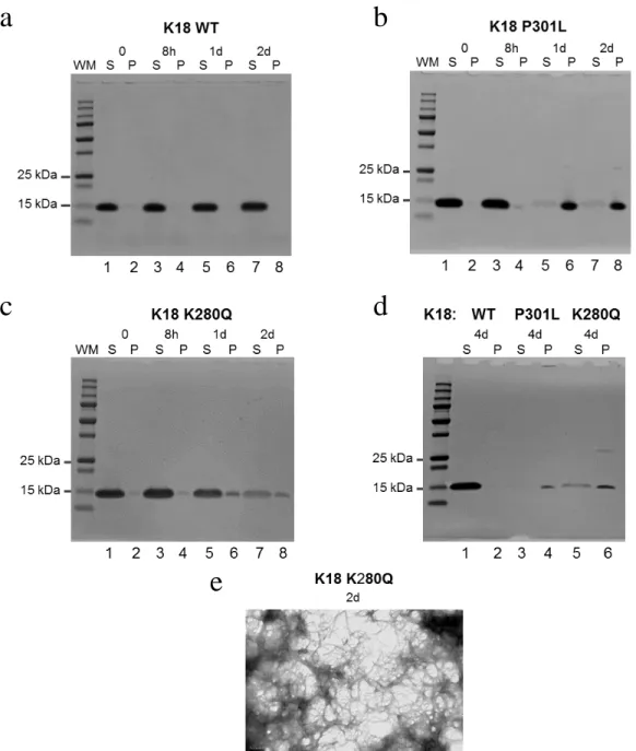
We first performed the tau-K18 fibrillization reactions without heparin, a polyanionic factor often used in these reactions to induce and expedite aggregation. The initial non-heparin fibril reactions and subsequent sedimentation assay and electron microscopy was a success (Fig. 2). The sedimentation assay begins with a centrifugation step that allows for isolation of a supernatant tau fraction containing largely soluble tau, and a pelleted tau fraction which largely contains tau fibrils. After isolating the two fractions, they are resolved by SDS-PAGE and stained with Coomassie Blue (Fig. 2a-d). As anticipated, the WT tau-K18 never transitioned to the pellet fraction over the 4 day time course. We also used a tau mutation, P301L, that we anticipated would be driven into the pellet fraction since the mutation is known to be an
aggregation-prone mutation associated with frontotemporal dementia. Results for P301L mutant tau-K18 also met expectations, with tau transitioning to the pellet fraction between 8 hours and 1 day (Fig. 2b). Our results on K280Q, the acetylation mimic, are somewhere on the spectrum between WT tau and P301L tau, showing a slight transition to the pellet fraction starting at 8 hours and growing by 1-2 days, and finally beginning to shift completely to the pellet fraction at ~4 days (Fig. 2c,d). We were also able to confirm we had robust K280Q tau fibrillization at 2 days via electron microscopy (Fig 2e). While this data does indicate that K280Q tau-K18 has the propensity to form fibrils, compared to WT tau-K18, we were unable to generate these same results on a consistent basis. This lack of consistency was unfortunate because the presence of heparin, at least the type we used, appeared to be problematic in neuron fibril treatment
experiments.
K18 was almost fully pelleted at 1 hour (Fig. 3b). Most importantly, the K280Q mutant tau-K18 also was fully pelleted by 1 hour (Fig. 3c), while the K280R mutant behaved similarly to WT tau-K18 (Fig. 3d). To confirm that K280Q was promoting tau aggregation, we used a Thioflavin T (ThT) fluorescence assay to detect amyloid structure over a time course of
fibrillization reactions. Our results mirrored those of the sedimentation assay. The ThT signal for both WT and K280R tau-K18 were low until 8 hours, at which time it rose slightly (Fig. 3e). In contrast, the ThT signal for both P301L and K280Q tau-K18 sharply increased (almost 5-6 fold that of WT and K280R) at 1 hour and held this signal level over the 8-hour time course (Fig. 3e).
These tau-K18 results were promising, and we decided to test if the K280Q mutation in full-length tau would have a similar effect on tau aggregation. In this next set of fibrillization reactions, we used the full-length tau-T40 isoform. We observed that the aggregation kinetics for K280Q is similar to P301L and is accelerated compared to WT tau-T40, showing increased pellet fraction at 1 day (Fig 4a-c). The sedimentation assay appears to indicate that the
aggregation dynamics of WT tau-T40 catches up to K280Q at 2 days for 1 of the 2 independent fibril reactions. This accelerated protein aggregation behavior is also mirrored in the ThT results (Fig. 4d), but the data also indicates amyloid structure in the P301L fibril reactions is by far the most robust.
4-hour time point, and this data also showed a paucity of WT fibrils while the K280Q and P301L fibrils were still in numerous, dense networks (Fig. 5b, bottom row).
The tau pathology observed in AD are rich is β-sheet secondary structure (Cohen et al., 2011) and this pathology is also positive for ac-K280 (Cohen et al., 2011). We decided to test if K280Q fibrils also were rich in β-sheet secondary structure using circular dichroism (CD), and secondarily whether CD could be used as another measure of the kinetics of tau aggregation. CD is a spectroscopic technique that detects differences between left and right-handed polarized light, and it is a helpful tool in predicting the secondary structure of protein molecules. The technique can distinguish whether a protein has adopted a conformation that is largely (1) disordered/random coil or contains (2) α-helix or (3) β-sheet structure. CD has previously been used to assess the secondary structure of tau protein (Kumar et al., 2014). With our fibrillization reactions, we expected to see a transition of tau secondary structure from random coil (0-hour time point) to β-sheet structure (1-hour pellet fraction time point). With a shift in minimum absorbance to around 215 nm by 1 hour, we observed that both K280Q and P301L pellet
fractions had CD profiles that indicate a transition to β-sheet structure (Fig. 6b,c). Furthermore, this minimum wavelength shift is consistent with other reports for aggregate-prone fibrils
(Barghorn et al., 2000; von Bergen et al., 2000). By 1 hour, neither WT (Fig. 6a) or K280R (Fig. 6d) pellet fractions displayed as strong a shift to β-sheet structure as K280Q, which is in
could just be the product of the faster kinetics of the fibril reaction. A more distinct difference between WT and K280Q or P301L could be achieved with a lower concentration of tau in the fibrillization reaction used for circular dichroism. At the 20 µM tau concentration used, the WT is seemingly catching up to K280Q quicker than the 10 µM concentrations used in the
sedimentation assays, ThT, and EM. Taken together, these results indicate that a single K280Q mutation is sufficient to accelerate tau aggregation in vitro.
Testing the effect of methylene blue on tau solubility
paper indicated that the oxidation of tau cysteines (i.e. C291, C322) would result in the inhibition of monomeric tau fibril formation when all four of tau’s microtubule-binding tandem repeats are present (4R-tau), but result in dimerization and high MW species formation when only tandem repeats 1, 3, and 4 are present (3R-tau).
points was that a previous study reported that giving MB prior to cognitive decline reduced insoluble tau in a mouse model (Hochgrafe et al., 2015). However, in this same study gels were cut off above 80 kDa and were very bright so it is impossible to independently ascertain whether higher MW tau was present upon MB treatment. In subsequent experiments, (1) earlier time points were added, (2) MB concentration and pre-incubation time before heparin was added was increased, (3) tau concentration was decreased, and (4) no agitation was used in the fibril
reactions. It is difficult to say what the clinical implications are of the observation of enhanced presence of dimer and high MW tau species upon MB treatment. A potential concern related to the dimer and other high MW tau species formation is that some in the field have labeled oligomeric and other high molecular weight tau as potentially toxic (Ward et al., 2012). Additionally, even if it were true that MB caused higher MW species of tau only in 3R-tau (Crowe et al., 2013), this still negatively could impact AD patients, since 3R-tau is present in adult brain. This data showing an increase in higher MW species of tau upon exposure to MB suggests that MB may be counterproductive in the treatment of tau aggregates in AD at more advanced disease states.
mice (PS19), there was a resultant appearance of acetylated K280 tau (Iba et al., 2013).
WT in the insoluble fraction and the other showed only that WT tau-T40 was insoluble and that K280Q and K280R were not.
Since we had previous indication that using P301L tau-T40 would reliably drive the formation of insoluble (SDS-soluble) tau, we decided to test whether preventing K280
acetylation would reduce the amount of insoluble tau. Using the QBI-293 cells, we performed transfections with WT, K280R-P301L (KR-PL), and P301L tau-T40 and then introduced K280Q fibrils (and P301L fibrils) into the cells. We planned to include a K280Q-P301L plasmid as well, but this plasmid did not generate tau protein in previous transfection experiments. In the first trial of the experiment, we saw that all three plasmids produced insoluble AT8 and T46 tau when treated with either K280Q or P301L fibrils, but that P301L tau-T40 was trending higher than either WT and K280R-P301L tau (Fig. 10b,c). In the soluble AT8 fraction, upon increasing the contrast setting, it should be noted that the KR-PL and P301L plasmids with K280Q fibril treatment had very faint bands of high MW signal that did not seem to run on the gel, with the KR-PL showing a bit higher signal than P301L (Fig. 10a). The no fibril controls showed very minimal presence of insoluble tau, with only a very faint band visible for P301L plasmid using the T46 total tau antibody and tau-1 antibody detecting small amounts with each plasmid (Fig. 10c,d). This low amount of insoluble tau in the no fibril treatment is good because it indicates the seeding assay is working. Tau-1 might have a higher affinity than T46 at the dilutions we used so that may be why it detects a small amount whereas T46 is not showing anything.
P301L fibrils, and injected these treatments into the hippocampus of the mice. We chose to perform this initial injection at 6 months of age since this is before the mouse develops mature tau pathology. We removed and fixed the brains at one-month post-injection, again since this was still prior to formation of mature tau pathology known for this PS19 mouse line.
Subsequently, we performed immunohistochemistry on brain slices using the AT8 phospho-tau antibody (Fig. 11). We saw intense AT8 signal in the ipsilateral (injected) hippocampus of 1 of 3 mice injected with K280Q fibrils, and there was less AT8 signal in the contralateral
hippocampus indicative of seeding. The other two mice injected with K280Q fibrils had less intense AT8 staining on the ipsilateral side relative to the contralateral side, but there might still be a trend. Two of the three mice injected with P301L fibrils showed some hints of seeding on the ipsilateral side but one P301L fibril injected mouse did not show any signs of seeding. In the PBS injected mice, the level of AT8 signal on the ipsilateral side look very similar to the two weak AT8 signal K280Q fibril injected mice, with PBS-1 contralateral even looking higher in intensity. We would need to quantify staining intensity to adjudicate if there are any statistical significance differences. Also, the non-injected side hippocampus was rotated out of plane (or disfigured) in two of the PBS control brains. Additional serial sectioning could determine if more information can be gleaned from these two contralateral PBS control hippocampus. Additional in vivo injection experiments are necessary to help make the case that K280Q is inducing seeding. Along with the preceding QBI-293 experiments, this initial in vivo experiment hints that K280Q fibrils are sufficient to induce the conversion of aggregrate-prone full length tau to an aberrantly phosphorylated, aggregated form of tau.
As stated previously, we injected tau fibrils into a line of mice expressing P301S, an aggregate-prone tau mutation linked to familial dementia. To study the effect of the K280Q tau mutation, we generated mice that express both a P301S and a K280Q mutation (referred to as PS-KQ).
We began characterizing this line by evaluating its seeding potential and tau protein expression using primary neuron culture from these PS-KQ mutant mice. We performed two seeding experiments with the PS-KQ mutant mice using cortical neurons, and I have performed cursory biochemical characterization from the first seeding experiment. We seeded the neurons at DIV5 with tau fibrils grown for the specified time: WT (8 hours), P301L (2 hours), and K280Q (4 hours). At DIV15, we harvested the neurons and separated them into a soluble and insoluble fraction, like we did for the QBI-293 cell seeding experiments. Others have reported being able to run their fibril treatments for 18 days, but we ended our treatments after 10-14 days of fibril treatment since we observed swelling of the neuron processes. We attributed this
swelling to residual high molecular weight heparin species in the fibril treatments, despite having centrifuged and re-suspended the tau fibrils in sodium acetate buffer. Neurons treated with the sodium acetate buffer alone were not observed to have the swelling processes. Interestingly, I saw what appeared to be insoluble, potentially endogenous mouse phospho-S396 tau in these neuron cultures, even in the non-fibril treated condition (Fig. 12a). The S396 residue is one of the residues known to be aberrantly phosphorylated in AD. If indeed it is endogenous mouse tau, seeing insoluble p-S396 is paradoxical since it is thought that endogenous mouse tau is not normally insoluble. This caused me to ask if this was unique to our PS-KQ mouse line.
Discussion
Prior to discovering that the K280 residue was acetylated in brains containing tauopathy, all the field knew that linked this residue to disease was a genetic deletion of K280 (∆K280) seen in FTDP-17 patients and a single case of AD (Momeni et al., 2009). At face value, the
acetylation of K280 and its deletion leading to neurodegenerative disease appear to be at odds. One unifying proposal is that the acetylation of K280 or its deletion, ∆K280, can bring about loss of normal tau function to stabilize microtubules and promote the formation of tau aggregates. Both events reduce the positive charge in that region of the protein. We also know that K280 and K281 residues mediate MT interactions (Goode and Feinstein, 1994). The shared
phenotypes between acetylated K280 and ∆K280, and the lack of proven association of causal tau mutations with AD, allows for the possibly that acetylation of K280 is one contributor to the aggregation of wild-type tau in AD.
tissue from PS-KQ mice. My observations when imaging tissues from one line of these mice indicated a lack of AT8 signal in all PS-KQ mice examined and a low level of total tau (tau 12) signal in some mice while other mice largely lacked tau 12. The IHC signal intensity also was reduced for PS-KQ mice compared to slices of PS19 tissue on the same slide.
It is possible that the additional K280Q mutation is serving to dephosphorylate the AT8 epitope. The observations from our early IHC work somewhat correlated with my early
biochemical characterization of PS-KQ primary neuron cultures, where there was a lack of AT8 in the insoluble fraction in the blots I have performed, resulting in blank blots, even after a more sensitive enhanced chemiluminescence (ECL) incubation. Further attempts at the AT8 western will need to include a known positive control for AT8, such as a Triton fraction from one of the QBI-293 experiments. Furthermore, in a blot for pS396 (Fig. 12b), the lack of a slightly higher MW band just above the two darker bands may also indicate a hypo-phosphorylation at this other site in our PS-KQ line, since the established P301S line showed this slightly higher MW band.
Performing additional IHC characterization on all of our PS-KQ lines of mice and adding appropriate size cohorts of nTg mice and mice expressing human WT tau would provide an even stronger case for these observations. In our three PS-KQ mouse lines, we should also confirm expression of the human tau in brain regions of interest (cortex and hippocampus) by western blot and compare this to age-matched human WT and P301S mutant tau transgenic controls.
endogenous tau and transgenic human tau into somatodendritic compartments of the cell, a translocation which is thought to be part of the reason why tau becomes toxic in AD. Similarly, we could also examine the interplay between this modeling of acetylation and tau-MT
interactions, since their group saw some MT dynamics effects with the acetylation mimicry (Sohn et al., 2016). Lastly, we could study how our K280Q mutation impacts tau’s pattern of phosphorylation, propensity to aggregate, synaptic function, and overall mouse behavioral phenotypes.
By expressing human tau in our mice, one critique might be that we are introducing another variable into the model. One way to address this is to continue to compare our results to nTg mice and mice expressing human WT tau. Outside of our approach, no way yet exists to target and keep single tau lysines acetylated in vivo since endogenous deacetylases (i.e. HDAC6) would act on the tau. Furthermore, knocking deacetylases down or out of a living organism would introduce even more variables since these enzymes act on a myriad of proteins. Other alternate approaches being considered in our lab are generating mice that over-express mutant K280Q human tau by itself or that express K280Q endogenous mouse tau. Previous attempts of the latter approach have been made with the aggregate-prone frontotemporal dementia tau mutation, P301L (Gilley et al., 2012). However, with both these acetylation mimic methods, we would still have to contend with the critique that the acetylation mimic approach keeps the tau in a permanently charge neutral state on that lysine.
binding assay, and we could also generate double acetylation mimic K280Q/K281Q mice. Continued biochemical characterization in cell culture and in AD brain to determine
stoichiometry of acetylation of tau lysines (especially K280 and K281), and to discover novel sites of acetylation would also be important contributions to the field.
Within the context of this work, our modeling of K280 and K281 acetylation, both in vitro and in vivo has further implicated the acetylation of these residues, and perhaps the local charge character of this region of tau, in the loss of tau function, acceleration of tau aggregation, and propagation of tau pathology. Our results begin to tie K280 acetylation to the initiation, as well as propagation, of AD tau pathology and merit further study of the effects of acetylation of this residue as well as ways in which to mitigate these effects pharmacologically. It also
Figures
Figure 1. Tau acetylation mimetics K280Q and K280Q/K281Q impairs tubulin polymerization. (a-b) Light scattering assay in the presence of tau-K18 proteins detecting tubulin polymerization, as determined by absorbance readings at 350 nm (No Tau, (N=7); WT, (N=9); K280Q, (N=8); 2KQ, (N=4); K280R, (N=7); 2KR, (N=6)). Error bars indicate standard deviation (s.d.) of the mean. GraphPad t-test results are as follows: WT v K280Q (***, p < 0.0001); WT v K280R (ns, p = 0.36); K280Q v K280R (***, p < 0.0001); WT v 2KR (**, p =
a
Figure 2: Tau acetylation mimetic K280Q displays accelerated aggregation compared to wild type tau and dense fibril formation without aid of heparin. (a-d) Coomassie blue staining of supernatant (S) and fibrillar pellet fractions of tau-K18 fibril reactions without heparin for (a) WT, (b) P301L, (c) K280Q protein from 0 – 2 day and (d) 4 day time points. One independent experiment. (e) Negative stain electron microscopy of the 2 day K280Q fibrillization reaction. One independent experiment.
a
b
c
d
Figure 3. K280Q tau enhances tau-K18 aggregation in vitro in presence of heparin. (a-d) Coomassie blue staining of supernatant (S) and fibrillar pellet fractions of tau-K18 fibril reactions with heparin for (a) WT, (b) P301L, (c) K280Q, and (d) K280R protein from 0 – 8 h time points. Coomassie blue staining was repeated using a second set of fibril reactions. (e) Thioflavin T (ThT) fluorescence of tau-K18 fibril reactions at the indicated time points from 0 to 8 h. Error bars indicate standard deviation (s.d.) of the mean. N=1 for each protein from three
b
a
d
c
Figure 4. K280Q tau enhances full-length tau-T40 aggregation in vitro in presence of
heparin. (a-c) Coomassie blue staining of supernatant (S) and fibrillar pellet fractions of tau-T40 fibril reactions with heparin for (a) WT, (b) P301L, and (c) K280Q protein from 0 to 3 day fibril reactions. Coomassie blue staining for the 0, 1d, and 3d time points was also performed using a second set of fibril reactions. Results repeated in time points except that T40 PL 1 d for second set of fibril reactions is a bit slower to aggregate than the T40 PL 1 d that appears in above figure. (d) ThT fluorescence of tau-T40 fibril reactions at the indicated time points from 0 to 3 d. Error bars indicate standard deviation (s.d.) of the mean. N=1 for each protein from two sets of fibril reactions.
a
b
Figure 5. K280Q tau-K18 also enhances fibril formation in vitro in presence of heparin. (a-b) Transmission electron microscopy of tau-K18 protein fibril reactions at (a) 2 h and ((a-b) 4 h.
a
Figure 6. K280Q tau-K18 promotes pellet fraction rich in beta sheet structure. (a-d) CD spectra of tau-K18 at the indicated time points from 0 to 1 h (0 h, solid black line; 1 h, grey line) were recorded from 190 to 250 nm for (a) WT, (b) P301L, (c) K280Q, and (d) K280R. 0 h time points reflect soluble stock tau dialyzed against CD buffer and 1 h time points reflect pelleted fraction of sedimentation assays resuspended in CD buffer. Error bars indicate standard deviation (s.d.) of the mean. N=3 for each time point.
a
b
Figure 7. Effects of methylene blue on tau-K18 solubility in vitro. (a-b) Coomassie blue staining of supernatant (S) and fibrillar pellet (P) fractions of tau-K18 fibril reactions plus or minus methylene blue for (a) WT and (b) K280Q. One independent experiment.
a
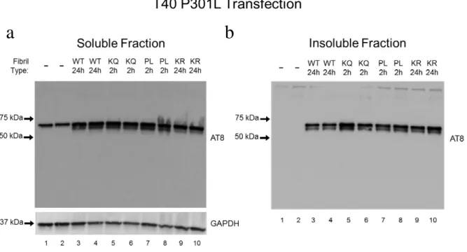
Figure 8. Effects of insoluble tau-K18 fibrils on tau phosphorylation and solubility using QBI-293 cell seeding assay. (a-b) Western blot analysis of (a) Triton X-100 soluble protein fraction and (b) SDS soluble protein fraction with phospho-tau (AT8) antibody. Cells were over-expressing tau-T40 mutant P301L and treated with tau-K18 fibrils (WT 24 h, K280Q 2 h, P301L 2 h, K280R 24 h) or a no fibril control. N=2 per treatment, one independent experiment.
Figure 9. Effects of K280Q fibrils on full-length WT, K280Q and K280R full-length tau seeding in QBI-293 cells. (a-b) Western blot analysis of (a) soluble and (b) insoluble QBI-293 cell lysate fractions with indicated antibodies of cells expressing tau T40 (WT, K280Q, or K280R) and treated with tau-K18 mutant K280Q fibrils. Monomeric tau is between 50 kDa and 75 kDa. Asterisks in insoluble T46 blot indicate faint high molecular weight protein bands positive for T46. N=3 per tau T40 plasmid transfection, one independent experiment.
Figure 10. Effects of K280Q and P301L fibrils on full-length WT, K280R-P301L, and P301L tau seeding in QBI 293 cells. (a-d) Western blot analysis of (a) soluble AT8, (b) insoluble AT8, (c) insoluble T46, and (d) insoluble tau-1 from QBI-293 lysate fractions of cells expressing tau T40 (WT, K280R-P301L, or P301L) and treated with tau-K18 fibrils (K280Q, N=2 or P301L, N=1) or a no fibril control, N=1. Molecular weights to left side of blots are in kDa. One independent experiment.
a
c
b
Figure 11. Effects of K280Q and P301L fibrils upon injection into PS19 mice.
Figure 12: Early characterization of phospho-tau in mice containing P301S and K280Q mutant human tau. (a) Western blot analysis of P301S-K280Q primary neuron (DIV15) lysate with phospho-S396 tau antibody. Lysate was from either untreated or fibril treated neurons as indicated. (b) Western blot analysis of CD-1, P301S, and P301S-K280Q primary neurons with phospho-S396 tau antibody. The lysates were not treated with fibrils. CD-1 and P301S lysates were from an unknown DIV and P301S-K280Q lysates were from DIV15. CD-1 and P301S were kindly provided by Jui-Heng Tseng from my lab. One independent experiment.
a
REFERENCES
Akoury, E., Pickhardt, M., Gajda, M., Biernat, J., Mandelkow, E., & Zweckstetter, M. (2013). Mechanistic basis of phenothiazine-driven inhibition of Tau aggregation. Angew Chem Int Ed Engl 52, 3511-3515.
Barghorn, S., Zheng-Fischhofer, Q., Ackmann, M., Biernat, J., von Bergen, M.,
Mandelkow, E. M., & Mandelkow, E. (2000). Structure, microtubule interactions, and paired helical filament aggregation by tau mutants of frontotemporal dementias.
Biochemistry 39, 11714-11721.
von Bergen, M., Friedhoff, P., Biernat, J., Heberle, J., Mandelkow, E. M., & Mandelkow, E. (2000). Assembly of tau protein into Alzheimer paired helical filaments depends on a local sequence motif ((306)VQIVYK(311)) forming beta structure. Proceedings of the National Academy of Sciences of the United States of America 97, 5129-5134.
von Bergen, M., Barghorn, S., Li, L., Marx, A., Biernat, J., Mandelkow, E. M., & Mandelkow, E. (2001). Mutations of tau protein in frontotemporal dementia promote aggregation of paired helical filaments by enhancing local beta-structure. J Biol Chem. 276, 48165-74.
Biernat, J., Gustke, N., Drewes, G., Mandelkow, E. M., & Mandelkow, E. (1993). Phosphorylation of Ser262 strongly reduces binding of tau to microtubules: distinction between PHF-like immunoreactivity and microtubule binding. Neuron. 11, 153-63. Bloom, G. S. (2014). Amyloid-β and tau: the trigger and bullet in Alzheimer disease
pathogenesis. JAMA Neurol. 71, 505-8.
Bos, J. L., Fearon, E. R., Hamilton, S. R., Verlaan-de Vries, M., van Boom, J. H., van der Eb, A. J., & Vogelstein B. (1987). Prevalence of ras gene mutations in human colorectal cancers. Nature 327, 293–297.
Braak, H., & Braak, E. (1997). Frequency of stages of Alzheimer-related lesions in different age categories. Neurobiol Aging 18, 351–357.
Braak, H., & Del Tredici, K. Neuroanatomy and Pathology of Sporadic Alzheimer's Disease, (2015). Springer. Adv Anat Embryol Cell Biol. 215, 1-162.
neurofibrillary tangles in Alzheimer brain: a fluorescence resonance energy transfer study. J Neuropathol Exp Neurol. 59, 966-71.
Cleveland, D. W., Hwo, S.-H., & Kirschner, M. W. (1977). Purification of tau. A microtubule associated protein that induces assembly from purified tubulin. J. Mol. Bio. 116, 207-226. Cohen, T. J., Guo, J. L., Hurtado, D. E., Kwong, L. K., Mills, I. P., Trojanowski, J. Q., &
Lee, V. M. (2011). The acetylation of tau inhibits its function and promotes pathological tau aggregation. Nature communications 2, 252.
Cohen, T. J., Friedmann, D., Hwang, A. W., Marmorstein, R., & Lee, V. M. (2013). The microtubule-associated tau protein has intrinsic acetyltransferase activity. Nat Struct Mol Biol 20, 756-762.
Congdon, E. E., Wu, J. W., Myeku, N., Figueroa, Y. H., Herman, M., Marinec, P. S., Gestwicki, J. E., Dickey, C. A., Yu, W. H., & Duff, K. E. (2012). Methylthioninium chloride (methylene blue) induces autophagy and attenuates tauopathy in vitro and in vivo. Autophagy 8, 609-622.
Cook, C., Stankowski, J. N., Carlomagno, Y., Stetler, C., & Petrucelli, L. (2014a).
Acetylation: a new key to unlock tau's role in neurodegeneration. Alzheimers Res Ther. 6, 29. Cook, C., Carlomagno, Y., Gendron, T. F., Dunmore, J., Scheffel, K., Stetler, C., Davis, M.,
Dickson, D., Jarpe, M., Deture, M., & Petrucelli, L. (2014b). Acetylation of the KXGS motifs in tau is a critical determinant in modulation of tau aggregation and clearance. Human molecular genetics 23, 104-116.
Crowe, A., James, M. J., Lee, V. M., Smith, A. B., 3rd, Trojanowski, J. Q., Ballatore, C., & Brunden, K. R. (2013). Aminothienopyridazines and methylene blue affect Tau fibrillization via cysteine oxidation. The Journal of biological chemistry 288, 11024-11037.
Dawson, H. N., Ferreira, A., Eyster, M. V., Ghoshal, N., Binder, L. I., & Vitek, M. P. (2001). Inhibition of neuronal maturation in primary hippocampal neurons from tau deficient mice. J. Cell Sci. 114, 1179–1187.
Ding, H., Dolan, P. J., Johnson, G. V. (2008). Histone deacetylase 6 interacts with the microtubule-associated protein tau. J Neurochem. 106, 2119-30.
Drubin, D.G., Kobayashi, S., & Kirschner, M. (1986). Association of tau protein with microtubules in living cells. Ann. NY Acad. Sci. 466, 257-268.
Flaherty, D. B., Soria, J. P., Tomasiewicz, H. G., & Wood, J. G. (2000). Phosphorylation of human tau protein by microtubule-associated kinases: GSK3beta and cdk5 are key
Flament, S., Delacourte, A., Hemon, B., & Defossez, A. (1989). Characterization of two pathological tau protein, variants in Alzheimer brain cortices. J Neurol Sci 92, 133-141. Forrester, K., Almoguera, C., Han, K., Grizzle, W. E., & Perucho, M. (1987). Detection of
high incidence of K-ras oncogenes during human colon tumorigenesis. Nature 327, 298–303. Frost, B., Jacks, R. L., & Diamond, M. I. (2009). Propagation of tau misfolding from the
outside to the inside of a cell. J Biol Chem. 284, 12845-52.
Ghetti, B., Oblak, A. L., Boeve, B. F., Johnson, K. A., Dickerson, B. C., & Goedert, M. (2015). Invited review: Frontotemporal dementia caused by microtubule-associated protein tau gene (MAPT) mutations: a chameleon for neuropathology and neuroimaging. 41, 24-46. Gilley, J., Seereeram, A., Ando, K., Mosely, S., Andrews, S., Kerschensteiner, M., Misgeld,
T., Brion, J. P., Anderton, B., Hanger, D. P., & Coleman, M. P. (2012). Age-dependent axonal transport and locomotor changes and tau hypophosphorylation in a "P301L" tau knockin mouse. Neurobiol Aging. 33, 621.
Goedert, M., Wischik, C. M., Crowther, R. A., Walker, J. E., & Klug, A. (1988). Cloning and sequencing of the cDNA encoding a core protein of the paired helical filament of
Alzheimer disease: identification as the microtubule-associated protein tau. Proc. Natl. Acad. Sci. USA 85, 4051-4055.
Goedert, M., Spillantini, M. G., Jakes, R., Rutherford, D., & Crowther, R. A. (1989a). Multiple isoforms of human microtubule-associated protein tau: sequences and localization in neurofibrillary tangles of Alzheimer's disease. Neuron 3, 519-526.
Goedert, M., Spillantini, M. C., Potier, M. C., Ulrich, J., & Crowther, R. A. (1989b).
Cloning and sequencing of the cDNA encoding an isoform of microtubule-associated protein tau containing four tandem repeats: differential expression of tau protein mRNAs in human brain. EMBO J. 8, 393-399.
Goode, B. L., & Feinstein, S. C. (1994). Identification of a novel microtubule binding and assembly domain in the developmentally regulated inter-repeat region of tau. The Journal of cell biology 124, 769-782.
Goode, B. L., Denis, P. E., Panda, D., Radeke, M. J., Miller, H. P., Wilson, L., & Feinstein, S. C. (1997). Functional interactions between the proline-rich and repeat regions of tau enhance microtubule binding and assembly. Molecular biology of the cell 8, 353-365. Gong, C. X., Liu, F., Grundke-Iqbal, I., & Iqbal, K. (2005). Post-translational modifications
of tau protein in Alzheimer's disease. J Neural Transm (Vienna). 112, 813-38.
Götz, J., Chen, F., van Dorpe, J., & Nitsch, R. M. (2001). Formation of neurofibrillary tangles in P301l tau transgenic mice induced by Abeta 42 fibrils. Science. 293, 1491-1495.
Grundke-Iqbal, I., Iqbal, K., Tung, Y-C., Quinlan, M., Wisniewski, H. M., & Binder, L. I. (1986). Abnormal phosphorylation of the microtubule-associated protein tau (tau) in
Alzheimer cytoskeletal pathology. Proc Natl Acad Sci 83, 4913-4917.
Grundke-Iqbal, I., Vorbrodt, A.W., Iqbal, K., Tung, Y-C., Wang G.P., & Wisniewski, H.M. (1988). Microtubule-associated polypeptides tau are altered in Alzheimer paired helical filaments. Molecular Brain Res. 464, 43-52.
Gu, G. J., Lund, H., Wu, D., Blokzijl, A., Classon, C., von Euler, G., Landegren, U.,
Sunnemark, D., & Kamali-Moghaddam, M. (2013). Role of individual MARK isoforms in phosphorylation of tau at Ser²⁶² in Alzheimer's disease. Neuromolecular Med. 15, 458-69. Guo, J. L., & Lee, V. M. (2011). Seeding of normal Tau by pathological Tau conformers drives
pathogenesis of Alzheimer-like tangles. J Biol Chem. 286, 15317-31.
Guo J. L., & Lee, V. M. (2013). Neurofibrillary tangle-like tau pathology induced by synthetic tau fibrils in primary neurons over-expressing mutant tau. FEBS Lett. 587, 717-23. Erratum in: FEBS Lett. 2013 587, 2484.
Gustke, N., Trinczek, B., Biernat, J., Mandelkow, E. M., & Mandelkow, E. (1994). Domains of tau protein and interactions with microtubules. Biochemistry. 33, 9511-22.
Hochgräfe K., Sydow, A., Matenia, D., Cadinu, D., Konen, S., Petrova, O., Pickhardt, M., Goll, P., Morellini, F., Mandelkow, E., & Mandelkow, E. M. (2015). Preventive
methylene blue treatment preserves cognition in mice expressing full-length pro-aggregant human Tau. Acta Neuropathol Commun 3, 25.
Hosokawa, M., Arai, T., Masuda-Suzukake, M., Nonaka, T., Yamashita, M., Akiyama, H., & Hasegawa, M. (2012). Methylene blue reduced abnormal tau accumulation in P301L tau transgenic mice. PloS one 7, e52389.
Hurtado, D. E., Molina-Porcel, L., Iba, M., Aboagye, A. K., Paul, S. M., Trojanowski, J. Q., & Lee, V. M. (2010). Abeta accelerates the spatiotemporal progression of tau pathology and augments tau amyloidosis in an Alzheimer mouse model. Am J Pathol. 177, 1977-1988. Iba, M., Guo, J. L., McBride, J. D., Zhang, B., Trojanowski, J. Q., & Lee, V. M. (2013).
Synthetic tau fibrils mediate transmission of neurofibrillary tangles in a transgenic mouse model of Alzheimer's-like tauopathy. The Journal of neuroscience: the official journal of the Society for Neuroscience 33, 1024-1037.
Iqbal, K., Grundke-Iqbal, I., editors: Iqbal, K., McLachlan, D.R.C., Winblad, B., & Wisniewski, H. M. (1991). Alzheimer’s Disease: From Cytoskeletal Protein Pathology to Neuronal Degeneration. Alzheimer’s Disease: Basic Mechanisms, Diagnosis and
Therapeutic Strategies. 173-180.
Irwin, D. J., Cohen, T. J., Grossman, M., Arnold, S. E., McCarty-Wood, E., Van Deerlin, V. M., Lee, V. M., & Trojanowski, J. Q. (2013). Acetylated tau neuropathology in sporadic and hereditary tauopathies. The American journal of pathology 183, 344-351.
Irwin, D. J., Cohen, T. J., Grossman, M., Arnold, S. E., Xie, S. X., Lee, V. M., &
Trojanowski, J. Q. (2012). Acetylated tau, a novel pathological signature in Alzheimer's disease and other tauopathies. Brain 135, 807-818.
Ittner, L. M., Ke, Y. D., Delerue, F., Bi, M., Gladbach, A., van Eersel, J., Wölfing, H., Chieng, B. C., Christie, M. J., Napier, I. A., Eckert, A., Staufenbiel, M., Hardeman, E., Götz, J. (2010). Dendritic function of tau mediates amyloid-beta toxicity in Alzheimer’s disease mouse models. Cell 142, 387–397.
Jin, N., Yin, X., Yu, D., Cao, M., Gong, C. X., Iqbal, K., Ding, F., Gu, X., & Liu, F. (2015). Truncation and activation of GSK-3β by calpain I: a molecular mechanism links to tau hyperphosphorylation in Alzheimer's disease. Sci Rep. 5, 8187.
Klein, C., Kramer, E. M., Cardine, A. M., Schraven, B., Brandt, R., & Trotter, J. (2002). Process outgrowth of oligodendrocytes is promoted by interaction of fyn kinase with the cytoskeletal protein tau. J. Neurosci. 22, 698–707.
Köpke, E., Tung, Y. C., Shaikh, S., Alonso, A. C., Iqbal, K., & Grundke-Iqbal, I. (1993). Microtubule-associated protein tau. Abnormal phosphorylation of a non-paired helical filament pool in Alzheimer disease. J Biol Chem 268, 24374-24384.
Kumar S., Tepper K., Kaniyappan S., Biernat J., Wegmann S., Mandelkow E. M., Müller D. J., & Mandelkow E. (2014). Stages and conformations of the Tau repeat domain during aggregation and its effect on neuronal toxicity. J Biol Chem. 289, 20318-32.
Lee, G., Neve, R. L., & Kosik, K. S. (1989). The microtubule binding domain of tau protein. Neuron. 2, 1615-24.
Lewis, J., Dickson, D. W., Lin, W-L., Chisholm, L., Corral, A., Jones, G., Yen, S. H.,
Sahara, N., Skipper, L., Yager, D., Eckman, C., Hardy, J., Hutton, M., & McGowan, E. (2001). Enhanced neurofibrillary degeneration in transgenic mice expressing mutant tau and APP. Science. 293, 1487-1491.
Luna-Muñoz, J., Harrington, C. R., Wischik, C. M., Flores-Rodrı́guez, P., Avila, J., Zamudio, S. R., De la Cruz, F., Mena, R., Meraz-Rı́os, M. A., & Floran-Garduño, B. (2013). Phosphorylation of Tau Protein Associated as a Protective Mechanism in the Presence of Toxic, C-Terminally Truncated Tau in Alzheimer's Disease, Understanding Alzheimer's Disease, Prof. Inga Zerr (Ed.), InTech, DOI: 10.5772/54228. Available from: http://www.intechopen.com/books/understanding-alzheimer-s-disease/phosphorylation-of-tau-protein-associated-as-a-protective-mechanism-in-the-presence-of-toxic-c-termi.
Min, S. W., Cho, S. H., Zhou, Y., Schroeder, S., Haroutunian, V., Seeley, W. W., Huang, E. J., Shen, Y., Masliah, E., Mukherjee, C., Meyers, D., Cole, P. A., Ott, M., & Gan, L. (2010). Acetylation of tau inhibits its degradation and contributes to tauopathy. Neuron 67, 953-966.
Min, S. W., Chen, X., Tracy, T. E., Li, Y., Zhou, Y., Wang, C., Shirakawa, K., Minami, S. S., Defensor, E., Mok, S. A., Sohn, P. D., Schilling, B., Cong, X., Ellerby, L., Gibson, B. W., Johnson, J., Krogan, N., Shamloo, M., Gestwicki, J., Masliah, E., Verdin, E., & Gan, L. (2015). Critical role of acetylation in tau-mediated neurodegeneration and cognitive deficits. Nat Med 21, 1154-1162.
Morris, M., Maeda, S., Vossel, K., & Mucke, L. (2011). The many faces of tau. Neuron. 70, 410-26.
Rankin, C.A., Sun, Q., & Gamblin, T.C. (2005). Pseudo-phosphorylation of tau at Ser202 and Thr205 affects tau filament formation. Mol Brain Res. 138, 84-93.
Roberson E. D., Scearce-Levie, K., Palop, J. J., Yan, F., Cheng, I. H., Wu, T., Gerstein, H., Yu, G. Q., & Mucke, L. (2007). Reducing endogenous tau ameliorates amyloid beta-induced deficits in an Alzheimer's disease mouse model. Science 316, 750-4.
Rodenhuis S., van de Wetering, M. L., Mooi, W. J., Evers, S. G., van Zandwijk, N., & Bos, J.L. (1987). Mutational activation of the K-ras oncogene. A possible pathogenetic factor in adenocarcinoma of the lung. N Engl J Med. 317, 929–935.
Sanders, D. W., Kaufman, S. K., DeVos, S. L., Sharma, A. M., Mirbaha, H., Li, A., Barker, S. J., Foley, A. C., Thorpe, J. R., Serpell, L. C., Miller, T. M., Grinberg, L. T., Seeley, W. W., & Diamond, M. I. (2014). Distinct tau prion strains propagate in cells and mice and define different tauopathies. Neuron. 82, 1271-88.
Schneider, A., Biernat, J., von Bergen, M., Mandelkow, E., & Mandelkow, E. M. (1999). Phosphorylation that detaches tau protein from microtubules (Ser262, Ser214) also protects it against aggregation into Alzheimer paired helical filaments. Biochemistry. 38, 3549-58. Shirazi, S. K., & Wood, J. G. (1993). The protein tyrosine kinase, fyn, in Alzheimer's disease
Singleton, A. B. (2005). Altered alpha-synuclein homeostasis causing Parkinson's disease: the potential roles of dardarin. Trends Neurosci. 28, 416-21.
Smit, V. T., Boot, A. J., Smits, A. M., Fleuren, G. J., Cornelisse, C. J., & Bos, J. L. (1988). KRAS codon 12 mutations occur very frequently in pancreatic adenocarcinomas. Nucleic Acids Res. 16, 7773–7782.
Sohn, P. D., Tracy, T. E., Son, H. I., Zhou, Y., Leite, R. E., Miller, B. L., Seeley, W. W., Grinberg, L. T., & Gan, L. (2016). Acetylated tau destabilizes the cytoskeleton in the axon initial segment and is mislocalized to the somatodendritic compartment. Mol Neurodegener. 11, 47.
Song, L., Lu, S. X., Ouyang, X., Melchor, J., Lee, J., Terracina, G., Wang, X., Hyde, L., Hess, J. F., Parker, E. M., & Zhang, L. (2015). Analysis of tau post-translational modifications in rTg4510 mice, a model of tau pathology. Mol Neurodegener 10, 14. Taddei, K., Fisher, C., Laws, S. M., Martins, G., Paton, A., Clarnette, R. M., Chung, C.,
Brooks, W. S., Hallmayer, J., Miklossy J., Relkin N., St George-Hyslop P. H., Gandy S. E., & Martins R. N. (2002). Association between presenilin-1 Glu318Gly mutation and familial Alzheimer's disease in the Australian population. Molecular Psychiatry 7, 776–781. Taniguchi, S., Suzuki, N., Masuda, M., Hisanaga, S., Iwatsubo, T., Goedert, M., &
Hasegawa, M., (2005). Inhibition of heparin-induced tau filament formation by phenothiazines, polyphenols, and porphyrins. J Biol Chem. 280, 7614-23.
Tanimukai, H., Grundke-Iqbal, I., & Iqbal, K. (2005). Up-regulation of inhibitors of protein phosphatase-2A in Alzheimer's disease. The American journal of pathology 166, 1761-1771. Tracy, T. E., Sohn, P. D., Minami, S. S., Wang, C., Min, S. W., Li, Y., Zhou, Y., Le, D., Lo,
I., Ponnusamy, R., Cong, X., Schilling, B., Ellerby, L. M., Huganir, R. L., & Gan, L. (2016). Acetylated Tau Obstructs KIBRA-Mediated Signaling in Synaptic Plasticity and Promotes Tauopathy-Related Memory Loss. Neuron 90, 245-260.
Wang, Y., Yang, R., Gu, J., Yin, X., Jin, N., Xie, S., Wang, Y., Chang, H., Qian, W., Shi, J., Iqbal, K., Gong, C. X., Cheng, C., & Liu, F. (2015). Cross talk between
PI3K-AKT-GSK-3β and PP2A pathways determines tau hyperphosphorylation. Neurobiol Aging. 36, 188-200.
Ward, S. M., Himmelstein, D. S., Lancia, J. K., & Binder, L. I. (2012). Tau oligomers and tau toxicity in neurodegenerative disease. Biochem Soc Trans. 40, 667-71.