GERM CELL IMMORTALITY IN C. ELEGANS: REDUCED INSULIN SIGNALING RESTORES GERMLINE IMMORTALITY IN PIWI ARGONAUTE PRG-1 MUTANTS
Matthew Alan Simon
A dissertation submitted to the faculty at the University of North Carolina at Chapel Hill in partial fulfillment of the requirements for the degree of Doctor of Philosophy in the Curriculum of Genetics and
Molecular Biology in the School of Medicine.
Chapel Hill 2014
iii ABSTRACT
Matthew Alan Simon: GERM CELL IMMORTALITY IN C. ELEGANS: REDUCED INSULIN SIGNALING RESTORES GERMLINE IMMORTALITY IN PIWI ARGONAUTE PRG-1 MUTANTS
(Under the direction of Shawn Ahmed)
A key distinction between germ and somatic cells is the capacity for indefinite self-renewal. Somatic cells typically exist for a single generation, whereas germ cells are effectively immortal as they proliferate from one generation to the next. Thus, germ cells must overcome damage that can
contribute to aging of the soma, such as telomere erosion, DNA damage or proteotoxicity. It is unknown how many pathways contribute to germ cell immortality and what connections there might be between germline and somatic aging. Understanding the role of genes that maintain germ cell immortality is of interest to the fields of aging and cancer, as it may provide insight into the aging of disposable somatic tissues and understanding into how normally mortal cells might enter into the immortal state, as in the case of various cancers.
To gain insight into germ cell immortality, I report here the role of the C. elegans PIWI Argonaute paralog PRG-1 in maintaining the immortal state. In the Chapters 1-3, I demonstrate that dysfunction of prg-1 results in a mortal germline phenotype. We provide evidence that increased misexpression of repetitive elements results in toxic stress that causes sterility. Additionally, I show that decreased signaling in the DAF-2/IGF-1 insulin signaling somatic longevity pathway restores germline immortality to prg-1 mutant animals and decreases repetitive element misexpression.
iv
starvation-induced phenotype known as adult reproductive diapause (ARD). Through genetic
characterization, I demonstrate the prg-1 atrophy phenotype is, indeed, the ARD respond. Furthermore, we describe a new role for the FOXO transcription factor, DAF-16, in prg-1-induced ARD. Continued exploration of the daf-16-mediated rescued identified a critical role for the dsRNA uptake channel, SID-1, in both the rescue of prg-1’s mortal germline (mrt) phenotype and in normal prg-1-induced ARD. I go on to show that components of the rrf-3 endogenous RNAi pathway are required for daf-16-mediated rescue of the mrt phenotype in prg-1. I conclude that prg-1 functions to protect the integrity of the germline and that the insulin signaling somatic longevity pathway can also function in this capacity. I conclude that daf-16-mediated rescue of prg-1 progressive sterility and its role in prg-1-induced ARD requires a previously unidentified endogenous RNAi pathway.
The fourth section covers a collaborative project in which I played a significant role, focusing on the role of the RNAi spreading genes, rsd-2 and rsd-6 in germline immortality. We found that these two genes were responsible for maintaining germline immortality at the stressful temperature of 25ºC through a small RNA-mediated transcriptional silencing pathway. Similar to prg-1, stressed rsd mutants displayed desilencing of repetitive loci, as well as misregulation of spermatogenesis genes and
chromosome missegregation.
In the fifth and final section, I summarize the major findings of my work. I cover the advances my research has contributed to the field of germline immortality, aging, and small RNA biology and their implications on future work. I also describe ongoing work and proposed future experiments for
v
ACKNOWLEDGMENTS
I would like to thank:
My thesis advisor Shawn Ahmed for his guidance and enthusiasm for my work throughout the course of my doctoral research
vi
TABLE OF CONTENTS
LIST OF TABLES... ix
LIST OF FIGURES ... x
LIST OF ABBREVIATIONS ... xi
Chapter: I: REDUCED INSULIN/IGF-1 SIGNALING RESTORES GERM CELL IMMORTALITY TO CAENORHABDITIS ELEGANS PIWI MUTANT ... 1
INTRODUCTION ... 2
RESULTS ... 4
Deficiency for prg-1 results in progressive sterility ... 4
Deficiency for daf-2 suppresses progressive sterility of prg-1 mutants ... 5
Progressive sterility of prg-1 does not result from transposition ... 9
Transgene silencing is separable from germ cell immortality ... 10
Disrupted silencing of repetitive loci in prg-1 mutants ... 11
A small RNA pathway restores germ cell immortality prg-1 mutants ... 20
DISCUSSION ... 23
MATERIALS AND METHODS ... 27
SUPPORTING MATERIAL ... 31
II: ADULT REPRODUCTIVE DIAPAUSE IN RESPONSE TO A HERITABLE EPIGENETIC STRESS ... 40
INTRODUCTION ... 41
vii
Adult Reproductive Diapause in sterile prg-1 mutants ... 42
Stress response transcription factors promote germ cell atrophy ... 45
Germ cell atrophy represents a form of reversible sterility ... 50
DISCUSSION ... 53
MATERIALS AND METHODS ... 55
SUPPORTING MATERIAL ... 57
III: DAF-2/DAF-16 INSULIN SIGNALING FUNCTIONS THROUGH A SMALL RNA PATHWAY TO DIRECT GERMLINE HEALTH IN PRG-1 MUTANTS ... 59
INTRODUCTION ... 60
RESULTS ... 62
sid-1 facilitates daf-16 rescue of prg-1 mutants ... 62
daf-16 rescue functions through an RNAi pathway... 63
Multiple somatic tissues contribute to daf-16 signal input ... 64
DISCUSSION ... 65
MATERIALS AND METHODS ... 68
IV: C. ELEGANS RSD-2 AND RSD-6 PROMOTE GERM CELL IMMORTALITY BY MAINTAINING SMALL INTERFERING RNA POPULATIONS ... 69
INTRODUCTION ... 70
RESULTS ... 72
RSD-2 and RSD-6 promote germ cell immortality at stressful Temperatures ... 72
RSD-2 and RSD-6 may function in germ cells to promote germ cell immortality... 72
viii
Small RNAs targeting spermatogenesis genes are perturbed in
rsd mutants ... 77
Desilencing of repetitive loci in stressed late-generation rsd mutants ... 81
RSD-6 acts via small RNA-mediated transcriptional silencing ... 84
DISCUSSION ... 86
MATERIALS AND METHODS ... 89
SUPPORTING MATERIAL ... 96
V: SUMMARY AND FUTURE DIRECTIONS ... 108
ix LIST OF TABLES
x
LIST OF FIGURES
1-1. Progressive sterility of prg-1 mutants………5
1-2. daf-2 signaling can suppress fertility defects of prg-1 and modulates germline remodeling at sterility……….………8
1-3. piRNA loss affects expression of few genes targeted by piRNAs………13
1-4. Derepression of repetitive elements in prg-1 mutants is cured by daf-2………...15
1-5. Altered 22G-RNA frequencies and gene expression changes in prg-1 mutants………19
1-6. Model of parallel small RNA silencing pathways that can repress transgenerational fertility defects……….……….…22
2-1. Germline atrophy in sterile prg-1 animals………41
2-2. Cell death potentiates germline remodeling of sterile prg-1 animals………..………43
2-3. daf-16 translocalizes to intestinal nuclei in sterile prg-1 animals………46
2-4. Reduced daf-2 signaling restores fertility to sterile prg-1 adults………....50
3-1. sid-1 is required for daf-2 rescue and ARD in prg-1 mutants………..……61
3-2. An endogenous RNAi pathway functions to enable somatic tissue-derived daf-16 rescue of prg-1 progressive sterility………..62
3-3. DAF-16 functions through a cell non-autonomous RNAi pathway to rescue prg-1 dysfunction……….63
4-1. rsd-2 and rsd-6 promote germ cell immortality………..….72
4-2. Germ cell proliferation defects and non-disjunction in late-generation rsd Mutants……….……….74
4-3. Gene expression changes in rsd mutants associated with loss of small RNAs………..……….78
xi
LIST OF ABBREVIATIONS ARD Adult Reproductive Diapause
DAPI 4',6-diamidino-2-phenylindole
mrt mortal germline
IIR Insulin/IGF Receptor RNA Ribonucleic Acid DNA Deoxyribonucleic Acid siRNA Small interfering RNA piRNA Piwi-interacting RNA cDNA Complimentary DNA
CGH Comparative Genome Hybridization NGM Nematode Growth Medium
1
CHAPTER I: REDUCED INSULIN/IGF-1 SIGNALING RESTORES GERM CELL IMMORTALITY TO CAENORHABDITIS ELEGANS PIWI MUTANTS1
SUMMARY
Defects in the Piwi/piRNA pathway lead to transposon desilencing and immediate sterility in many organisms. We found that the C. elegans Piwi mutant for prg-1 became sterile after growth for many generations. This phenotype did not occur for RNA interference mutants with strong transposon silencing defects and was separable from the role of PRG-1 in transgene silencing. Brief periods of starvation extended the transgenerational lifespan of prg-1 mutants by up-regulating the DAF-16/FOXO longevity transcription factor. Constitutive activation of DAF-16 via reduced daf-2 insulin/IGF-1 signaling immortalized prg-1 strains via the activities of RNA interference proteins and histone H3 lysine 4
demethylases. In late-generation prg-1 mutants, desilencing of repetitive segments of the genome occurred, and silencing of repetitive loci was restored in prg-1; daf-2 mutants. This study reveals an unexpected interface between aging and transgenerational maintenance of germ cells, where somatic longevity is coupled to a genome silencing pathway that promotes germ cell immortality in parallel to the Piwi/piRNA system.
__________________________
1
2 INTRODUCTION
Somatic cells accumulate stress that limits proliferation within a single generation, whereas germ cells are effectively immortal as they proliferate from one generation to the next. Genetic studies of C. elegans have revealed that telomerase-mediated telomere maintenance is essential for germ cell immortality (Meier et al., 2006) and that several histone modification enzymes contribute to germline maintenance over generations (Andersen and Horvitz, 2007; Buckley et al., 2012; Katz et al., 2009; Xiao et al., 2011). Although deficiency for telomerase in humans is likely to contribute to proliferative aging of somatic cells (Armanios and Blackburn, 2012), other pathways that promote germ cell immortality could be specific to the germ cells, or could reveal new connections between the germline and somatic aging.
Piwi is an Argonaute protein that associates with a diverse class of small RNAs that are abundant in germ cells termed Piwi-Interacting RNAs (piRNAs) (Juliano et al., 2011). Conserved functions for Piwi include suppression of transposons and self-renewal of germline or meristematic stem cells. Deficiency for Piwi and Piwi-like genes in Drosophila, Arabidopsis and in vertebrate males results in immediate sterility (Juliano et al., 2011). Further, mating of Drosophila females that lack piRNAs targeting a
3
C. elegans has two closely related Piwi homologs, 1 and 2, but only deficiency for PRG-1 has phenotypic consequences (Bagijn et al., 20PRG-12; Batista et al., 2008; Das et al., 2008). PRG-PRG-1 is expressed in germ cells, and prg-1 mutants were previously reported to display temperature-sensitive sterility accompanied by transposition of the Tc3 transposon, but not other DNA transposons (Bagijn et al., 2012; Batista et al., 2008; Das et al., 2008). These phenotypes could conceivably be related to hybrid dysgenesis in Drosophila (Juliano et al., 2011; Kidwell et al., 1977). In addition, PRG-1 was recently shown to initiate silencing of foreign transgenes (Ashe et al., 2012; Luteijn et al., 2012; Shirayama et al., 2012), though silencing was then maintained by a number of factors, including small interfering RNA proteins that are responsible for silencing many active transposons in C. elegans.
4 RESULTS Deficiency for prg-1 results in progressive sterility
To study the effects of Piwi on fertility in C. elegans, we backcrossed three alleles of prg-1 and four alleles of prg-2 (Batista et al., 2008; Wang and Reinke, 2008), thereby removing unlinked mutations and/or epigenetic effects of the parental backgrounds. In contrast to previous findings (Bagijn et al., 2012; Batista et al., 2008; Das et al., 2008), we did not generally observe strong defects in fertility at high temperature for outcrossed prg-1 mutants. Instead, slightly reduced brood sizes occurred for maternally depleted F3 prg-1 homozygotes at both 20oC and 25oC in comparison to N2 wild-type controls (Figure 1-1A), and a minority of prg-1 mutants displayed >80% embryonic lethality at 25oC (Figures 1A and 1-S1A). We saw robust levels of fertility during propagation of prg-1 and prg-2 for 8 generations at either 20oC or 25oC based on Mortal Germline (Mrt) assays, where 6 L1 larvae were transferred to freshly seeded plates once per week (Ahmed and Hodgkin, 2000; Meier et al., 2006). No strain displayed even a moderate reduction in fertility during this period, based on complete consumption of the E. coli lawn (Ahmed and Hodgkin, 2000; Meier et al., 2006). Continued propagation of prg-1 but not prg-2 strains resulted in drops in fertility and ultimately complete sterility (Figures 1-1B and 1-S1B).This drop in fertility occurred both at 20oC and 25oC. Deficiency for three of four prg-2 alleles failed to exacerbate the fertility defects of prg-1 (Figure 1-1C). Although accelerated sterility was observed for 1; prg-2(ok1328) double mutants, this likely reflects an effect of a background mutation linked to ok1328. Thus loss of function of prg-1 leads to progressive loss of fertility over several generations, independent of prg-2.
We confirmed that prg-1 mutations were the cause of progressive sterility in the above
5
Slicer-dead PRG-1 (Bagijn et al., 2012; Batista et al., 2008; Das et al., 2008), which also rescued the progressive sterility phenotype caused by prg-1 deficiency (Figure 1-S1C). Consistently, Slicer nuclease activity is not required for piRNA-dependent genome silencing by PRG-1 (Bagijn et al., 2012; Batista et al., 2008; Das et al., 2008).
Figure 1-1. Progressive sterility of prg-1 mutants. (A) Levels of fertility for different prg-1 mutants at 20°C and 25°C (mean±s.d., n=10 lines per strain). Individuals from independently derived lines were singled and their progeny counted. (B) prg-1 exhibits progressive sterility at both 20°C and 25°C. Animals at 25°C appear more robust than siblings at 20°C (Mantel-Cox log-rank test, P=0.001 for tm872, P=0.030 for pk2298, P=0.144 for n4357) (n=12). (C) Progressive sterility of prg-1(tm872); prg-2 double mutants (n=5 strains per genotype). (D) Starvation extends transgenerational lifespan of prg-1 strains propagated at 25oC. Extension by starvation is dependent on daf-16.
Deficiency for daf-2 suppresses progressive sterility of prg-1 mutants
6
that prg-1 mutant strains reproduce prior to becoming sterile). The term ‘transgenerational lifespan’ reflects the proliferative capacity of germ cells across generations, and was inspired by studies of ‘replicative lifespan’ that assess cellular aging in yeast or mammalian cells (Polymenis and Kennedy, 2012; Smelick and Ahmed, 2005). In contrast, ‘adult lifespan’ concerns aging that occurs in a single generation (Kenyon, 2010).
While studying the transgenerational lifespan of prg-1 mutants, we noticed that propagation at 25oC led to frequent starvation of the plates prior to transfer, which was rare at 20oC. We therefore repeated the assay ensuring that prg-1 strains did not starve at 25oC, and found that this eliminated the extension of transgenerational lifespan that was observed for prg-1 mutants that starve transiently (Figure 1-1D). Typically, C. elegans stocks are subjected to long periods of starvation and are
infrequently outcrossed. We hypothesize that these conditions suppress the Mrt phenotype of prg-1 mutants and instead cause a distinct and possibly related epigenetic defect that is manifest as temperature-sensitive sterility (Batista et al., 2008; Wang and Reinke, 2008).
Many effects of starvation in C. elegans are triggered by activation of the DAF-16/FOXO
transcription factor that promotes stress resistance and longevity (Kenyon, 2010; Lin et al., 1997; Ogg et al., 1997). We therefore constructed prg-1 daf-16 double mutants and found that their
transgenerational lifespan was not extended by starvation (Figure 1-1D), implying that transient
7
daf-2 mutations promote stress resistance and dauer formation through DAF-16/FOXO (Kenyon, 2010). prg-1 daf-16; daf-2 triple mutants became progressively sterile, indicating that daf-2 deficiency suppresses the fertility defects of prg-1 by activating DAF-16 (Figure 1-2B). Transgenerational lifespan of prg-1 was reduced by ~30% for prg-1 daf-16 or prg-1 daf-16; daf-2 strains (P=5.05E-03 and 1.56E-03, respectively, Mantel-Cox log-rank test) (Figure 1-2B). We confirmed these observations using
independent mutations in daf-18, which functions upstream of DAF-16 to promote longevity in response to reduced DAF-2 signaling (Ogg and Ruvkun, 1998), and found that prg-1; daf-18 double mutants also displayed shortened transgenerational lifespan (Figure 1-2B). Neither daf-16 nor daf-18 single mutants become sterile in Mortal Germline assays (Ahmed, 2006).
Reduced daf-2 activity is associated with an enhanced response to exogenous RNA interference and a soma-to-germline transformation (Curran et al., 2009; Wang and Ruvkun, 2004). However, these phenotypes also occur when lin-15B is deficient (Wang et al., 2005), and lin-15B did not suppress progressive sterility of prg-1 (Figure 1-2B).
Taken together these data show that a specific response downstream of inactivation of the daf-2 pathway allows animals to remain fertile in the absence of prg-1. As prg-1; daf-daf-2 lines can be
propagated indefinitely, we wondered whether these animals accumulate defects that cause them to become sterile immediately when daf-2 activity is restored - in other words whether daf-2 protects against accumulation of damage or simply allows animals to tolerate high levels of damage. To test this we crossed late-generation prg-1; daf-2 double mutants with early-generation prg-1 mutant males and selected 16 prg-1 -/-; daf-2 +/+ lines descended from prg-1 -/-; daf-2 +/- F1 animals (Figure 1-S2E). These lines maintained fertility for 10 generations and then became progressively sterile in a manner similar to prg-1 single mutants (Figure 1-2C), suggesting that daf-2 deficiency directly prevents damage
8
Figure 1-2. daf-2 signaling can suppress fertility defects of prg-1 and modulates germline remodeling at sterility. (A) Reduced daf-2 signaling suppresses prg-1 mediated progressive sterility. (B) lin-15B does not suppress transgenerational lifespan of prg-1 (n=30 strains per genotype). daf-16 and daf-18 are required for suppression of prg-1 by daf-2. Four prg-1 daf-16 double mutant strains were studied where prg-1 alleles tm872 and n4357 were combined with daf-16 alleles mgDf50 or mu86 (n=10 strains per genotype). Four prg-1; daf-18 double mutant strains were studied where prg-1 alleles tm872 and n4357 were combined with daf-18 alleles e1375 and ok480 (n=10 strains per genotype). For prg-1 daf-16; daf-2 triple mutants, prg-1(tm872) daf-16(mgDf50) and prg-1(n4357) daf-16(mu86) were each combined with three daf-2 alleles m41, e1368 and e1370 (n=10 strains scored per triple mutant genotype) and
examined for progressive sterility. Four prg-1; daf-2(e1368); daf-18 lines were constructed from the four allelic combinations of prg-1; daf-18 and examined for progressive sterility (n=10 strains scored per triple mutant genotype). Data for all independent alleles was combined to show transgenerational lifespan for strains of the same genotype. (C) Lack of immediate sterility upon removal of daf-2 suggests suppression of the heritable epigenetic defect that causes sterility in prg-1 mutants. prg-1; daf-2(+/+) strains represent cross-progeny of late-generation prg-1; daf-2 double mutants where the daf-2
9
unc-58 and unc-54. (F) Treatment of sterile late-generation prg-1 adults with daf-2 or age-1 RNAi restores fertility, and fertility can be maintained on these RNAi strains for at least 10 generations while RNAi is maintained.
Progressive sterility of prg-1 does not result from transposition
10
despite causing ~6- to 12-fold increases in the frequency of spontaneous mutation (Figures 1-2D, 1-2E and Table 1-S1). Third, we did not observe any evidence of chromosomal instability as measured by DAPI staining in late-generation prg-1 animals compared to either early-generation prg-1 or wild-type animals (Figure 1-S1D) (Ahmed and Hodgkin, 2000).
Finally, if the prg-1 defects were caused by increased transposition or associated genomic instability we would not expect them to be reversible. We therefore treated late-generation sterile prg-1 adults with RNAi against either daf-2 or age-prg-1, which also negatively regulates daf-prg-16, and found that either RNAi treatment allowed some infertile adults to produce viable offspring again (Figure 1-2F). Further, fertility could be maintained for at least 10 additional generations by maintaining lines with RNAi treatment. The reversibility of the sterility phenotype makes it highly unlikely that sterility is caused by accumulation of deleterious transposon insertions. Our results instead suggest that the sterility phenotype of prg-1 mutants is likely due to accumulated epigenetic changes.
Transgene silencing is separable from germ cell immortality
11 Disrupted silencing of repetitive loci in prg-1 mutants
PRG-1 interacts with ~16,000 21 nucleotide (nt) RNAs possessing 5´ uracil that represent the C. elegans piRNA repertoire and silence many segments of the genome (Bagijn et al., 2012; Batista et al., 2008; Das et al., 2008). PRG-1 has been implicated previously in small RNA-mediated silencing pathways (Batista et al., 2008; Das et al., 2008) and indeed can trigger transgenerational silencing that can persist in the absence of prg-1 (Ashe et al., 2012; Luteijn et al., 2012; Shirayama et al., 2012). Therefore to search for possible determinants of the epigenetic defects responsible for sterility, we examined the functional consequences of prg-1 deficiency on small RNA-mediated silencing pathways. We searched for changes in small RNAs or their targets that would become more severe over continued propagation and could be suppressed by deficiency in daf-2.
In Drosophila, piRNAs can be inherited through the female germline (Brennecke et al., 2008). We therefore asked first whether a progressive loss of piRNAs occurred during propagation of prg-1 mutants. We prepared small RNA libraries from wild-type animals and prg-1 mutant animals at different generations. We found that piRNAs are essentially absent from prg-1 strains at generations 4 and 8, as well as generation 12, which was close to sterility for both alleles examined (Figure 1-3A). As piRNA annotation is based on a genomic motif likely associated with piRNA biogenesis (Ruby et al., 2006), the few remaining small RNAs that fit our piRNA criteria are likely misannotated rather than persistent piRNA species. Progressive loss of piRNAs is therefore unlikely to explain the delay in loss of fertility in prg-1 animals.
12
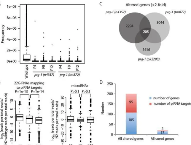
to these genes in later generation prg-1 animals (Figure 1-3B). No such progressive reduction was seen in microRNA levels. We tested the hypothesis that expression of 22G-RNA target genes might be deregulated by preparing RNA from early- and late-generation prg-1 strains and using cDNA created from this RNA to perform tiling microarrays that interrogate the entire C. elegans genome. Analysis of microarrays for three independent alleles of prg-1 showed little consistent change in the expression of genes targeted by 22G-RNAs (data not shown). Instead, we identified a set of 205 genes whose expression changed by more than 2-fold in late-generation prg-1 strains (Figure 1-3C), an intersection significantly greater than that predicted by 1000 simulations of random overlap (Z=35) and all but 3 of these genes changed in the same direction in all alleles, more than 10-fold greater than expected by chance (P<2e-16, 2 test, two-tailed). However, the expression changes of only 20 of the 205 genes were significantly reduced by more than 1.4-fold (0.5 Log2 units) in late-generation prg-1; daf-2 mutants
13
Figure 1-3. piRNA loss affects expression of few genes targeted by piRNAs. (A) Boxplots are based on read frequencies for 4,839 piRNAs sequenced in one or more libraries. Boxes indicate interquartile ranges, horizontal bars medians, whiskers extend to the most extreme data points with distance from the box no more than 1.5 times the interquartile range, crosses indicate outliers. (B) Boxplots showing reduced median levels of 22G-RNAs for piRNA targets in later generation prg-1 animals. Levels of microRNAs do not show the same progressive reduction. P values are for Wilcoxon signed rank tests. (C) Analysis of genes whose expression changed more than 2-fold in late- versus early-generation prg-1 strains reveals few common genes are altered for prg-1 alleles. (D) Few altered genes that are cured by daf-2 are predicted piRNA targets.
As mentioned above, piRNAs in a number of organisms, including C. elegans, have been shown to target transposons for silencing (Bagijn et al., 2012; Batista et al., 2008; Das et al., 2008; Juliano et al., 2011). Thus, the epigenetic defect of prg-1 germ cells could be due to derepression of repetitive
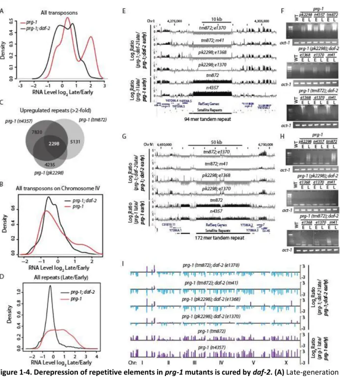
genomic loci that are targeted by PRG-1 piRNAs. Using genome-wide tiling arrays, we observed an increase in the expression of a subset of transposons in late-generation prg-1 mutants (Figures 1-4A and 1-4B). The most up-regulated were the Mariner class transposons, which include the previously
14
15
Figure 1-4. Derepression of repetitive elements in prg-1 mutants is cured by daf-2. (A) Late-generation prg-1 mutants show a mean increase in transposon expression, compared to the expression of
16
repeat tracts defined by visually scanning the C. elegans genome in 70 kb sliding windows. Typically, tandem repeats display increased expression in late-generation prg-1(tm872) and prg-1(n4357) single mutants but silencing in prg-1 double mutants with daf-2 alleles e1368, e1370 or m41.
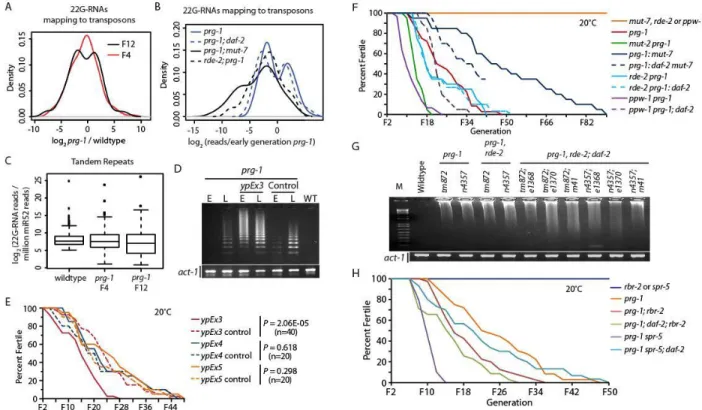
We asked whether progressive loss of 22G-RNAs might account for loss of silencing of transposons and tandem repeats. Overall levels of secondary 22G-RNAs mapping with up to two mismatches to transposon consensus sequences, normalized to the levels of a somatic microRNA, were reduced in early-generation prg-1 strains (P=0.0008, 1 sample T-test, two-sided) (Figures 5A and 1-S4G). In later generations, RNAs targeting some transposons were further reduced, whereas 22G-RNAs for other transposon classes were restored to high levels, reflected by an apparently bimodal distribution of read differences relative to N2 wildtype (Figure 1-5A). Transposons with increased 22G-RNAs in late-generation prg-1 animals were enriched for the Tc4 family of transposons whilst
17
Outcrossed mut-7 and rde-2 Mutator mutants are not Mrt (Figure 5F), despite being required for general amplification of 22G-RNAs in response to exogenous and endogenous primary siRNAs (Zhang et al., 2011). By comparing the abundance of 22G-RNAs from the prg-1 and mut-7 single mutants with prg-1; mut-7 double mutants, the prg-1; mut-7 double mutant were much more similar to the mut-7 single mutant than to the prg-1 single mutant (Figure 1-S5A). Importantly out of all the transposon consensus sequences, there were no transposons with more than 5 antisense 22G-RNA reads per million in mut-7 that had fewer than 5 reads per million in the prg-1 single mutant, showing that prg-1 is
unlikely to operate in parallel to mut-7 at transposons. There were some isolated cases where there were fewer reads in prg-1; mut-7 than in mut-7; however, because these had a larger number of reads in prg-1, it is not straightforward to argue that loss of these small RNAs is related to the Mrt phenotype. Overall, the vast majority of PRG-1-dependent 22G-RNAs are also dependent on MUT-7 and are
dispensable for germ cell immortality.
Several upregulated repeat tracts are also predicted targets of at least one piRNA (Tables 1-S5 and 1-S6). We therefore examined 22G-RNAs against simple repeats in prg-1 mutants. The number of 22G-RNAs against simple repeat regions was smaller in prg-1 mutants than in wild-type animals (P=0.002, 1 sample T-test, two-sided), and overall 22G-RNA levels decreased further in late-generation prg-1 animals (P=0.00026, 1 sample T-test, two-side) (Figure 1-5C). Furthermore, similarly to
18
repetitive regions of the genome in late-generation prg-1 mutants is accompanied by progressive dysfunction of 22G-RNAs targeting these regions, reflecting both loss of 22G-RNAs downstream of piRNAs, and potentially, upregulation of a prg-1-independent 22G-RNA pathway that can silence a subset of transposons and simple repeats.
19
Figure 1-5. Altered 22G-RNA frequencies and gene expression changes in prg-1 mutants. (A) 22G-RNAs targeting transposons in prg-1 versus wild-type strains. (B) Increased 22G reads in late-generation prg-1 strains mapping to transposons are reduced in prg-1 mutants deficient for the Mutator pathway genes rde-2 or mut-7. (C) 22G-RNAs targeting tandem repeats in prg-1 versus wild-type strains. Note that it is easier to clearly identify transposon 22G-RNAs, because many permutations of each tandem repeat are found, so tandem repeat 22G-RNA data are almost certainly underestimated. (D) RT-PCR reveal
increased expression of 174 mer tandem repeat for strains carrying the ypEx3 extrachromosomal array in comparison to sibling control strains lacking this array or prg-1 single mutant controls. (E) Repetitive extrachromosomal arrays containing CeRep59 and a 174 mer tandem repeats (ypEx3 array) or a histone locus cluster containing his-13, his-14, his-15, and his-16 genes (ypEx4 array) or a Helitron transposon (ypEx5 array) reveal that ypEx3 array accelerates the progressive sterility phenotype of prg-1. (F) rde-2 and ppw-1 are required for suppression of prg-1 by daf-2. Two ppw-1 prg-1 double mutant strains were studied where prg-1 alleles tm872 and n4357 were combined with ppw-1(pk1425) (n=10 strains per genotype). Four ppw-1 prg-1; daf-2 triple mutants were studied where both ppw-1 prg-1 mutants were combined with daf-2 alleles e1370 or e1368 (n=5 strains per genotype). Two rde-2 prg-1 double mutant strains were studied where prg-1 alleles tm872 and n4357 were combined with rde-2(ne221) (n=10 strains per genotype). Six rde-2 prg-1; daf-2 triple mutants were studied where both rde-2 prg-1
20
mutant was combined with daf-2(e1370) and prg-1(n4357) spr-5 was combined with daf-2(e1368) (n=10 strains per genotype).
A small RNA pathway restores germ cell immortality prg-1 mutants
Having established that progressive loss of 22G-RNAs against tandem repeats and transposons occurred in prg-1, we tested whether daf-2 might suppress increased repeat expression via siRNAs. We examined small RNA libraries from prg-1; daf-2 mutants at early- and late-generations. As for prg-1 single mutants, piRNAs were lost from both early- and late-generation prg-1; daf-2. We therefore examined protein-coding genes, transposons and simple repeats showing decreased small RNAs in late-generation prg-1 relative to early-late-generation for suppression of translate-generational decrease by daf-2. The number of suppressed genes was statistically significant for 22G-RNAs mapping to genes and simple repeats but not transposons, although individual examples of transposons in this category were found (Tables 1-1 and 1-S6). In all cases, this suppression involved the Mutator pathway, as the majority of suppressed genes, transposons and repeats showed reduced 22G-RNA reads mapping to them in rde-2, prg-1; daf-2 triple mutants compared to early-generation prg-1 mutants (Table 1-1). In addition, sequencing of small RNAs from progeny of sterile prg-1 mutants that were restored by daf-2 RNAi revealed that 22G-RNAs targeting repetitive sequences were partially restored in comparison to late-generation prg-1 controls, though not to early-late-generation levels (P<1e-16 to late late-generation, two-tailed paired T-test) (Figure 1-S5H). We therefore hypothesized that daf-2 may suppress activation of
repetitive loci by upregulating an alternative prg-1-independent silencing pathway.
We therefore tested whether the suppression of prg-1 fertility defects by daf-2 might be
21
required for efficient germline RNAi, PPW-1 (Tijsterman et al., 2002), is required for suppression of prg-1 by daf-2 (Figure 1-5F). We conclude that an endogenous RNA interference pathway that requires RDE-2, MUT-7 and PPW-1 can restore germ cell immortality to prg-1 mutants (Figure 1-6).
Small RNA pathways can promote gene silencing by degrading RNA in the cytoplasm or by silencing loci within the nucleus. We tested the hypothesis that the histone H3 lysine 4 (H3K4) demethylase RBR-2, which removes trimethyl H3K4 marks and promotes transcriptional silencing (Christensen et al., 2007), mediates the effects of transgenerational epigenetic marks that regulate somatic longevity in C. elegans (Greer et al., 2010). Further, the rbr-2 locus is regulated by reduced daf-2 signaling (Lee et al., 2003). We found that rbr-2 is required for suppression of prg-1 by daf-2 mutation (Figure 1-5H). Late-generation prg-1; daf-2; rbr-2 strains expressed high levels of RNA from repetitive loci (Figures 1-S5L-N), implying that RBR-2 demethylase suppresses the transgenerational fertility defects of prg-1 mutants by silencing these loci in response to reduced daf-2 signaling. We then tested a second demethylase, SPR-5, which removes dimethyl H3K4 marks. Although one allele of spr-5 has been reported to be Mrt at 20oC (Katz et al., 2009), we tested another null allele of spr-5, by134, which does not display fertility defects at 20oC. We then used spr-5 to confirm that H3K4 demethylation is required for suppression of prg-1 by daf-2 (Figure 1-5H).
22
Figure 1-6. Model of parallel small RNA silencing pathways that can repress transgenerational fertility defects. (A) Wild-type PRG-1 maintains transgenerational fertility by silencing repetitive RNA expression, and functions separately to initiate silencing of foreign transgenes and most transposons. (B) Deficiency for prg-1 results in transgenerational desilencing of repetitive loci and sterility. Transposon and
transgene silencing is maintained by Mutator proteins. (C) Increased DAF-16 signaling via daf-2 mutation suppresses progressive sterility and desilencing of repetitive loci when prg-1 is mutant. Mutations are indicated by a red X. Light color tints indicate pathway dysfunction for prg-1 mutants or low levels of DAF-16 activity in response to wild-type DAF-2 signaling.
Table 1-1. Effect of daf-2 on transgenerational alterations in 22G-RNA levels in prg-1.
2-fold down late prg-1 vs early
prg-1
Suppressed daf-2 Significance (Fisher's exact test)
Suppressed dependent on rde-2
Transposons 54 4 1 4
Genes 356 71 5.80E-29 50
Simple
23
DISCUSSION
Here we demonstrate that C. elegans prg-1 is required for germ cell immortality. This phenotype shows two clear distinctions from the role of Piwi proteins in promoting fertility in other organisms. First, prg-1 mutant animals do not become sterile immediately, and indeed maintain wild-type levels of fertility for several generations before becoming progressively sterile. Second, we show that the sterility of prg-1 animals is not due to increased transposition. Instead, our data supports a transgenerational epigenetic cause of sterility in prg-1 mutants.
A role for PRG-1 in transgenerational epigenetic maintenance of fertility in C. elegans connects to recent data showing that prg-1 acts upstream of epigenetic silencing of foreign transgenes in C. elegans germ cells (Ashe et al., 2012; Luteijn et al., 2012; Shirayama et al., 2012). In the case of transgenes however, the silent state is maintained independently of PRG-1 activity by proteins that mediate downstream 22G-RNA production, such as RDE-2, by nuclear RNAi factors, and by chromatin silencing proteins (Figure 1-6A-B) (Ashe et al., 2012; Luteijn et al., 2012; Shirayama et al., 2012). Several nuclear RNAi factors were recently reported to promote germ cell immortality (Buckley et al., 2012), and PRG-1 and associated piRNAs could function upstream of these proteins to direct silencing of
endogenous nuclear loci. However, we show that PRG-1 is also required continuously for germ cell immortality, suggesting that ultimately prg-1 is indispensable for silencing of some endogenous loci. A second contrast with transgene silencing is that Mutator mutants such as rde-2 and mut-7, which display a strongly reduced secondary siRNA response, are wild-type for germ cell immortality at low
24
evidence for a distinction between transgene silencing and the Mrt phenotype of prg-1 by showing that transgene silencing is not disrupted in sterile late-generation prg-1 mutant adults (Figure 1-S2N). We suggest that at least two classes of ‘non-self’ DNA exist: recently introduced foreign transgenes whose permanent silencing can rapidly become independent of PRG-1 (Ashe et al., 2012; Luteijn et al., 2012; Shirayama et al., 2012), and a distinct class of loci, possibly rapidly evolving tandem repeats or a recently introduced transposable element, whose long-term silencing requires the activity of PRG-1.
Despite the continuous requirement for PRG-1 in a wild-type background for maintenance of silencing of repetitive loci (Figures 1-4 and 1-S4D-E), an alternative PRG-1-independent silencing pathway can be activated by reduced insulin signaling (Figure 1-2). Analysis of small RNA populations in a variety of prg-1 mutant backgrounds suggested that a small RNA silencing pathway could be the mechanism by which reduced insulin signaling suppresses the transgenerational fertility defects of prg-1 mutants (Figures 1-5A-C, 1-S4G-I and 1-S5B-D). This led us to define components of an endogenous small RNA silencing pathway that are required for daf-2 to suppress prg-1 (Figures 1-5F and 1-6C, Table 1-S2). This small RNA pathway may act upstream of a chromatin-based silencing pathway involving rbr-2- and spr-5-mediated demethylation of histone H3 K4 (Figure 1-5H). Thus our results indicate that activation of a small RNA genome silencing pathway that protects germ cell immortality, in addition to its known role in initiating anti-aging gene expression (Balch et al., 2008; Kenyon, 2010), is a significant consequence of daf-2 deficiency. Piwi proteins promote silencing of transposable elements,
transcriptional activation, imprinting, heterochromatin formation and modulation of protein function via Heat Shock Proteins (Juliano et al., 2011; Rangan et al., 2011; Watanabe et al., 2011), and our results suggest that a genome silencing function of Piwi that is independent of suppression of many
25
We found that daf-16 and daf-18 mutations shortened the transgenerational lifespan of prg-1 mutants and prg-1; daf-2 double mutants (Figure 1-2B). Thus, basal levels of DAF-16 activity contribute to the transgenerational lifespan of prg-1 mutants, revealing an intriguing parallel with the established role for low levels of DAF-16 activity in promoting the adult lifespan of wildtype animals (Kenyon et al., 1993; Larsen et al., 1995). Although many mutations in the small RNA silencing pathway that functions downstream of DAF-16 to suppress deficiency for prg-1 also resulted in reduced transgenerational lifespan when combined with prg-1, this effect did not occur for all such mutations (Table 1-S2). The reason for the shortened transgenerational lifespan of prg-1 daf-16 double mutants therefore remains uncertain.
Our observations defy a prediction of the antagonistic pleiotropy theory of aging, which suggests that prolonged lifespan might result in compromised fertility (Williams, 1957). Instead, some interventions that repress aging in somatic cells may be beneficial to germ cells. Whether the heritable epigenetic defects that result from prg-1 deficiency impact somatic lifespan, and if these are related to the germline function of RBR-2 that can extend adult lifespan (Greer et al., 2011), and how DAF-16 regulates the small RNA pathway that suppresses deficiency for prg-1, are intriguing questions raised by this study.
26
mammalian cells undergo senescence (De Cecco et al., 2013) and also in the aging-related disorder cancer (Ting et al., 2011; Zhu et al., 2011). Further, increased expression of Alu retrotransposons may contribute to adult-onset macular degeneration as well as proliferative aging of human stem cells grown in vitro (Kaneko et al., 2011; Wang et al., 2011). We speculate that Piwi-dependent regulation of
27
MATERIALS AND METHODS Germline Mortality assays
Worms were assessed for the Mortal Germline phenotype using the assay previously described (Ahmed and Hodgkin, 2000). Once per week, 6 L1 or L2 animals would be placed on fresh NGM plates seeded with OP50 E. coli bacteria. Each passage would be recorded and plates that yielded no additional L1 animals were marked as sterile. Mantel-Cox Log Rank Analysis was used to determine differences of transgenerational lifespan between strains.
Small RNA Sequence Analysis
High-throughput sequencing of whole animal small RNAs was performed after generating 5’ independent 18-30 nucleotide small RNA libraries as described previously (Das et al., 2008). Alignment to the genome and matching to known piRNA loci was performed as described previously (Bagijn et al., 2012, see Extended Experimental Procedures). For analysis of 22G-RNAs mapping to transposons or simple repeats, 22G-RNAs were first selected from the libraries using a custom Perl script. Transposon consensus sequences were downloaded from RepBase (version 17.05) and repeat sequences were extracted from the ce6 genomic sequence using the genomic coordinates of simple repeats,
downloaded from the UCSC genome browser website (simpleRepeats.txt). Mapping was carried out using Bowtie reporting the best single match with up to two mismatches, to avoid the problem of multiple matches potentially exaggerating differences between samples. With these parameters if there are multiple “best” matches, as is likely to be the case for repeats, Bowtie will report one match
28
uniquely to the ce6 genome with no mismatches were selected from the libraries using custom Perl scripts, and those mapping antisense to genes were identified by comparing coordinates with the coordinates of genes (sangerGene.txt) downloaded from the UCSC genome browser website. For comparison, sequences perfectly matching C. elegans mature microRNA sequences downloaded from miRbase (http://www.mirbase.org/) (Kozomara and Griffiths-Jones, 2011) were selected using a custom Perl script.
Microarray analysis of RNA expression and CGH
Total RNA was harvested from whole plates of worms that had been allowed to deplete their food supply, but not enter starvation. Standard phenol/chloroform RNA extraction was used to recover whole animal RNA. DNA for CGH was harvested from animals grown on NGM agar plates using Qiagen DNeasy Blood and Tissue Kit. cDNA and genomic DNA was processed and used for tiling microarray analysis following the Nimblegen protocols for HX1 microarrays (Ikegami et al., 2010). Scanning of arrays was performed using calibrations for repeat elements.
Microarray data analysis
C. elegans tiling arrays normalized to gene models were used to compare gene expression changes in prg-1 and prg-1; daf-2. Repeat expression was analyzed by mapping the probes to repeat positions downloaded from the UCSC genome browser as described above. Control regions for each chromosome were generated using a custom script in R. First, a set of 1000 repeat sequences were sampled from the total complement of repeats on each chromosome (with replacement) and the length of these sequences stored. A random number generator was used to provide 1000 starting positions across the chromosome and these starting positions were paired with the lengths at random to
29
repeats was generated for each allele by sampling N members of the total set of either genes or repeats, where N is the number of altered genes or repeats for the allele in question. The overlap for the three random samples was then calculated and stored. The entire process was repeated 1000 times and a mean and standard deviation of random overlap computed. The size of the observed overlap could thus be compared to simulated random overlap using the Z statistic.
CGH analysis
Comparison between early- and late-generation prg-1 alleles n4357 and tm872 for copy number across the genome was used to specifically interrogate regions mapping to genes, transposons and tandem repeats downloaded from Wormbase (ce6 assembly; WS190). Tandem repeat copy number changes were compared to randomly selected genomic regions generated as for expression analysis above.
Extrachromosomal arrays
Primers were designed to amplify repetitive sequences directly from the genome. For an experimental array consisting of direct repeats, sequences from two CeRep59 loci (chrI:4281435, chrIII:7405366) and a 174-mer simple repeat (chrIV:6682640) were used. An additional experimental array consisting of a Helitron transposon sequence (chrIV:16880484). A control array was created using primers flanking a stretch of genomic sequence containing the H2A, H2B, H3 and H4 histone locus his-13, his-14, his-15, and his-16.
RNA Fluorescence In Situ Hybridization
Freshly outcrossed alleles of
prg-1
,
n4357
and
tm872
, and
prg-1
lines containing the
extrachromosomal array
ypEx3
were created, and RNA FISH was performed on F4 animals to
visualize repetitive RNA expression. A DNA oligonucleotide probe coupled with a 5' Cy5
fluorophore was designed to detect RNA transcripts of
CeRep59
on Chromosome
I
(located at
30
RNA FISH was performed with mixed stage animals from non-starved plates. Animals
were washed off plates into microcentrifuge tubes then washed once in 1mL M9 buffer
followed by three washes in 1mL of 1x DEPC-treated PBS. Animals were then fixed for 45
minutes at room temperature in 1 mL of fixation buffer (3.7% formaldehyde in 1x DEPC-treated
PBS). Following fixation, animals were washed twice in 1mL 1x DEPC-treated PBS and
permeabilized overnight at 4 degrees in 1mL of 70% ethanol in DEPC-treated H
2O.
The following day, hybridization buffer was prepared (0.2 g dextran sulfate, 200 mL 20x
RNAse-free SSC, 200 mL deionized formamide, 1.5mL DEPC-treated H
2O). Dry probes were
diluted in RNAse-free TE buffer to a concentration of 25 mM. Probes were then further diluted
by mixing into hybridization buffer for a final concentration of 1.25 μM. Permeabilized animals
were washed for 5 minutes at room temperature in 1mL wash buffer (10% formamide in 2x
RNAse-free SSC). Wash buffer was removed and 100 mL of probe in hybridization buffer was
added to each sample. Then the samples were incubated overnight at 30 degrees. The next day,
samples were washed once in 1mL wash buffer for 30 minutes at room temperature then a
second time in 1mL wash buffer with 25 ng/mL DAPI counterstained for 30 minutes at room
temperature. Animals were mounted on glass slides using VECTASHIELD mounting media
(Vector Laboratories, Inc.) and imaged by epifluorescence microscopy using the same exposure
31
SUPPORTING MATERIAL
32
nrde-33
1 strain reveals transgene expression in a nrde-1 nuclear RNAi-deficient background. (M) DIC image for L. (N) The silenced GFP piRNA sensor transgene remains silent in for fertile prg-1 mutant adults in early and late generations as well as for sterile late generation prg-1 adults (shown in M). (O) DIC image for N. The GFP piRNA sensor transgene shown in panels L and N was initially created in a prg-1 mutant
background, then silenced by crossing away into a wildtype prg-1(+) background, and then crossed back into either nrde-1 or prg-1 mutant backgrounds. Dotted lines outline the germline. Signal outside the germlines is due to autofluorescence from the intestine.
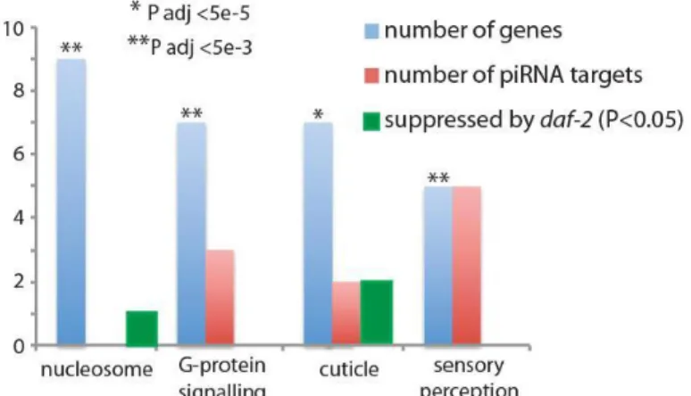
Figure 1-S3. Altered gene expression can reflect piRNA targeting and can be affected by daf-2. For genes that changed more than 2-fold in late-generation prg-1 strains, these were not significantly enriched for germline-specific genes (P>0.1, Fisher’s Exact Test). Gene Ontology analysis showed the set to be highly enriched (P<0.005, Fisher’s exact test after Benjamani and Hochberg multiple test
34
Figure 1-S4. Supplementary analysis of transposon families, tandem repeat tracts and 22G RNAs. (A) Transposon categories enriched or depleted for increased expression in late-generation prg-1 relative to early-generation. (B) Transposon categories enriched or depleted for increased 22G reads in
35
primers were used to confirm the microarray results shown in panel D by RT-PCR. (F) Silencing to tandem repeat loci restored when late-generation sterile prg-1 adults are rescued by RNAi of daf-2 in comparison to late-generation strains fed on OP50 bacteria. (G) Progressively reduced median
36
37
38
Figure 1-S6. Increased FISH staining CeRep59 transgenic animals carrying the ypEx3 array. Antisense Cy5 probe against the CeRep59 repeat was used to evaluate the expression levels and pattern of the sequence encoded by the array injected into prg-1 mutants. (A, B) Low expression observed in N2 wild-type animals observed in the embryos. (C-F) Probe staining in prg-1(n4357) shows expression in embryos and low, diffuse staining throughout the animals. (G-J) Increased CeRep59 expression in all tissues and embryos of prg-1(n4357) animals containing the ypEx3 array encoding copies of the CeRep59 repetitive sequence. Animals also displayed strong staining in the pharynx (H, arrows), due to the
incorporation of a Pmyo-2::mCherry plasmid to track the injected array, where the myo-2 promoter drives expression in the pharynx. (K-N) Probe staining for prg-1(tm872). (O-R) Increased staining in prg-1(tm872) lines containing ypEx3 array. All paired images were taken in the same focal plane. DAPI images and Cy5 images all taken with equal exposure. When compared to wildtype, prg-1 mutant lines displayed a 1.23-fold and 1.02-fold increase in staining in the embryos and body, respectively. prg-1 lines containing ypEx3 displayed a 5.05-fold increase in embryo staining over prg-1 controls and a 5.35-fold increase for general body staining.
Tables 1-S1 through 1-S8 can be viewed at:
39
CHAPTER II: ADULT REPRODUCTIVE DIAPAUSE IN RESPONSE TO A HERITABLE EPIGENETIC STRESS 2
SUMMARY
Environmental cues such as starvation or seasonal change can transiently suspend reproduction in response to stress. Deficiency for the C. elegans Argonaute Piwi prg-1 results in strains that can reproduce for many generations but then become sterile in response to a heritable epigenetic defect. Late-generation prg-1 mutants matured into sterile adults that displayed germ cell atrophy. This developmental program was reversible and depended on the apoptosis protein CED-3 and the nuclear hormone receptor NHR-49, which remodel the germline in response to starvation. Germ cell atrophy also required CEP-1, whose mammalian homolog p53 regulates senescence in the context of
proliferative aging, and the DAF-16/Foxo stress response transcription factor. We propose that prg-1 mutants transmit a heritable, epigenetic stress that induces Adult Reproductive Diapause, a
developmental program where germ cells enter a reversible state of suspended animation that could reflect an ancestral interface between aging and senescence.
__________________________
2
40 INTRODUCTION
Diapause is a state of developmental arrest that promotes survival in response to harsh environmental conditions such as seasonal change or limited nutrient availability and has been documented for a variety of invertebrates (Schiesari and O’Connor, 2013; Saunders et al., 2002). Vertebrates, possibly including humans, can also enter a state of suspended animation in response to anoxia (Blackstone and Roth, 2005). One form of diapause in insects is Adult Reproductive Diapause (ARD), where larvae mature into sterile adults that can become fertile when environmental conditions improve. While environmental cues can modulate ARD, it is unclear if this developmental program can be triggered in response to an endogenous stress.
41 RESULTS Adult Reproductive Diapause in sterile prg-1 mutants
Late-generation prg-1 mutant populations were identified by scoring for populations where all young adults present were sterile and younger sibling animals, likely to become sterile, were available for analysis. In both early generation and sterile generation animals, germline development was
comparable to wildtype for L4 larvae, the stage immediately preceding adulthood (Figure 2-1A, B, Figure 2-2A, B, and Figure 2-S1A). Although a fraction of sterile prg-1 adults contained germlines of normal sizes (Figure 2-S1F), germline remodeling events occurred in most sterile prg-1 worms during the L4 to adult transition, resulting in severe germ cell atrophy or lack of germ cells altogether (Figures 2-1D, E). Additionally, a small fraction of the population displayed a shortened germline, considerably more flush and populated than the atrophy phenotype, but not as robust as those displaying a “normal” phenotype (Figure 2-1F). Extensive scoring revealed that the majority of sterile young adults displayed germline atrophy, where small populations of germ cells remained in distal arms of the germline and mature oocytes were absent (Figure 2-2B). While germ cells of early-generation prg-1 mutant adults displayed normal progression from mitosis to meiosis, germ cell atrophy in sterile prg-1 adults resulted in germ cell nuclei with condensed chromosomes, suggesting mitotic arrest, and lacked a transition zone where chromosomes pair during initiation of meiosis or more differentiated meiotic pachytene cells
42
Figure 2-1. Germline atrophy in sterile prg-1 animals. DAPI-stained photos of (A) an N2 wildtype L4, (B) a late generation prg-1 L4 likely to become sterile, (C) an N2 wildtype adult, (D) a late generation prg-1 mutant sterile adult with germline atrophy, (E) a late generation prg-1 mutant sterile adult with two empty germline arms, (F) a late generation prg-1sterile adult with shortened germline arms, (G) the mitotic region of an early generation prg-1 adult, (H) the meiotic region of an early generation prg-1 adult, (I) mitotic cells in a late generation prg-1 worm with germline atrophy, (J) meiotic cells in a sterile prg-1, daf-16 adult.
43
rearrangement during the transition into adulthood. This suggests a programmed developmental response to the heritable epigenetic defect that causes sterility in prg-1 mutants. Additionally, the prevalent phenotype of atrophied germlines in sterile animals resembles a germline remodeling event that occurs when C. elegans L4 larvae undergo starvation and enter a state of adult reproductive diapause, termed ARD (Angelo et al., 2009). Starvation-induced ARD requires the apoptosis gene ced-3 and the nuclear hormone receptor nhr-49 (Angelo et al., 2009). A role for apoptotic cell death in the dramatic reduction in germ cell numbers that occurs as most sterile prg-1 mutants was assessed using 3 and 4 mutations that eliminate apoptosis (Metzstein et al., 1998). Deficiency for 3 or ced-4 failed to rescue the progressive sterility phenotype of prg-1 (Figure 2-2C). However, sterile prg-1; ced-3 and prg-1; ced-4 double mutants displayed reduced levels of germline atrophy in comparison to prg-1 single mutant controls (P=1.0E-04 for ced-3, P=2.0E-08 for ced-4) (Figure 2-2D). Thus, apoptotic cell death contributes to germline atrophy in sterile prg-1 animals.
44
Figure 2-2. Cell death potentiates germline remodeling of sterile prg-1 animals.(A) Phenotypes of fertile, early generation (F4) germlines in L4 larva and young adults. (B) Phenotypes of sterile, late-generation germlines in L4 larva and young adults. Sterile animals were isolated as L4s from sterile sister plates for 24 hours to confirm sterility, DAPI stained, and then scored. (C) prg-1, cep-1,prg-1; ced-3,prg-1; ced-4, and dpy-5, prg-1, nhr-49 double mutants become progressively sterile. prg-ced-3,prg-1; ced-3 and prg-ced-3,prg-1; ced-4 double mutants were made for three prg-1 alleles, pk2298, n4357 and tm872 (n=10 lines were propagated per genotype). Additionally, prg-1, cep-1 and dpy-5, prg-1, nhr-49 double mutants were made for two prg-1 alleles, n4357 and tm872 (n=10 lines were propagated per genotype). Generational lifespan for combined prg-1 alleles is shown in comparison to prg-1 single mutant controls. (D) Effects of starvation-induced ARD genes and stress response transcription factors on sterile germline phenotypes of prg-1
Stress response transcription factors promote germ cell atrophy
45
proteotoxicity, oxygen radicals, pathogenic bacteria and high metal concentrations (Zhou et al., 2011). Based on these results, we hypothesized that DAF-16 activity likely plays a role in prg-1-induced ARD. A role for DAF-16 in germline atrophy of prg-1 mutants was directly evaluated by examining sterile late-generation prg-1, daf-16 double mutant adults, which displayed strongly reduced frequencies of germline atrophy (Figure 2-S2A). Although germline arms of sterile prg-1, daf-16 double mutants were primarily underproliferated (short) in comparison to those of wildtype, most contained many more cells than atrophied germlines of prg-1 single mutants (Figure 2-S1G). Short prg-1, daf-16 germline arms also displayed hallmarks normally observed during meiosis such as a transition zone (chromosome pairing) and synapsed homologous chromosomes (Dernburg et al., 1998), as well as mature oocytes at the proximal end of the germline, despite a lack of fertilized embryos (Figure 2-1J and Figure 2-S1G). These phenotypes were recapitulated in prg-1, daf-16; daf-2 triple mutants (Figure 2-S2A). Thus, DAF-16 promotes proper germline remodeling in sterile late-generation prg-1 animals via the daf-2/daf-16 insulin signaling pathway.
46
Having established a role for daf-16 in prg-1-induced ARD, we set out to further elucidate the role of DAF-16 in the germline remodeling program of ARD. DAF-16 can translocate from the cytoplasm into the nucleus in response to various stresses as well as reduced daf-2 signaling (Friedman, 1998; Kenyon, 1993)). We crossed independent DAF-16::GFP transgenes into prg-1 mutant backgrounds, and found that DAF-16::GFP was primarily cytoplasmic for early generation and fertile late-generation adults (Figure 2-3A-C, G-I), indicative of low DAF-16 activity. In sterile late-generation adults, DAF-16::GFP translocated into nuclei of intestinal cells (Figure 2-3D-F, J-L), which supports the observation that reduced daf-2 signaling promotes germ cell remodeling in sterile prg-1 adults.
47
Figure 2-3. daf-16 translocalizes to intestinal nuclei in sterile prg-1 animals. (A-C, G-I) DAF-16::GFP is primarily cytoplasmic in early generation prg-1 animals, with little to no nuclear localization. (D-F, J-L) DAF-16::GFP localizes to intestinal nuclei in sterile, ARD animals. No apparent activity is seen in the germline. CF1407 animals were imaged with a 5 sec exposure, while TJ356 animals were imaged with 2 second exposure.
48
germline rearrangement in sterile prg-1 animals, suggesting a large role for stress response pathways in ARD.
The involvement of ced-3 and ced-4 in germline rearrangement in sterile prg-1 animals suggests a strong role for the apoptotic cell death pathway. Mammalian p53 are well as the C. elegans homolog of p53, CEP-1, can promote cell death or cell cycle arrest in response to a variety of stresses, including DNA damage, heat shock, oxidative, anoxia, osmotic stresses (Gartner et al. 2008). CEP-1 acts upstream of CED-3 and CED-4 to promote germ cell apoptosis in response to genotoxic stress as well as other stresses (Gartner et al., 2000). Based on this information, we hypothesized that cep-1 may play a large role in facilitation germline rearrangement, perhaps more so than either ced-3 or ced-4 by themselves. cep-1 failed to rescue progressive sterility of prg-1 and significantly enhanced the onset of sterility (p = 6.35E-05) (Figure 2-2C). Additionally, prg-1, cep-1 sterile animals displayed a dramatically altered ARD response, with a substantial increase in germlines of normal sizes, though these animals lacked embryos (Figure 2-2D). This robust shift favoring larger germlines was a significantly larger percent of the
population of sterile animals than found in either ced-3 or ced-4 double mutants with prg-1 (p <1.0E-06 vs. prg-1; ced-3, p<4.32E-04 vs. prg-1; ced-4). Thus, cep-1 strongly promotes germline rearrangement during ARD.
49
germlines, this data indicates that cep-1 functions upstream of daf-18 activity and suggests that it may be epistatic to the input from the insulin signaling pathway. Finally, we looked at the germlines of prg-1, nhr-49; ced-4 animals and found that the distribution did not favor either of the double mutants (Figure 2-S2B). While not as informative as a clear epistatic relationship, this result (in conjunction the
discrepancies in the other phenotypes observed for the other triple mutants) suggests that the
interaction between these genes and their corresponding pathways is complex and may involve several genes interactions that affect each pathway at several intersections.
Germ cell atrophy represents a form of reversible sterility
Germline remodeling of sterile prg-1 L4 larvae that mature into adults resembles starvation-induced ARD. For starvation-starvation-induced ARD, addition of food can restore fertility to animals by inducing the germline to exit ARD, demonstrating that ARD is a facultative stress response that suspends reproduction in response to starvation until favorable nutritional cues are encountered (Angelo et al., 2009). We previously showed that strong reductions in daf-2 signaling can abolish the progressive sterility phenotype of prg-1 mutants, and that RNA interference (RNAi)-mediated knockdown of daf-2 or the downstream PI3 kinase subunit age-1 can restore fertility to sterile prg-1 adults (See Chapter I). To determine if daf-2 RNAi can restore fertility to animals possessing the germ cell atrophy phenotype, sterile prg-1 adults were sorted into two pools: those with germlines of normal size and those with atrophied. A fraction of animals from each pool became fertile when treated with daf-2 RNAi (Figure 2-4A). Thus, germline atrophy represents a state of reversible sterility akin to ARD, in which fertility could be restored in sterile animals containing either normal or atrophied germlines.
50
became fertile upon treatment with daf-2 RNAi (Figure 2-4A). These results are consistent with restoration of fertility to a small fraction of aged adults subjected to starvation-induced ARD.
51
52 DISCUSSION
Harsh environmental conditions are known to cause a reversible form of sterility termed ARD (Angelo et al., 2009; Ragland et al., 2010). The timing of the germline remodeling events of prg-1 mutants at the L4/adult transition, the genetic requirements of ced-3 and nhr-49, and restoration of fertility to old sterile prg-1 adults, match previously established criteria for starvation-induced ARD (Angelo et al., 2009). We conclude that the heritable epigenetic defect of prg-1 mutants triggers a developmental response that also occurs in response to severe exogenous stresses. We found that fertility could be restored for sterile prg-1 mutants that displayed germlines of normal size or dramatic germ cell atrophy. Thus, germline atrophy is not necessary for animals to enter ARD and instead represents one form of reversible reproductive diapause.
DAF-16/Foxo activity has previously been shown to promote a 60% increase in somatic longevity of adults in response to germline ablation (Berman et al., 2006; Hsin, 1999), and our data demonstrate that DAF-16 can reprogram development of the adult germline (Figure 2-S2A). We found that nuclear DAF-16 was observed in intestinal cells of sterile prg-1 adults which accompanied strong increases in adult lifespan in comparison to fertile late-generation siblings (Figure 2-4B). We also found that three major stress response transcription factors, CEP-1/p53, HSF-1 and DAF-16/Foxo promote germ cell atrophy in sterile prg-1 adults. Together, these results suggest that prg-1 mutants transmit a form of ‘heritable stress’ that accumulates transgenerationally to cause sterility and that triggers somatic longevity extension when it becomes severe.
53
cause sterility in prg-1 mutants. Repetitive elements play major roles in genome evolution. For example, rapid evolution of tandem repeats that specify vertebrate centromeres promotes genomic conflict and speciation (Malik, 2002). We propose that ARD could occur in the context of genomic conflict, or when transposons or retroviruses that enter the germline in horizontal transfer events and create bursts of genomic stress. Thus, ARD may integrate development with genome evolution, possibly allowing time for the genome to adapt to or shape genomic incidents (Figure 2-4C). Consistently, we show that DAF-16/Foxo is stimulated to promote entry into ARD, and that a further increase in DAF-16 activity via daf-2 RNAi restores fertility to prg-1 mutants. Thus, the activity of at least one pathway that promotes ARD is capable of resolving the stress that causes ARD.
We propose that ARD represents a reversible state of germ cell arrest or quiescence that can occur in response to heritable stress. Our results imply that prg-1 mutants become ‘stressed to sterility’ as their germ cells proliferate over the generations. Consistently, increased lifespans were observed in post-mitotic sterile adults, likely due to the presence of DAF-16 in intestinal nuclei. Thus, the heritable stress transmitted by prg-1 mutants can affect post-mitotic aging as well as aging as cells proliferate, suggesting that it could be relevant for understanding how gametes influence aging in mammals (De Cecco et al. 2013). ARD can be triggered by exogenous stresses (Angelo et al., 2009; Ragland et al., 2010), whereas Lamarckian inheritance can also occur in response to environmental cues. These
observations suggest a possible interface between stress-induced decisions that affect transgenerational inheritance and ARD, which could represent a cornerstone of the evolutionary biology of aging in
54
MATERIALS AND METHODS Strains
Unless noted otherwise, all strains were cultured at 20°C on Nematode Growth Medium (NGM) plates seeded with E. coli OP50. Strains used include Bristol N2 wild type, dpy-5(e61) I, unc-13(e450) I, prg-1(n4357) I, prg-1(tm876), prg-1(pk2298), unc-55(e402) I, unc-29(e193) I, 16(mg50) I, 16(mu86) I, hsf-1 (sy441) I, kri-1 (ok1251) I, nhr-49 (nr2041) I, nhr-49 (ok2165) I, 2 (e1368) III, daf-2(e1370) III, daf-2(m41) III, ced-4(n1162) III, 17(e164) III, daf-18(e1375), daf-18(ok480) IV, dpy-9(e12) IV, unc-24(e120) IV, ced-3(n717) IV.
prg-1 mutations were outcrossed versus an outcrossed stock of dpy-5(e61) unc-13(e450), and freshly isolated homozygous F2 lines were established for analysis. dpy-5, unc-55; daf-2 triple mutant were crossed with prg-1 / dpy-5(e61) unc-13(e450) males which were then selected based on Dauer phenotype at 25oC and loss of dpy-5 unc-55 to create prg-1; daf-2. Analogous crosses using marker mutations dpy-17 for ced-4, or dpy-9 for daf-18, or unc-24 for ced-3, as balancers to create double mutant strains. To create the linked prg-1, daf-16 double mutant, prg-1, dpy-24 and unc-13, daf-16 double mutants were first created, then progeny of prg-1, dpy-24 / unc-13, daf-16 heterozygotes that lost unc-13 were identified, and the resulting putative prg-1, daf-16 recombinant chromosomes were made homozygous and PCR genotyped to verify the presence of prg-1 or daf-16 deletions. prg-1, daf-16 doubles were crossed with unc-13 dpy-24; daf-2 / + +; + heterozygous males and then selected for Daf and against Dpy Unc phenotypes to create prg-1, daf-16; daf-2 triples.