A Critical Role for Muscle Ring Finger-1 in Acute Lung
Injury–associated Skeletal Muscle Wasting
D. Clark Files1,2, Franco R. D’Alessio1, Laura F. Johnston1, Priya Kesari1, Neil R. Aggarwal1, Brian T. Garibaldi1, Jason R. Mock1, Jessica L. Simmers3, Antonio DeGorordo1, Jared Murdoch1, Monte S. Willis4, Cam Patterson4, Clarke G. Tankersley5, Maria L. Messi6, Chun Liu2,
Osvaldo Delbono6, J. David Furlow7,8, Sue C. Bodine7,8, Ronald D. Cohn3, Landon S. King1, and Michael T. Crow1
1Division of Pulmonary and Critical Care Medicine, Johns Hopkins Asthma and Allergy Center, Baltimore, Maryland;3McKusick-Nathans Institute of
Genetic Medicine, Baltimore, Maryland;4McAllister Heart Institute, University of North Carolina, Chapel Hill, North Carolina;5Department of Environmental Health Sciences, School of Public Health, Johns Hopkins University, Baltimore, Maryland;6Department of Internal Medicine-Gerontology and2Internal Medicine-Pulmonary, Critical Care, Allergy and Immunology, Wake Forest University School of Medicine, Winston-Salem, North Carolina; and7Department of Neurobiology, Physiology, and Behavior, and8Department of Physiology and Membrane Biology, University of California, Davis, California
Rationale: Acute lung injury (ALI) is a debilitating condition associated with severe skeletal muscle weakness that persists in humans long after lung injury has resolved. The molecular mechanisms underlying this condition are unknown.
Objectives: To identify the muscle-specific molecular mechanisms re-sponsible for muscle wasting in a mouse model of ALI.
Methods: Changes in skeletal muscle weight, fiber size,in vivocontractile performance, and expression of mRNAs and proteins encoding muscle atrophy–associated genes for muscle ring finger-1 (MuRF1) and atro-gin1 were measured. Genetic inactivation of MuRF1 or electroporation-mediated transduction of miRNA-based short hairpin RNAs targeting either MuRF1 or atrogin1 were used to identify their role in ALI-associated skeletal muscle wasting.
Measurements and Main Results: Mice with ALI developed profound muscle atrophy and preferential loss of muscle contractile proteins as-sociated with reduced muscle functionin vivo. Although mRNA expres-sion of the muscle-specific ubiquitin ligases, MuRF1 and atrogin1, was increased in ALI mice, only MuRF1 protein levels were up-regulated. Consistent with these changes, suppression of MuRF1 by genetic or biochemical approaches prevented muscle fiber atrophy, whereas sup-pression of atrogin1 exsup-pression was without effect. Despite resolution of lung injury and down-regulation of MuRF1 and atrogin1, force gen-eration in ALI mice remained suppressed.
Conclusions: These data show that MuRF1 is responsible for mediating muscle atrophy that occurs during the period of active lung injury in ALI mice and that, as in humans, skeletal muscle dysfunction persists de-spite resolution of lung injury.
Keywords:skeletal muscle atrophy; intensive care unit–acquired weakness; critical illness myopathy; muscle atrophy genes; proteasomal-mediated protein degradation
Acute lung injury (ALI) is a syndrome characterized by the acute onset of pulmonary infiltrates and respiratory failure often lead-ing to the need for mechanical ventilation (1). Approximately 200,000 people per year develop ALI in the United States, with mortality high at 30–40% (2). A common complication associ-ated with ALI is skeletal muscle weakness. Weakness in these patients results in decreased long-term mobility and functional status. Skeletal muscle weakness is initiated early in the course of ALI, and has been shown to persist in a large percentage of patients for up to 5 years after resolution of lung injury and hospital discharge (3–6). Although multiple factors may con-tribute to ALI-induced muscle atrophy including reduced nutri-tion, inactivity caused by bed rest, and systemic inflammanutri-tion, the etiology of ALI-associated skeletal muscle atrophy remains incompletely understood.
Skeletal muscle weakness is a common finding not only among patients with ALI, but also in patients with other critical illnesses. Clinically apparent weakness is present in 20–50% of patients with critical illness and has been shown to be an inde-pendent risk factor for mortality in these patients (3, 5, 7, 8). A variety of terms have been used in the literature to describe the myopathic weakness in these patients including acute quadri-plegic myopathy, critical illness myopathy, and thick filament (Received in original form June 29, 2011; accepted in final form January 12, 2012)
Supported by NIH NIAMS F32AR057637 (D.C.F.); NIH NHLBI HL089346 (L.S.K.); pilot grant (NIH P30 DK072488) (M.T.C.); AHA Biomedical Research Grant (M.T.C.); and Eudowood Foundation Bauernschmidt Fellowship Award (D.C.F.).
Author Contributions: D.C.F., F.R.D., L.S.K., and M.T.C. conceived and designed experiments; D.C.F., F.R.D., L.F.J., P.K., N.R.A., B.T.G., J.R.M., J.L.S., A.D., M.L.M., C.L., J.M., and O.D. performed experiments and analysis; M.S.W. and C.P. provided transgenic mice; J.D.F. and S.B. provided research reagents; C.G.T. and R.D.C. pro-vided equipment and oversight for physiologic experiments; and D.C.F. and M.T.C. wrote the manuscript and provided creative input.
Correspondence and requests for reprints should be addressed to Michael T. Crow, Ph.D., Johns Hopkins University School of Medicine, Division of Pulmonary and Crit-ical Care Medicine, The Johns Hopkins Asthma and Allergy Center, 5A.58, 5501 Hopkins Bayview Circle, Baltimore, MD 21224. E-mail: [email protected]
This article has an online supplement, which is accessible from this issue’s table of contents at www.atsjournals.org
Am J Respir Crit Care Med Vol 185, Iss. 8, pp 825–834, Apr 15, 2012 Copyrightª2012 by the American Thoracic Society
Originally Published in Press as DOI: 10.1164/rccm.201106-1150OC on February 3, 2012 Internet address: www.atsjournals.org
AT A GLANCE COMMENTARY
Scientific Knowledge on the Subject
Muscle weakness contributes to the mortality and long-term morbidity of acute lung injury (ALI) in patients. There are currently no reported animal models of ALI-associated skeletal muscle wasting or established molecular mechanisms of how such wasting occurs.
What This Study Adds to the Field
myopathy, the latter referring to the preferential loss of myosin observed in the muscles of these patients (9–11).
Studies over the last 12 years have defined important roles for muscle-specific genes that regulate muscle wasting in well-defined models of skeletal muscle atrophy, including immobili-zation, denervation, and hindlimb suspension (12). Prominent among these are the genes Fbx032(atrogin1 or MAFbx) and Trim63(muscle ring finger protein-1 [MuRF1]), both of which function as ubiquitin E3 ligases in the proteasome-mediated degradation of skeletal muscle proteins (12, 13). Up-regulation of MuRF1 and atrogin has also been observed in the peripheral muscles of patients with chronic obstructive pulmonary disease and in the diaphragms of mechanically ventilated brain-dead patients (14, 15).
In this study, we used a mouse model of ALI to define the molecular mechanisms responsible for ALI-induced muscle wasting. This model of direct lung injury is highly reproducible and proceeds through distinct injury and resolution phases over a relatively short time course (16). Our data show that there is a marked up-regulation of MuRF1 and atrogin1 gene expres-sion in ALI mice within 2–3 days after inducing lung injury and, as in human ALI, contractile dysfunction triggered by ALI that persists despite down-regulation of MuRF1 and
atrogin1 gene expression and complete resolution of lung in-jury. We tested the hypothesis that MuRF1 is necessary for the ALI-associated decrease in muscle mass and loss of contractile proteins during the period of active lung injury. Some of the results of these studies have been previously reported in ab-stract form (17–19).
METHODS
Animals
All animal experiments were conducted under protocols approved by the Johns Hopkins Animal Care and Use Committee. Eight-week-old male C57BL/6 mice (The Jackson Laboratory, Bar Harbor, ME) were housed in a pathogen-free facility. To induce lung injury, mice were anesthetized with an intraperitoneal (IP) injection of 150 mg/kg ket-amine and 13.5 mg/kg acetylpromazine and the trachea exposed. Escherichia coli LPS (O55:B5 L2880; Sigma-Aldrich, St. Louis, MO) at 3mg/g mouse or an equivalent volume of sterile H2O (sham
control) was then instilled intratracheally using a 20-gauge catheter. Animals were not mechanically ventilated and oxygenation or activity levels were not monitored. For IP-LPS studies, mice were given 3mg/g LPS IP and harvested at subsequent time points. Breeding pairs of MuRF11/2mice were provided by Drs. Cam Patterson and Monte
lar lavage fluid (BALF) of ALI (intratracheal [IT]-LPS) and sham (IT-H2O) mice revealed an increase in total cell count (A) and total protein (B) that peaked between Days 3 and 4 in ALI mice and returned to baseline by Day 10. *P,0.03 versus baseline val-ues; n¼5–6. Based on these data and the accompanying histology of the lungs (see Figure E1), we refer to the period between Days 1 and 4 after IT-LPS as the “lung in-jury phase” and between Days 4 and 10 as the “lung resolution phase.” (C) ALI mice have an approximate 20% drop in body weight (BW) 3 days after IT-LPS. yP ,
Willis of the McAllister Heart Institute at the University of North Carolina.
Bronchoalveolar Lavage and Analysis
At specified times after instillation, mice were anesthetized, blood or bronchoalveolar lavage fluid (BALF) removed, and the animals then killed by exsanguination. BALF counts and protein analyses were per-formed as previously described (16).
Lung Histology
Lungs were inflated to 25 cm H2O with 1% low-melting agarose
(Invi-trogen, San Diego, CA); fixed in 10% formalin; then embedded in paraffin and sectioned for histologic evaluation by hematoxylin and eosin staining (20).
Measurement of Plasma Levels of LPS
Blood was collected retroorbitally from mice at 6, 24, and 72 hours after intratracheal (IT) H2O, IT-LPS, or IP-LPS administration and then
placed in ethylenediaminetetraacetic acid–coated tubes. Mice were
then killed by administration of ketamine and acepromazine followed by cervical dislocation. The blood was prepared for measurements of LPS (Limulus assay) levels, according to the manufacturer’s instruc-tions (Hycult Biotech, Plymouth Meeting, PA).
Mouse Grip Strength
Mouse grip strength was measured using a grip strength meter (Co-lumbus Instruments, Co(Co-lumbus, OH) and the peak force generated from one of five successive trials was recorded as described previously (21).
Statistical Evaluation of Data
Data are expressed as the mean6SEM or median and interquartile ranges. Unpaired Studentt test, analysis of variance, or the Mann-Whitney test was used for statistical comparisons when appropriate. Differences were considered significant atPless than 0.05.
Additional methods for reverse transcriptase quantitative polymer-ase chain reaction, Western blotting,in vivoforce measurements, plas-mid generation, and skeletal muscle electroporation are provided in the online supplement.
RESULTS
IT-LPS Causes ALI and Reduced Muscle Function
As described previously (16, 22, 23) and shown in Figure E1 in the online supplement, IT-LPS induced within 3 days a pattern of lung injury that was distinguished by severe alveolar consoli-dation with cellular infiltrates and interstitial thickening. At 7 days post–IT-LPS, lung injury had nearly resolved, and by Day 10 the lungs appeared normal. Total BALF cell counts and protein levels (Figures 1A and 1B) followed a similar time course, rising during the period of active lung injury (Days 1–4) and returning to base-line during the lung recovery and resolution phases (Days 5–10). Sham mice receiving IT-H2O and then killed at Day 3 showed no evidence of lung injury (sham data points, Figures 1A and 1B). Data in the literature and experiments described in Figure E2 suggest that this IT-LPS model differs significantly in its pharma-cokinetics from IP-LPS models of sepsis (22, 23).
Within 3 days after the administration of IT-LPS to mice (here-after referred to as ALI mice), there was an approximate 20% loss in body weight (Figure 1C). Because loss of body weight could be caused in part or in whole by the marked reduction in food intake by ALI mice (Figure 1D), we generated pair-fed (PF) mice to control for this effect (Figure 1E). Importantly, despite similar weight loss profiles in PF and ALI mice (Figure 1F), only
ALI mice exhibited a significant drop in muscle force production as measured by a grip strength meter (Figure 1G).
Reduced Muscle Function Is Associated with Skeletal Muscle Atrophy
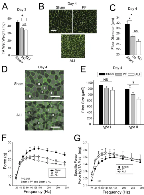
To assess muscle atrophy in sham, PF, and ALI mice in response to lung injury, we first measured the wet weights of the tibialis anterior (TA) muscles. TA wet weights in PF and ALI mice were significantly lower than in sham mice (Figure 2A). To elucidate the mechanisms underlying these losses, we measured the size distribution of muscle fibers sorted by diameter as described in the online supplement. Figure 2B shows images of cryosections with the sarcolemmae delineated byg-laminin immunofluores-cence chemistry. Figure E3 shows a histogram of the size distri-bution of fiber diameters and Figure 2C the mean fiber diameters from each of the three groups. These data reveal that mean fiber diameters of ALI and PF mice were statistically different from time-matched controls but not from each other (Figures 2B and 2C).
To determine if different muscle fibers are differentially af-fected by ALI, mean fiber diameters of types I and II muscle fibers were assessed using the soleus muscle, because the TA muscle has few type I fibers. Staining of the muscles to identify type I (light
gray) and II (dark gray) fibers is shown in Figure 2D and the mean fiber distribution of types I and II fibers is summarized in Figure 2E. The results show that type II muscle fibers are differ-entially affected by ALI-induced muscle wasting.
Because grip strength measurements reflect not only the contrac-tile properties of the muscle, but also neurogenic and behavioral effects, we also measured muscle force production elicited byin situstimulation of the peritoneal nerve. Day 4 PF and ALI mice showed a similar reduction in absolute force generation compared with sham mice (Figure 2F). Specific force generation (absolute force normalized to muscle mass), however, was the same for all three groups (Figure 2G), suggesting that decreased force gener-ation in ALI and PF mice at Day 4 may be directly caused by loss of muscle mass alone (Figure 2A).
MuRF1 Protein Levels Are Differentially Regulated in ALI and PF Mice
Although loss of muscle mass and mean fiber diameter were sim-ilar in PF and ALI mice during the period of active lung injury
(Days 2–4), an analysis of the molecular changes occurring in these mice revealed substantial qualitative differences. Figure 3A shows that the expression of sarcomeric muscle myosin heavy chain (MHC) proteins was suppressed in PF mice and even more so in ALI mice. Because striated MHCs are well-described targets for degradation by the ubiquitin-proteasomal system (24), we measured levels of polyubiquinated proteins in the three experimental groups (Figure 3B) and found that they were increased in ALI but not PF mice. The blot in Figure 3B shows extensive ubiquitination of proteins coincident with sar-comeric MHCs.
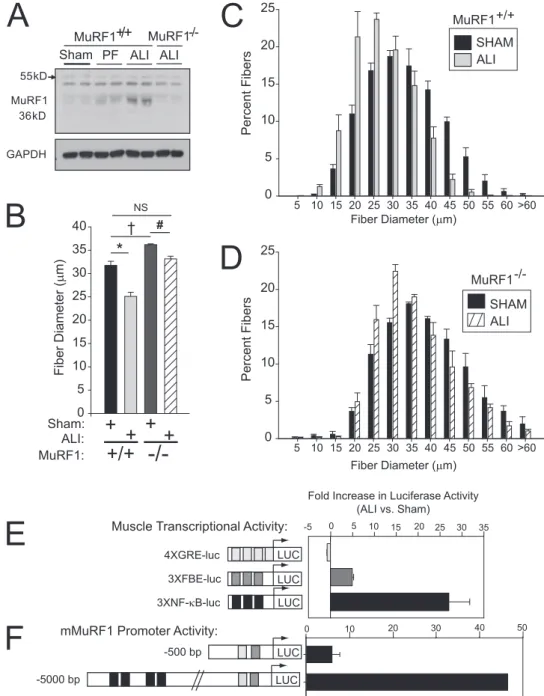
Two of the most common genes up-regulated in muscle wast-ing areTrim63(MuRF1) andFbxo32(atrogin1) (12, 13, 25–27). These proteins function as muscle-specific ubiquitin E3-ligases that tag proteins for the ubiquitin-proteasomal pathway. Figures 3C and 3D show that MuRF1 and atrogin1 mRNA expression were mark-edly up-regulated in the TA muscles of ALI mice, but only modestly up-regulated in PF muscles. Expression of MuRF1 protein followed a pattern similar to its mRNA, higher in ALI muscles (Figure 3F) and lower in PF muscles (Figure 3E), whereas the expression of Figure 4. Muscle wasting in muscle ring finger-1 (MuRF1)2/2mice and increased
nuclear factor (NF)-kB activity in acute lung injury (ALI) wild-type (WT) mouse muscles. (A) Western blot of MuRF1 pro-tein expression shows up-regulation in ALI MuRF1 WT but no up-regulation in ALI MuRF12/2mouse muscles. GAPDH¼ glyceraldehyde phosphate dehydroge-nase; PF¼pair fed. (B) Mean fiber diame-ters for WT and MuRF12/2tibialis anterior (TA) muscles show a moderate increase in mean muscle fiber diameters in sham con-trol MuRF12/2 mice. Exposure to ALI
markedly reduces mean muscle fiber di-ameter in WT muscles mice but only a slight reduction in the MuRF12/2
mus-cle. *P¼0.004;yP¼0.007;#P¼0.03. (C) Histograms of the fiber distribution of sham and ALI-treated MuRF11/1 (WT)
muscles reveal a large shift to the left (smaller size). (D) Histograms of the fiber distribution of sham and ALI-treated MuRF12/2(knockout) muscles show only
atrogin1 protein was not affected by any condition. Data in Figure E4 show that MuRF1 and atrogin1 mRNA expression were also up-regulated in the diaphragms from Day 3 ALI mice.
MuRF1 Is Necessary for Skeletal Muscle Wasting during the Period of ALI
Because the previously mentioned data reveal a strong association of MuRF1 mRNA and protein expression with ALI-associated skeletal muscle wasting, we tested MuRF1’s role in ALI-associated muscle wasting in MuRF12/2 (knockout [KO]) mice. We probed muscle extracts from sham, PF, and ALI wild-type (WT) mice and ALI KO mice and found, as expected, that MuRF1 expression increased only in ALI WT mice (Figure 4A). The mean fiber diameter of the TA muscle in untreated MuRF1 KO mice at baseline was slightly larger than in untreated WT mice (Figure 4B). Although ALI resulted in a reduction of the mean fiber diameter of greater than 25% in WT mice, the reduc-tion in mean muscle fiber diameter in MuRF1 KO ALI mice was less than 5% and not significantly different from the mean muscle fiber diameter in untreated WT mice. These data show that the up-regulation of MuRF1 is required for ALI-induced skeletal muscle wasting.
We then searched for signaling pathways that are preferentially activated during lung injury and capable of driving increased MuRF1 transcription in ALI muscles. Other studies have impli-cated signaling through glucocorticoid receptors, FOXO proteins, and nuclear factor-kB (NF-kB) transcriptional activity as impor-tant regulators of muscle wasting (28–31). We introduced plasmids reporting on each of these pathways into TA muscles and assessed their activity 3 days after either sham or ALI treatment. The data in Figure 4E show that reporter activity for glucocorticoid signal-ing (4XGRE-luc) was not up-regulated in ALI mice, whereas FOXO (3XFBE-luc) and NF-kB (3XNF-kB-luc) signaling were
increased by more than 5- and 25-fold, respectively. To determine if the marked up-regulation of NF-kB activity in ALI mice was linked to MuRF1 gene transcription, we compared the ALI-induced activation of two different MuRF1 promoter constructs, one lacking the all NF-kB binding sites (2500 MuRF1) and the other containing multiple NF-kB sites (25,000 MuRF1). The data in Figure 4F show that the 5,000 MuRF1 reporter construct was activated nearly 50-fold in ALI muscles, more than 10 times that of the 500 MuRF1 reporter. These results show that NF-kB ac-tivity is markedly and selectively up-regulated in ALI muscles and that this up-regulation drives increased MuRF1 transcription.
Suppression of Muscle Wasting in ALI Mice by Plasmid-based shRNAs
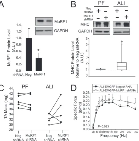
To control for potential developmental effects of congenitally decreased MuRF1 expression in MuRF1 KO mice, we also trans-duced plasmids coexpressing Emerald GFP (EmGFP) and miRNA-based short hairpin (sh) RNAs directly into TA muscles by electroporation. Figures E5 and E6 show the relative effec-tiveness of these shRNAs in cells expressing mouse MuRF1 or atrogin, respectively. One week later, the mice were randomly assigned to sham, PF, or ALI treatment groups and the muscles harvested 4 days later for analysis. From EmGFP expression, we determined that 70–80% of the muscle fibers in the mouse TA were transduced using our modification of the muscle electro-poration technique (seeFigure E7). Biochemical analyses of the entire muscle revealed an approximate 70% reduction in MuRF1 protein levels in MuRF1 shRNA-transduced muscles compared with muscles transduced with the EmGFP and a neg-ative control shRNA (Figure 5A). With an overallin vivo trans-duction efficiency of 70–80%, MuRF1 protein levels in MuRF1 shRNA-transduced muscle fibers should be reduced by approx-imately 75–85%.
Using this approach, we found that the MuRF1 shRNA 520 effectively prevented muscle wasting in ALI muscles. First, we found that MuRF1 shRNA treatment in ALI mice significantly increased MHC expression but had no effect on MHC expression in PF mice (Figure 5B). In four out of five ALI mice, treatment with MuRF1 shRNA showed a trend toward increased TA mus-cle weight compared with the contralateral (Neg-shRNA) limb. In PF mice, the trend was toward a decrease in muscle weight with MuRF1 shRNA (Figure 5C). We also measured force pro-duction in ALI-MuRF1 shRNA and ALI-Neg shRNA mice. Although we found no change in force production at Day 4 post–IT-LPS (data not shown), we did observe a modest but significant restoration of force production with MuRF1 shRNA at Day 10 post–IT-LPS (Figure 5D). In addition, although the mean diameter of EmGFP-positive muscle fibers was unaffected in MuRF1 shRNA transduced muscles from sham and PF mice, there was a large rightward shift in fiber size distribution (creased muscle fiber diameter) and a statistically significant in-crease in mean fiber diameter in MuRF1 shRNA transduced
muscles from ALI mice (Figures 6A, 6B, and 6D). We repeated this series of experiments using a second MuRF1 shRNA (610; seeFigure E5) targeting a different region of the mouse MuRF1 coding sequence and obtained near-identical results.
We also constructed two plasmids encoding EmGFP and one of two atrogin1 shRNAs (384 and 534), which were transduced into the TA muscles of ALI mice (seeFigure E6). In contrast to the results seen with the MuRF1 knockdown, however, knock-down of atrogin1 had no effect on ALI-associated muscle atro-phy (Figures 6C and 6D). Together, these results show that inhibition of MuRF1, but not atrogin1, abrogates muscle fiber atrophy in ALI mice.
ALI Muscle Wasting Persists Despite Resolution of Lung Injury
(Figures 7B and 7C). Wet weights of the PF and ALI muscles were, however, similar to sham control levels (Figure 7A). Al-though data in Figures 3E and Figure 7D show that Day 10 MuRF1 protein levels have been at baseline for several days, MHC protein levels remain depressed (Figure 7D). Absolute force production from the TA at Day 10 is similar in sham and PF mice, whereas force production in ALI mice is markedly suppressed (Figure 7E). When force measurements are normal-ized to muscle weight (specific force), the differences between PF and ALI persisted (Figure 7F). These data show that recov-ery of skeletal muscles from ALI is markedly delayed or sup-pressed despite complete resolution of lung injury and the down-regulation of MuRF1 mRNA and protein expression. They also show that the restoration of muscle mass alone (Fig-ure 7A) is not sufficient to restore muscle function, evoking the possibility of intrinsic changes to the contractile apparatus, the latter consistent with the low levels of MHC expression in ALI mice at Day 10.
DISCUSSION
It is increasingly recognized that ALI-associated skeletal mus-cle wasting represents a clinically significant complication that contributes to the morbidity and mortality of ALI (3–5).
Understanding the molecular mechanisms that promote and control skeletal muscle wasting associated with ALI is essential in designing therapeutic approaches targeting this syndrome. As an important step in that direction, we describe here a mouse model of ALI that displays some of the key features of human ALI-associated muscle wasting. These include skeletal muscle atrophy and weakness that is temporally tied to lung injury and the persistence of muscle weakness after resolution of lung in-jury, the later documented to be present in a large percentage of patients at least 5 years posthospitalization (3–6).
changes are associated with reductions in the intrinsic contrac-tility of the muscles (Figure 7F) and persistent suppression of striated muscle MHCs (Figure 7D).
The biochemical pathways most likely responsible for ALI-associated muscle wasting are the ubiquitin-mediated proteasomal and autophagy-lysosomal pathways. In our model, we uncovered evidence that both pathways were activated, the former shown in this report to be triggered by increased expression and activity of the muscle-specific E3 ligase, MuRF1, and the latter presumably by the marked reduction in food intake in ALI mice that triggered the up-regulation of selective autophagy-associated genes (33) and the need to include PF controls in the protocol (seeFigure E10). The use of PF controls is a common practice in the study of muscle-wasting disorders in which appetite is suppressed or access to food is limited. It is based on the assumption that the effects of reduced food intake on muscle mass and function are a separate, independent, and indirect component of the overall reduction in muscle mass caused by catabolic processes, such as the activation of the ubiquitin-mediated proteasomal pathway. If muscle wasting in ALI mice is a composite response of the effects of activating both proteasomal- and autophagy-mediated molecular and cellular deg-radation, then muscle wasting should be greater in ALI compared with PF muscle. Contrary to that prediction, our data show that mus-cle loss and reduction in musmus-cle fiber diameters were similar in ALI and PF mice (Figures 2A, 2C, and 4B;seeFigures E3, E8, and E9). Furthermore, suppressing MuRF1 expression through genetic inac-tivation or biochemical suppression in this model prevented the loss of muscle mass in ALI mice but had no effect in restoring muscle mass in PF muscles. These data show that all of the muscle wasting in ALI mice and none of it in PF mice was caused by increased MuRF1-driven proteasomal activity, excluding a direct contribution of autophagy to muscle wasting in ALI mice. These data are not consistent with the assumption that the secondary effects of pair-feeding are additive with the primary effects of ubiquitin-mediated proteolysis in muscle, but instead suggest that they may be interde-pendent and that crosstalk between the two degradative pathways decides which catabolic pathways are activated or suppressed. One likely candidate that might mediate this crosstalk is the protein p62 (sequestosome1) (34), which is a negative regulator of autophagy and a potential binding partner of MuRF1 (35).
In summary, our data demonstrate that ALI in mice produces marked skeletal muscle wasting and dysfunction similar to that ob-served in patients with ALI. Muscle wasting and dysfunction in this model is associated with markedly increased NF-kB activity and MuRF1 transcriptional activation and is suppressed by genetic inactivation or biochemical suppression of MuRF1. In contrast, muscle wasting in PF mice is not affected by suppressing MuRF1. It remains to be determined if MuRF1 is expressed in the skeletal muscles of humans with ALI and whether blockade of MuRF1 could prevent muscle wasting in these patients. Although recog-nizing that the results obtained in the mouse may have limited relevance to ALI in humans, MuRF1 seems to be an attractive therapeutic target for ALI-associated skeletal muscle wasting.
Author disclosuresare available with the text of this article at www.atsjournals.org.
Acknowledgment: The authors thank James Watkins and Andre Robinson in the Johns Hopkins Pulmonary Histology Core and the laboratory of Dr. Jeremy Walston for the use of their grip strength meter.
References
1. Ware LB, Matthay MA. The acute respiratory distress syndrome.N Engl J Med2000;342:1334–1349.
2. Rubenfeld GD, Caldwell E, Peabody E, Weaver J, Martin DP, Neff M, Stern EJ, Hudson LD. Incidence and outcomes of acute lung injury.N Engl J Med2005;353:1685–1693.
3. Ali NA, O’Brien JM Jr, Hoffmann SP, Phillips G, Garland A, Finley JC, Almoosa K, Hejal R, Wolf KM, Lemeshow S,et al; Midwest Critical Care Consortium. Acquired weakness, handgrip strength, and mortality in critically ill patients.Am J Respir Crit Care Med2008;178:261–268. 4. Fletcher SN, Kennedy DD, Ghosh IR, Misra VP, Kiff K, Coakley JH,
Hinds CJ. Persistent neuromuscular and neurophysiologic abnor-malities in long-term survivors of prolonged critical illness.Crit Care Med2003;31:1012–1016.
5. Herridge MS, Cheung AM, Tansey CM, Matte-Martyn A, Diaz-Granados N, Al-Saidi F, Cooper AB, Guest CB, Mazer CD, Mehta S,et al. Ca-nadian Critical Care Trials Group One-year outcomes in survivors of the acute respiratory distress syndrome.N Engl J Med2003;348:683–693. 6. Herridge MS, Tansey CM, Matte´ A, Tomlinson G, Diaz-Granados N,
Cooper A, Guest CB, Mazer CD, Mehta S, Stewart TE,et al; Canadian Critical Care Trials Group. Functional disability 5 years after acute respiratory distress syndrome.N Engl J Med2011;364:1293–1304. 7. De Jonghe B, Bastuji-Garin S, Durand MC, Malissin I, Rodrigues P, Cerf C,
Outin H, Sharshar T. Groupe de Re´flexion et d’Etude des Neuro-myopathies en Re´animation. Respiratory weakness is associated with limb weakness and delayed weaning in critical illness.Crit Care Med
2007;35:2007–2015.
8. Sharshar T, Bastuji-Garin S, Stevens RD, Durand MC, Malissin I, Rodriguez P, Cerf C, Outin H, De Jonghe B. Presence and severity of intensive care unit- acquired paresis at time of awakening are associated with increased intensive care unit and hospital mortality.Crit Care Med
2009;37:3047–3053.
9. al-Lozi MT, Pestronk A, Yee WC, Flaris N, Cooper J. Rapidly evolving my-opathy with myosin-deficient muscle fibers.Ann Neurol1994;35:273–279. 10. Norman H, Zackrisson H, Hedstrom Y, Andersson P, Nordquist J,
Eriksson LI, Libelius R, Larsson L. Myofibrillar protein and gene ex-pression in acute quadriplegic myopathy.J Neurol Sci2009;285:28–38. 11. Sher JH, Shafiq SA, Schutta HS. Acute myopathy with selective lysis of
myosin filaments.Neurology1979;29:100–106.
12. Bodine SC, Latres E, Baumhueter S, Lai VK, Nunez L, Clarke BA, Poueymirou WT, Panaro FJ, Na E, Dharmarajan K,et al. Identifi-cation of ubiquitin ligases required for skeletal muscle atrophy. Sci-ence2001;294:1704–1708.
13. Gomes MD, Lecker SH, Jagoe RT, Navon A, Goldberg AL. Atrogin-1, a muscle- specific f-box protein highly expressed during muscle atrophy.
Proc Natl Acad Sci USA2001;98:14440–14445.
14. Doucet M, Russell A, Le´ger B, Debigare´ R, Joanisse DR, Caron M, LeBlanc P, Maltais F. Muscle atrophy and hypertrophy signaling in patients with chronic obstructive pulmonary disease.Am J Respir Crit Care Med2007;176:261–269.
15. Hussain SN, Mofarrahi M, Sigala I, Kim HC, Vassilakopoulos T, Maltais F, Bellenis I, Chaturvedi R, Gottfried SB, Metrakos P,et al. Mechanical ventilation-induced diaphragm disuse in humans triggers autophagy.
Am J Respir Crit Care Med2010;182:1377–1386.
16. D’Alessio FR, Tsushima K, Aggarwal NR, West EE, Willett MH, Britos MF, Pipeling MR, Brower RG, Tuder RM, McDyer JF, et al. Cd41cd251foxp31Tregs resolve experimental lung injury in mice and are present in humans with acute lung injury.J Clin Invest2009; 119:2898–2913.
17. Files DC, DeGorordo A, Kesari P, Johnston L, Tsushima K, Aggarwal N, Sidhaye VK, D’Alessio F, King LS, Crow MT. Lung injury induces a skeletal muscle program that is qualitatively different from that induced by starvation. Oral presentation, ATS Annual Meeting, 2009, San Diego, CA.
18. Files DC, D’Alessio FR, Johnston L, Aggarwal NR, Garibaldi BT, Sidhaye VK, Chau E, Cohn R, King LS, Crow MT. Skeletal muscle atrophy in acute lung injury: differing mechanisms for wasting both during and after lung injury. Poster presentation, ATS Annual Meeting, 2010, New Orleans, LA.
19. Files DC, Johnston L, D’Alessio FR, Simmers J, Cohn RD, King L, Crow MT. Muscle RING-finger protein-1 (MuRF-1) is a critical mediator of acute lung injury-associated muscle wasting. Poster pre-sentation, ATS Annual Meeting, 2011, Denver, CO.
20. Halbower AC, Mason RJ, Abman SH, Tuder RM. Agarose infiltration improves morphology of cryostat sections of lung.Lab Invest1994;71: 149–153.
Prevention of LPS-induced acute lung injury in mice by mesenchymal stem cells overexpressing angiopoietin 1.PLoS Med2007;4:e269. 23. Rojas M, Woods CR, Mora AL, Xu J, Brigham KL. Endotoxin-induced
lung injury in mice: structural, functional, and biochemical responses.
Am J Physiol Lung Cell Mol Physiol2005;288:L333–L341. 24. Cohen S, Brault JJ, Gygi SP, Glass DJ, Valenzuela DM, Gartner C, Latres
E, Goldberg AL. During muscle atrophy, thick, but not thin, filament components are degraded by MuRF1-dependent ubiquitinylation. J Cell Biol2009;185:1083–1095.
25. Dehoux MJ, van Beneden RP, Fernandez-Celemin L, Lause PL, Thissen JP. Induction of mafbx and MuRF ubiquitin ligase mRNAs in rat skeletal muscle after LPS injection.FEBS Lett2003;544:214–217. 26. Lecker SH, Jagoe RT, Gilbert A, Gomes M, Baracos V, Bailey J, Price SR,
Mitch WE, Goldberg AL. Multiple types of skeletal muscle atrophy involve a common program of changes in gene expression.FASEB J
2004;18:39–51.
27. Sachek JM, Hyatt JP, Raffaello A, Jagoe RT, Roy RR, Edgerton VR, Price SR, Mitch WE, Goldberg AL. Rapid disuse and denervation at-rophy involve transcriptional changes similar to those of muscle wasting during systemic diseases.FASEB J2007;21:140–155.
28. Waddell DS, Baehr LM, van der Brandt J, Johnsen SA, Reichart HM, Furlow JD, Bodine SC. The glucocorticoid receptor and FOX01 synergistically activate the skeletal muscle atrophy-associated
to emerge yet much still points to the proteasome.Clin Cancer Res
2007;13:1356–1361.
30. Reed SA, Senf SM, Cornwell EW, Kandarian SC, Judge AR. Inhibition of IkappaB kinase alpa (IKKa) or IKKbeta (IKKb) plus Forkhead box O (Foxo) abolishes skeletal muscle atrophy.Biochem Biophys Res Com-mun2011;405:491–496.
31. Cai D, Frantz JD, Tawa NE Jr, Medendez PA, Oh BC, Lidov HG, Hasselgren PO, Frontera WR, Lee J, Glass DJ,et al. IKKbeta/NF-kappaB activation causes severe muscle wasting in mice.Cell2004; 119:285–298.
32. Moriscot AS, Baptista IL, Bogomolovas J, Witt C, Nirner S, Granzier H, Labeit S. MuRF1 is a muscle fiber type-II associated factor and together with MuRF2 regulates type II fiber trophicity and maintenance.J Struct Biol2010;170:344–353.
33. Masiero E, Agatea L, Mammucari C, Blaauw B, Loro E, Komatsu M, Metzger D, Reggiani C, Schiaffino S, Sandri M. Autophagy is required to maintain muscle mass.Cell Metab2009;10:507–515.
34. Komatsu M, Ichimura Y. Physiological significance of selective degrada-tion of p62 by autophagy.FEBS Lett2010;584:1374–1378.