Original Research Article
Effect of perinatal asphyxia on thyroid stimulating hormone and
thyroid hormone levels in a rural tertiary care center in
Mandya district of Karnataka, India
Sunil Kumar P.*, Haricharan K. R., Venugopala K. L.
INTRODUCTION
The thyroid hormones, thyroxine (T4) and triiodothyronine (T3), are tyrosine-based hormones produced by the thyroid gland. An important component in the synthesis is iodine. The thyronines act on the body to increase the basal metabolic rate, affect protein synthesis and increase the body's sensitivity to catecholamines (such as adrenaline) by permissiveness. The thyroid hormones are essential to proper development and differentiation of all cells of the human body. These hormones also regulate protein, fat, and carbohydrate metabolism, affecting how human cells use
energetic compounds. Numerous physiological and pathological stimuli influence thyroid hormone synthesis.1
The thyroid hormone, thyroxine binding globulin and thyroid stimulating hormone concentrations in term and preterm infants at birth, over the neonatal period, and during early infancy is in a state of flux during the perinatal period.2,3
However, very little data available which attempt to evaluate the possible effect of perinatal asphyxia on neonatal thyroid function.4,5
ABSTRACT
Background: Studies have shown a difference between serum concentrations of TSH, T4, T3 and FT4 in asphyxiated newborns than in normal newborns which suggesting abnormal thyroid function in asphyxia. We planned this study to assess and compare the effect of perinatal asphyxia on thyroid stimulating hormone and thyroid hormone levels.
Methods: This was a tertiary care teaching hospital based, prospective case control study conducted on normal full term and full term asphyxiated neonates delivered and admitted in the neonatal intensive care unit at Adichunchanagiri Institute of Medical Sciences, B. G. Nagara, Karnataka, India from December 2012 to May 2014.
Results: The means for thyroid hormones, in cord blood, were similar in both groups, except rT3are higher for non-asphyxiated new born as compared to asphyxiate ones. In newborns with 18-24 hours of life, lower levels of T4, T3, FT4, and TSH were observed in asphyxiated newborns, in which the basal levels (in cord blood) with exception of FT4, failed to increase.
Conclusions: Our study suggests that lower T4, free T4, and T3 levels are secondary to lower TSH levels in asphyxiated newborns; also, peripheral metabolism of T4 in asphyxiated infants can be altered due to low T3 and normal reverse T3 levels.
Keywords: Asphyxia, TSH, Thyroid hormone levels
Department ofPediatrics, Adichunchanagiri Institute of Medical Science, B. G. Nagara - 571448, Karnataka, India
Received: 26 October 2016
Accepted: 02 November 2016
*Correspondence:
Dr. Sunil Kumar P.,
E-mail: [email protected]
Copyright: © the author(s), publisher and licensee Medip Academy. This is an open-access article distributed under the terms of the Creative Commons Attribution Non-Commercial License, which permits unrestricted non-commercial use, distribution, and reproduction in any medium, provided the original work is properly cited.
The cord serum rT3 concentration was not influenced by maturity, birth-weight, or neonatal risk factors, whereas these factors did affect the concentrations of T3, T4, and TBG. There is no arteriovenous rT3 concentration difference across the placenta; therefore the cord rT3 reflects the systemic rT3 concentration in the baby at birth. As rT3 in the neonate largely, if not entirely, derives from thyroxine from the fetal thyroid, measurement of the cord rT3 concentration may be a good immediate screening test for neonatal hypothyroidism.5
Few studies have shown a difference between serum concentrations of TSH, T4, T3 and FT4 in asphyxiated newborns compared to normal newborn which suggests central hypothyroidism secondary to asphyxia. Moreover, asphyxiated newborns with moderate/severe hypoxic-ischemic encephalopathy present a greater involvement of the thyroid function and consequently a greater risk of death.6
We have planned this prospective institutional study of serum concentrations of thyroid hormones - T4, T3, free T4 (FT4) and reverse T3 (rT3) and thyroid-stimulating hormone (TSH) found in the umbilical cord blood of term newborns.
Objective of this study was the serum concentrations of thyroid hormones - T4, T3, free T4 (FT4) and reverse T3 (rT3) and thyroid-stimulating hormone (TSH) found in the umbilical cord blood of term newborns with and without asphyxia and those found in their arterial blood collected between 18 and 24 hours after birth.
METHODS
Place of this study was Adichunchanagiri institute of medical scienses, B. G. Nagar, Karnataka, India. And the duration of this study was December 2012 to May 2014. Prospective case control study was designed. And sample size was 50 neonates samples.
The sample size was estimated on the basis of a single proportion design. We assumed the confidence interval of 95% and confidence level of 14.5%. The sample size actually obtained for this study was 50 neonates.
Data collection
The study was a prospective case control study conducted on normal full term and full term asphyxiated neonates admitted in the Neonatal Intensive Care Unit. A total of 50 neonates on (n=25) normal full term and (n=25) full term asphyxiated neonates who were treated for the diagnosis of perinatal asphyxia were enrolled for the study.
Study tools
T4, T3, FT4, and TSH were measured by radioimmunoassay (Accu Bind VAST ELISA Kits, Monobind Inc., Ca., USA)
rT3 was dosed with reverse T3 kit, also by the radioimmunoassay method (ELISA Kit, CEC022 Ge, Monobind Inc., Ca., USA)
The values of FT4 and T3 were expressed in ng/dl, TSH in mU/ml, those of total T4 in mg/dl, and those of rT3, in ng/ml.
Study population consisted of full-term asphyxiated newborns with 1 and 5 minute Apgar scores < 7 were included in asphyxiated newborns and infants with Apgar score of 7 and above were included as non-asphyxiated group.
RESULTS
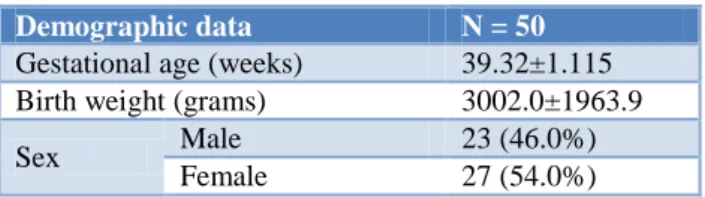
A total of 50 newborns were included and analyzed in our study. The mean gestational age of the infants was 39.32±1.115 months with average birth weight was 3002.0±1963.9 and in our study 54% of the newborns were females. In our study 40 (80%) were born to primipara and 10 (20%) to multipara.
Table 1: Demographic data of the newborns.
Demographic data N = 50
Gestational age (weeks) 39.32±1.115 Birth weight (grams) 3002.0±1963.9
Sex Male 23 (46.0%) Female 27 (54.0%)
Table 2: Distribution of APGAR score among all neonates.
APGAR score Median (minimum-maximum)
At 1 minute 6 (1 -10) At 5 minute 7.5 (1 -10)
APGAR score distribution of newborns at 1 min and at 5 min was 6 and 7.5 respectively. Among all neonates delivery methods of new born were 33 (66.0%) unassisted, 7 (14.0%) forceps, 6 (12.0%) emergency caesarean and 4 (8.0%) elective caesarean respectively.
The mean values in respect to umbilical cord blood and blood of newborns of T4 was 10.80±2.204 and 12.26±3.089 mg/dl respectively, T3 was 65.66±18.947 and 102.70±62.612 ng/dl respectively, FT4 was 1.36±0.273 and 2.344±0.5365 ng/dl respectively, rT3 was 1.728±0.5151 and 2.344±0.5365 ng/dl respectively and TSH was 12.16±3.316 and 11.98±3.473 mU/ml respectively for all newborn babies.
indices are also mentioned against each. Ongoing through the table we observe that while p values in respect of rT3
and TSH are significant (p<0.05) the same are non-significant in respect of T4, T3 and FT4 (p>0.05).
Table 3: Distribution T4, T3, FT4, rT3 and TSH of umbilical cord blood and blood of newborns.
No of newborn Umbilical cord blood Blood of newborns
Mean±SD Range Mean±SD Range
T4 (mg/dl) 50 10.80±2.204 (7 -15) 12.26±3.089 (7 - 19) T3 (ng/dl) 50 65.66±18.947 (41 -110) 102.70±62.612 (35 - 173) FT4 (ng/dl) 50 1.36±0.273 (1 - 2) 2.344±0.5365 (1.4 - 3.1) rT3 (ng/ml) 50 1.728±0.5151 (0.5 - 3.1) 1.422±0.2427 (0.8 - 1.8) TSH (m U/ml) 50 12.16±3.316 (5 -19) 11.98±3.473 (5 - 19)
Table 4: Plasma concentration of thyroid hormones in umbilical cord blood of asphyxiated and non-asphyxiated neonates.
Umbilical cord blood Group P value
Asphyxiated Non-asphyxiated
T4 (m g/dl) 11.12±2.088 10.48±2.312 0.309 T3 (ng/dl) 68.96±21.868 62.36±15.234 0.222
FT4 (ng/dl) 1.37±0.273 1.36±0.279 0.878
rT3 (ng/ml) 2.016±0.3671 1.440±0.4839 0.000 TSH (m U/ml) 14.12±2.804 10.20±2.566 0.000
Table 5: Distribution of plasma concentration of thyroid hormones in asphyxiated and non-asphyxiated newborns with 18-24 hours of life.
Newborns Group P value
Asphyxiated Non-asphyxiated
T4(m g/dl) 9.76±1.480 14.76±2.067 0.000
T3(ng/dl) 40.88±4.146 164.52±4.976 0.000
FT4(ng/dl) 1.856±0.2274 2.832±0.1994 0.000 rT3(ng/ml) 1.528±0.1671 1.316±0.2625 0.001
TSH(m U/ml) 9.32±2.155 14.64±2.289 0.000
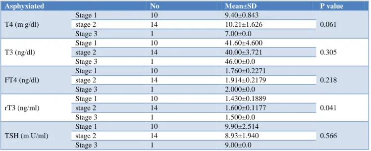
Table 6: Distribution of thyroid hormones levels in asphyxiated newborns with 18-24 hours of life.
Asphyxiated No Mean±SD P value
T4 (m g/dl)
Stage 1 10 9.40±0.843
0.061 stage 2 14 10.21±1.626
Stage 3 1 7.00±0.0
T3 (ng/dl)
Stage 1 10 41.60±4.600
0.305 stage 2 14 40.00±3.721
Stage 3 1 46.00±0.0
FT4 (ng/dl)
Stage 1 10 1.760±0.2271
0.218 stage 2 14 1.914±0.2179
Stage 3 1 2.000±0.0
rT3 (ng/ml)
Stage 1 10 1.430±0.1889
0.041 stage 2 14 1.600±0.1177
Stage 3 1 1.500±0.0
TSH (m U/ml)
Stage 1 10 9.90±2.514
0.566 stage 2 14 8.93±1.940
Among both the groups the plasma concentration of thyroid hormones in asphyxiated and non-asphyxiated newborns within 18-24 hours of life were recorded. We observed that the concentrations in respect of all the thyroid hormones except for rT3 are higher for non-asphyxiated new born as compared to non-asphyxiated ones. P values are significant in respect of all the hormones as indicated in the Table 5.
The thyroid hormones like T4 (mg/dl), T3 (ng/dl), FT4 (ng/dl), rT3 (ng/ml) and TSH (m U/ml) in cases of new born after 18-24 hours of life were studied. It has given a comparative value in respect of hypoxic ischemic encephalopathy for asphyxiated new born. Ongoing through the Table 6 we find that P value in respect of rT3 (ng/ml) are statistically significant whereas values are non-significant in respect of T4 (m g/dl), T3 (ng/dl), FT4 (ng/dl) and TSH (m U/ml) (p>0.05).
DISCUSSION
The effect of hypoxia on thyroid hormones has been long recognized. In animals, hypoxia reduces thyroid function and extra thyroidal metabolism of T4. Similarly, Moshang et al found an increase in the levels of rT3 in patients with acute hypoxemia, suggesting a reduction in its degradation.7 In the same study, patients with chronic hypoxemia showed decreased levels of T3, and increased rT3 levels, revealing alterations in extra-thyroidal metabolism. The action of these hormones on the synthesis of mitochondrial enzymes and structural elements is extremely important, in addition to participating in thermogenesis, water and electrolyte transportation, and in the growth and development of the central nervous system and skeleton. Low levels of thyroid hormones in non-thyroidal illnesses are associated with poor prognosis
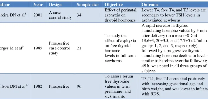
Table 7: Sample size, objective and outcomes.
Author Year Design Sample size Objective Outcome
Pereira DN et al8 2001 A
case-control study 34
Effect of perinatal asphyxia on thyroid hormones
Lower T4, free T4, and T3 levels are secondary to lower TSH levels in asphyxiated newborns
Borges M et al9 1985
Prospective case control study
21
To study the effect of asphyxia on free thyroid hormone levels in full term newborns
A rapid increase in
thyroid-stimulating hormone values by 5 min after delivery (to a mean±SD of 18.6±3, 20±3.5, and 17.7±5 uU/ml in groups 1, 2, and 3, respectively), followed by a progressive thyroid-stimulating hormone decline to levels similar to baseline over the following 48 h, was noted in all three groups of subjects.
Wilson DM et al10 1982 Prospective 96
To assess serum free thyroxine values in term, premature, and sick infants
T3, T4, free T4 correlated positively with increasing gestational age and birth weight, and was lower in infants with RDS.
The mean for thyroid hormones, in cord blood, were similar in both groups, except rT3 are higher for non-asphyxiated new born as compared to non-asphyxiated ones. This is similar to what has been reported by Pereira DN et al and Borges et al.8,9 they did not find differences in the concentration of FT4 and FT3 in cord blood, and those presented by Franklin et al which did not find statistical differences in the concentration of T4, T3, rT3, FT4, TBG, and TSH between normal and asphyxiated newborns.11 The increase of rT3 observed in cord blood may indicate an alteration in the peripheral metabolism of thyroid hormones through the inhibition of 5' - deiodinase activity, which is similar to what occurs in acute hypoxia.
On the other hand, in newborns with 18-24 hours of life, lower levels of T4, T3, FT4, and TSH were observed in asphyxiated newborns, in which the basal levels (in cord blood), with exception of FT4, failed to increase. Borges et al.8 found that FT4 and FT3 levels failed to increase within the first 48 hours of life in the group of asphyxiated newborns, even though their TSH levels were normal.
the role of T4 and/or T3 restoration in patients with subnormal hormonal levels should also be considered.
CONCLUSION
Our study suggests that lower T4, free T4, and T3 levels are secondary to lower TSH levels in asphyxiated newborns; also, peripheral metabolism of T4 in asphyxiated infants can be altered due to low T3 and normal reverse T3 levels.
Funding: No funding sources Conflict of interest: None declared
Ethical approval: The study was approved by the Institutional Ethics Committee
REFERENCES
1. Kliegman RM, Behrman RE, Jenson HB, Stanton BF, (eds.) Nelson textbook of Pediatrics. 19th ed. Philadelphia: Saunders; 2012.
2. Fisher DA, Dussault JH, Sack J, Chopra IJ. Ontogenesis of hypothalamic-pituitary-thyroid function and metabolism in man, sheep and rat. Recent Prog Horm Res. 1977;33:59-116.
3. Klein AH, Oddie TH, Parslow M, Foley TP, Fisher DA. Developmental changes in pituitary-thyroid function in the human fetus and newborn. Early Hum Dev. 1982;6:321-30.
4. Erenberg A. The effect of perinatal factors on cord thyroxine concentration. Early Hum Dev. 1978;X2:283-9.
5. Byfield PGH, Bird D, Yepez R, Land IM, Himsworth RL. Reverse tri-iodothyronine, thyroid hormone, and thyrotrophin concentrations inplacental cord blood. Arch Dis Child. 1978;53:620-4.
6. Pereira DN, Procianoy RS. Effect of perinatal asphyxia on thyroid-stimulating hormone and thyroid hormone levels. Acta Paediatr. 2003;92(3):339-45.
7. Moshang T, Chance KH, Kaplan MM, Utiger RD, Takahashi O. Effects of hypoxia on thyroid function tests. J Pediatr. 1980;97:602-4.
8. Pereira DN, Procianoy RS. Effect of perinatal asphyxia on thyroid hormones. J Pediatr (Rio J). 2001;77(3):175-8.
9. Borges M, Lanes R, Moret LA, Balochi D, Gonzalez S. Effect of asphyxia on free thyroid hormone levels in full term newborns. Pediatr Res. 1985;19:1305-7.
10. Wilson DM, Hopper AO, McDougall JR, Bayer MF, Hintz RL, Stevenson DK, et al. Serum free thyroxine values in term, premature and sick infants. J Pediatr. 1982;101:113.
11. Franklin R, O’Grady C. Neonatal thyroid function: effects of non-thyroidalillness. J Pediatr. 1985;107:599-602.