Engineering PLGA Particles for Advanced Drug Delivery
Elizabeth Enlow
A dissertation submitted to the faculty of the University of North Carolina at Chapel Hill in partial fulfillment of the requirements for the degree of Doctor of Philosophy in the
Department of Chemistry
Chapel Hill 2010
Approved by
Professor Joseph M. DeSimone
Professor Valerie Ashby
Professor Sergei Sheiko
Professor Nancy Allbritton
Abstract
Elizabeth Enlow: Engineering PLGA Particles for Advanced Drug Delivery (under the direction of Professor Joseph M. DeSimone)
The author would like to acknowledge those individuals who assisted the work herein: Dr. J. Christopher Luft, Dr. Patricia Ropp, and Dr. Shaomin Tian for in vitro work and biology advice, and specifically Dr. Luft for cell culture training; Charlene Ross and the animal core facility whose help was invaluable on all in vivo work; Dr. Warefta Hasan for countless hours of particle making on both chemotherapy and siRNA projects; William Hinson for assistance with the early biodistribution experiments; Dr. Victoria Madden and Dr. Wallace Ambrose for TEM analysis; and Dr. Chuanzhen Zhou at North Carolina State University for TOF SIMS analysis. A list of those who’ve assisted would not be complete without acknowledging Dr. Kevin Herlihy and Dr. Janine Nunes for innumerable scientific discussions over the years and for keeping me sane; Dr. Mary Napier for everything she does and whose door is always open; and Kelly Chang and Dr. Christine Conwell for their friendship in and out of lab.
Of course none of this would be possible without my advisor, Prof. Joseph DeSimone who took me in with no polymer or biology experience and taught me how to move forward in unfamiliar territory, to always keep the big picture in mind, and that diversity in expertise and experience is fertile grounds for innovation. Getting to participate in the development of this new and promising technology has been a privilege and I thank you.
Table of Contents
Chapter 1: Introduction to Advanced Drug Delivery ...1
1.1 Introduction to Advanced Drug Delivery ...2
1.2 Cancer and therapeutic options...2
1.3 Systemic administration of solubilized drug and drug conjugates ...3
1.4 Enhanced drug delivery with liposomes and polymeric micelles...5
1.5 Drug delivery using polymer particles...6
1.5.1 Bioabsorbable polymers...7
1.5.2 Fabricating PLGA particles ...9
1.5.2.1 Emulsion/solvent evaporation methods ...9
1.5.2.2 Supercritical solvent/anti-solvent methods ...9
1.5.2.3 Methods to non-spherical particles ...10
1.5.3 PLGA-PEG particles: A popular variation on solid PLGA particles ...11
1.5.4 Factors affecting encapsulation and release of therapeutics ...12
1.5.5 In vitro toxicity and internalization of PLGA particles…… ...14
1.5.6 Biodistribution ...18
1.5.7 In vivo internalization and tumor penetration ...20
1.6 Chemotherapeutic Delivery ...22
1.6.1 Docetaxel encapsulation ...23
1.6.2 Docetaxel release ...24
1.6.4 In vivo efficacy via intratumoral injection ...26
1.6.5 In vivo efficacy via intravenous injection ...27
1.6.6 Chemotherapeutic PLGA particles in clinical trials ...28
1.7 RNA interference and siRNA delivery ...29
1.7.1 Incorporating siRNA in cationic PLGA nanoparticles for effective transfection...31
1.7.2 The effect of N/P ratio on in vitro gene knockdown ...33
1.7.3 In vivo gene knockdown by PLGA nanoparticles...35
1.8 Future Directions ...36
1.8.1 Combination therapy...37
1.8.2 Higher Degree of Control ...37
References...39
Chapter 2: Fabrication and Characterization of Engineered PLGA PRINT Particles ...48
2.1 Introduction to the PRINT® Process...49
2.1.1 Control of Size, Shape, and Composition...50
2.1.2 Mold Fabrication...52
2.2 The solvent evaporation, pressure fill PRINT method ...53
2.2.1 Experimental ...54
2.2.1.1 Materials ...54
2.2.1.2 Particle visualization and characterization...54
2.2.1.3 Particle fabrication procedure ...54
2.2.2 Results...55
2.3.1.1 Materials ...64
2.3.1.2 Particle visualization...64
2.3.1.3 Particle fabrication procedure ...64
2.3.3 Results...65
2.4 Particle Harvesting...67
2.4.1 Mechanical Harvesting ...68
2.4.2 Sacrificial Layer Harvesting ...69
2.5 PRINT process scale-up...71
2.6 In vitro cytotoxicity of engineered PLGA PRINT nanoparticles ...74
2.6.1 Experimental ...74
2.6.1.1 Materials ...74
2.6.1.2 Particle fabrication and post-reaction with PEG...75
2.6.1.3 In Vitro cytotoxicity...75
2.6.2 Results...76
2.7 In vivo immune response to engineered PLGA PRINT nanoparticles ...77
2.7.1 Experimental ...77
2.7.1.1 Materials ...77
2.7.1.2 Particle fabrication and post-reaction with PEG...77
2.7.1.3 Measurement of immune response ...78
2.7.2 Results...78
2.8 Biodistribution of engineered PLGA PRINT nanoparticles ...80
2.8.1 Experimental ...81
2.8.1.2 Particle fabrication and characterization...81
2.8.1.3 Biodistribution ...81
2.8.2 Imaging agents ...82
2.8.3 Biodistribution of PLGA PRINT nanoparticles in a SKOV3 xenograft model ...83
2.8.4 Biodistribution of PLGA PRINT nanoparticles in an orthotopic ASPC-1 model...86
2.9 Conclusions...87
2.10 Future Work ...88
References...90
Chapter 3: Engineered PLGA PRINT Particles for Therapeutic Delivery...92
3.1 Introduction...93
3.2 RNAi therapy...93
3.2.1 Experimental ...94
3.2.1.1 Particle fabrication ...94
3.2.1.2 Particle characterization...94
3.2.1.3 In vitro assays ...95
3.2.1.4 Confocal imaging...95
3.2.1.5 TEM imaging ...95
3.2.2 Encapsulating siRNA in PLGA PRINT particles ...96
3.2.3 siRNA release from PLGA PRINT nanoparticles ...98
3.2.4 Cellular internalization and particle degradation...100
3.2.5 In vitro knockdown ...102
3.2.7 Future Work ...105
3.2.7.1 Decreased EC50 with lipid coated compositions...105
3.2.7.2 Therapeutic siRNA and in vivo knockdown ...107
3.3 Chemotherapy...108
3.3.1 Experimental ...109
3.3.1.1 Particle Fabrication ...109
3.3.1.2 Particle characterization...109
3.3.1.3 Docetaxel encapsulation and release by HPLC ...110
3.3.1.4 In vitro cytotoxicity...110
3.3.1.5 In vivo efficacy...111
3.3.1.6 PK determination ...111
3.3.2 High Drug Loading...112
3.3.3 Release Profiles...114
3.3.4 In vitro drug delivery efficacy ...116
3.3.5 In vivo drug delivery efficacy ...118
3.3.6 Pharmacokinetics of docetaxel in vivo...119
3.3.7 Conclusions...122
3.3.8 Future Work ...123
3.3.8.1 PK studies with more sizes, shapes and compositions ...123
3.3.8.3 Combinations of gene therapy and chemotherapy...125
List of Tables
Table 1.1 Examples advanced chemotherapy systems in clinical stages...6
Table 1.2 The encapsulation of docetaxel in PLGA/PLA particles ...24
Table 1.3 Examples of release from docetaxel loaded PLGA/PLA particles ...25
Table 2.1 Relevant characteristics of solvents ...58
Table 2.2 Calculated and observed d spacing values from TEM electron diffraction...62
Table 2.3 A demonstration of PVOH removal and particle concentration by tangential flow filtration...74
Table 2.4 Dynamic light scattering characterization...84
Table 3.1 The characterization of 80 x 320 nm PLGA PRINT nanoparticles coated with lipid ...106
Table 3.2 Encapsulation efficiency of PLGA PRINT nanoparticles at varying drug loadings (w/w) and physical characterization by DLS...112
Table 3.3 IC50 values for docetaxel loaded PLGA PRINT Nanoparticles and Taxotere ...117
Table 3.4 The physical properties of PLGA PRINT nanoparticles from different PLGA polymers...120
List of Figures
Figure 1.1 The chemical structure of biodegradable polyesters A) poly(glycolic acid), B) poly(lactic acid), C) poly(dioxanone), D) poly(valerolactone), E) poly(ε
-caprolactone), F) poly(β-hydroxybutyrate), and G) poly(β-hydroxyvalerate) ...7
Figure 1.2 The chemical structure of docetaxel...23
Figure 1.3 The RNAi pathway...30
Figure 1.4 The chemical structure of branched poly(ethylene imine) (PEI) ...31
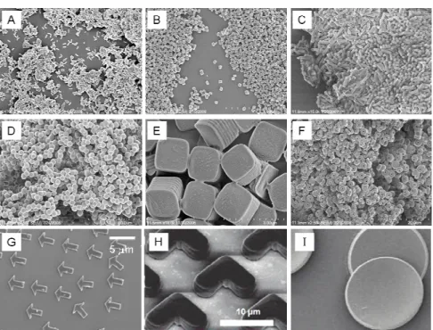
Figure 2.1 A variety of sizes and shapes generated using the PRINT process (A) 80nm x 360nm cylinders, (B) 200nm x 200nm cylinders, (C) 200nm x 600nm cylinders, (D) 1 µm sphere approximates, (E) 2 µm cubes with ridges on the sides, (F) 3 µm particles with center fenestrations, (G) 3 µm arrows, (H) 10 µm boomerangs, and (I) 7 µm discs...51
Figure 2.2 An example of the perfluorinated polyether oil structure. The chain length (n and m) can be varied as well as the end group functionalization ...52
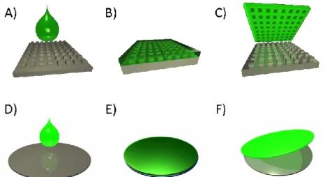
Figure 2.3 Mold and substrate fabrication (A) Fluorocur is poured onto a template, (B) The Fluorocur wets the template completely and crosslinks upon exposure to UV light, (C) The mold generated can be peeled from the template and now has the inverse pattern of the template, (D) Fluorocur is poured onto a flat silicon wafer, (E) The Fluorocur crosslinks upon exposure to UV light, (F) The substrate generated can be peeled from the template and is smooth on the wafer side...53
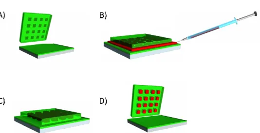
Figure 2.4 The solvent evaporation, pressure fill PRINT method (A) a mold and substrate are fabricated from Fluorocur, (B) a solution of polymer in organic solvent is injected between the mold and substrate, (C) pressure is applied to the sandwich which is placed under vacuum to remove the solvent solidifying the particles, and (D) the mold and substrate are separated revealing isolated particles in the mold ...55
Figure 2.5 PLLA particles from 1,4 dioxane solutions. (A) 1% (w/v) top view, (B) 1% (w/v) side view, (C) 5% (w/v) top view, and (D) 5% (w/v) side view ...57
Figure 2.6 PLLA particles from 5% (w/v) solutions from (A) dichloromethane top view, (B) dichloromethane side view, (C) 1,4-dioxane top view, and (D) 1,4-dioxane side view...57
Figure 2.8 A sample with both scum and free, isolated 200nm particles; typical
results for the solvent evaporation, pressure fill PRINT method of nanoparticles...63 Figure 2.9 The thermal, capillary fill PRINT process. (A) A film is cast on PET.
This sheet is termed the delivery sheet, (B) The delivery sheet in passed under a heated nip in contact with a mold, and (C) The mold is filled with isolated solid
polymer particles...65 Figure 2.10 80nm x 320nm PLGA nanoparticles ...66 Figure 2.11 An SEM image of the (A) the empty mold after particles are
removed, (B) the array of particles transferred out of the mold onto a substrate,
and (C) collected free particles ...68 Figure 2.12 Particle harvesting via the squeegee method: (A) the mold is placed
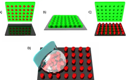
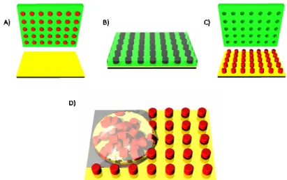
in contact with a solid substrate, (B) the mold and substrate are heated while in contact adhering the particles to the substrate, (C) when the mold is peeled back the particles are left in an array on the substrate, (D) a cell scraper (blue) and a bead of water containing stabilizer are used to mechanically collect the particles
using gentle lateral force...69 Figure 2.13 PVOH harvesting: (A) the mold is placed in contact with a solid
substrate coated with PVOH, (B) the mold and coated substrate are heated while in contact adhering the particles to the substrate, (C) when the mold is peeled back the particles are left in an array on the coated substrate, (D) a bead of water
is used to collect the particles without mechanical force...70 Figure 2.14 Advances in mold technology. Left: original master on a glass slide
with the mold size compared to a dime. Right: a roll of 200 nm mold supplied by
Liquidia Technologies ...71 Figure 2.15 Scaled-up PRINT process. (A) a mold is filled, (B) the particles
are transferred to a water soluble film, (C) the particles are collected in a
bead of water...72 Figure 2.16 Krosflow® MicroKros (X1-500S-200) tangential flow filtration
apparatus ...73 Figure 2.17 The 72 hour viability of H460 (blue) and SKOV3 (purple) cells
when treated with PVOH stabilized PLGA PRINT 200 nm cylindrical particles (solid bars), 90% PLGA / 10% PEI PRINT 200nm cylindrical particles (striped bars), and PEGylated (5K) 90% PLGA / 10% PEI PRINT 200nm cylindrical
Figure 2.19 The IL-12 levels in serum 6 hours after injection ...79 Figure 2.20 The release of DiD (inset structure) from PLGA PRINT
nanoparticles ...83 Figure 2.21 SEM images of (A) 80 x 320 nm particles and (B) 200 nm
particles ...83 Figure 2.22 The biodistribution of PVOH stabilized 200nm (blue) and 80 x 360
nm (purple) particles 24 hours after injection in SCID mice with xenograft
SKOV3 tumors...85 Figure 2.23 The biodistribution of PVOH stabilized 80 x 360 nm particles 4
hours (blue) and 24 hours (purple) after injection in nude mice with orthotopic
pancreatic tumors ...87 Figure 3.1 Examples of films cast from PLGA/PEI/siRNA solutions.
(A and B) heterogeneous films and (C) a homogeneous film ...96 Figure 3.2 The generation of homogeneous PLGA/PEI/siRNA films. (A) a
two-phase solution where yellow is DMF with siRNA/water and dark red is DMSO with PLGA/PEI, (B) the two phases are then mixed until completely
homogeneous, and (C) the film is then cast from this solution ...97 Figure 3.3 SEM of 200 nm PLGA PRINT particles containing 10% PEI
and 5% siRNA ...98 Figure 3.4 The release of siRNA from 200 nm PLGA/PEI particles over
72 hours...99 Figure 3.5 The internalization of (A) -24mV PLGA/siRNA particles and
(B) +34mV PLGA/PEI/siRNA particles. The nucleus is stained purple while
the particles contain Cy3-labeled (red) siRNA...100 Figure 3.6 TEM image of a HeLa cell internalizing PLGA/PEI/siRNA
particles ...101 Figure 3.7 The degradation of PLGA/PEI/siRNA particles over 72 hours ...102 Figure 3.8 Delivery of anti-luciferase siRNA. Expressed as a percent of
control (untreated) cells ...103 Figure 3.9 The internalization of lipid coated 80 x 320 nm PLGA PRINT
Figure 3.10 Knockdown of luciferase in HeLa-Luc cells by 80 x 320 nm
lipid coated PLGA/siRNA nanoparticles...107 Figure 3.11 SEM images of cylindrical 200nm x 200nm PLGA PRINT
nanoparticles containing varying amounts of docetaxel: A) 0%, B) 10%, C) 20%, D) 30%, E) 40%. Inset images are a magnification of a portion of
the image to the same scale for more detail...113 Figure 3.12 The HPLC trace of heat stressed docetaxel (blue) and
PLGA/docetaxel particles (red) ...114 Figure 3.13 Release profiles of PLGA nanoparticles containing 10-40%
docetaxel at pH 7.4 and 37oC...115 Figure 3.14 H460 cell viability after 72 hour exposure to Taxotere® and
PLGA PRINT nanoparticles containing various docetaxel weight percents: 0% docetaxel (x), 10% docetaxel (■), 20% docetaxel (▲), 30% docetaxel (●), 40% docetaxel (♦), and Taxotere® (□). Blank particles (0%) were dosed at equal particle concentrations to 10% docetaxel containing particles (i.e. the
highest particle dose) ...116 Figure 3.15 The in vivo efficacy of 40% docetaxel PLGA PRINT nanoparticles
in SKOV3 mouse model (*p<0.05, **p<0.01)...118 Figure 3.16 The release of docetaxel from two different PLGA polymers:
orange is a 50:50, 33kDa PLGA and purple is a 85:15, 50kDa PLGA ...120 Figure 3.17 The PK profiles of two PRINT compositions compared to
Taxotere over 24 hours. Concentrations in (A) plasma and (B) tumor ...121 Figure 3.18 A cutaway view of a PLGA/docetaxel film which is typically
300-500 nm in depth. The docetaxel is represented by the red color which is
List of Abbreviations ASPC-1 – human pancreas adenocarcinoma cell line AUC – area under the curve
DCM – dichloromethane
DiD – 1,1’-dioctadecyl-3,3,3’,3’-tetramethylindodicarbocyanine perchlorate DLS – dynamic light scattering
DMF – dimethylformamide DMSO – dimethyl sulfoxide
DOPE – 1,2-dioleoyl-sn-glycero-3-phophoethanolamine DOTAP – 1,2-dioleoyl-3-trimethylammonium-propane DSC – differential scanning calorimetry
EC50 – half maximal effective concentration
H460 – human non-small cell lung carcinoma cell line HeLa – human cervical adenocarcinoma cell line HPLC – high performance liquid chromatography IC50 – half maximal inhibitory concentration IL-12 – interleukin 12
I.T. – intratumoral I.V. – intravenous
kDa – kilodaltons (equivalent to 1,000 grams per mole) LNCaP – human prostate carcinoma cell line
PDI – polydispersity index
PDMS – poly(dimethyl siloxane) PEG – poly(ethylene glycol) PEI – poly(ethyleneimine)
PET – poly(ethylene terephthalate) PFPE – perfluoropolyether
PK – pharmacokinetics
PLGA – poly(lactic-co-glycolic acid) PLLA – poly(L-lactic acid)
PRINT – particle replication in non-wetting templates PVOH – poly(vinyl alcohol)
RNAi – ribonucleic acid interference SEM – scanning electron microscopy siRNA – small interfering ribonucleic acid
SKOV3 – human ovarian adenocarcinoma cell line TEM – transmission electron microscopy
Tg – glass transition temperature TNFα – tumor necrosis factor alpha
CHAPTER 1
1.1 Introduction to Advanced Drug Delivery
Advanced drug delivery is the growing effort to take existing therapeutics with low efficacy or highly undesirable side effects and improve their localization to the site of disease thereby limiting systemic exposure and increasing therapeutic concentrations. The most common treatments in today’s healthcare system are drugs administered orally or parenterally. Both result in the drug circulating in the bloodstream, reaching all tissues in the body. For some therapies this results in unwelcome side effects. This consequence is common for therapies where the drug is a cytotoxin, such as chemotherapies. Alternatively there are therapies where this exposure is not harmful for the body but degrades or destroys the activity of the therapeutic. This is common for therapeutics which are unstable or can be degraded by the body, such as RNA. For these reasons, the medical community is seeking new ways of delivering drugs. The most widespread application of these advanced therapies is in the treatment of cancer.
1.2 Cancer and therapeutic options
to kill the cells in the tumor. Unfortunately these cytotoxins reach not only the tumor, but the rest of the body as well, killing healthy cells and resulting in unwanted side effects. The reason this is effective to a degree is that chemotherapeutics tend to act more heavily on faster dividing cancer cells than healthy cells dividing at a normal rate. However, some healthy cells naturally have a high growth rate, such as hair, which is why people undergoing chemotherapy experience hair loss. Other common side effects include nausea, neuropathy, fluid retention, stomatitis, nail disorders, weakness, hypersensitivity reactions, and infection.1 These harsh side effects limit the amount of therapy that can be administered and the frequency of administration. This limits the effectiveness of the treatment. If drug could be delivered to the tumor with limited systemic exposure then higher concentrations of drug in the tumor could be maintained and efficacy could be increased. Another method being investigated for cancer treatment is gene therapy. The administration of RNA or DNA can alter the production of proteins which are abnormally expressed in tumor cells leading to drug resistance, rapid division, and metastasis. However, these therapies will need an effective delivery vehicle to have clinical applications. Gene therapy can be used alone or to sensitize cells to chemotherapeutics.
1.3 Systemic administration of solubilized drug and drug conjugates
escape the vasculature than can in healthy tissue. This vasculature is termed “leaky”. Second the lymphatic system which drains tissue is not properly formed which results in poor clearance. These combine to form the enhanced permeability and retention (EPR) effect. Particles accumulate in the tumor since more can exit the circulation and are not effectively cleared. Passive targeting is employed by some the most common cancer therapies in the clinic today; formulations designed to deliver hydrophobic chemotherapeutics which were initially passed over in preference to hydrophilic chemotherapeutics which could easily be administered intravenously. Stabilizers such as Cremphor EL and Tween 80 are mixed with the therapeutic in water with small amounts of solvent so that the drug can be administered through an infusion. These formulations tend to create nano-aggregates which enhance tumor accumulation via the EPR effect and as such are a step toward enhanced delivery. The stabilizers carry toxicities of their own, however, which cause side effects.
1.4 Enhanced drug delivery with liposomes and polymeric micelles
Liposomes, discovered in 19652 and proposed as drug delivery vehicles in 1976,3, 4 took the idea of drug delivery beyond simple targeting of single therapeutic molecules
care must be taken that this does not drop below the CMC. Finally, both micelles and liposomes are limited in the polymers/lipids that can be used for their formation as they require an amphiphile with specific subunit lengths, which restricts specific tailoring of delivery vehicles. Despite the disadvantages many therapeutics employing conjugate, liposome, and micelle technologies are approved by the FDA or are in clinical trials (Table 1.1).
Table 1.1 Examples advanced chemotherapy systems in clinical stages.7 Therapeutic
Name Platform Chemotherapeutic Clinical
Stage Smancs Polymer conjugate Neocarzinostatin Approved
PK1 Polymer conjugate Doxorubicin Phase 2
hT-101 Polymer conjugate Camptothecin Phase 2
Xyota Polymer conjugate Paclitaxel Phase 3
CT-2106 Polymer conjugate Camptothecin Phase 2 MAG-CPT Polymer conjugate Camptothecin Phase 1
Doxil Liposome Doxorubicin Approved
EndoTAG Liposome Paclitaxel Phase 2
LE-SN-38 Liposome SN-38 Phase 2
CPX-1 Liposome CPT-11, floxuridine Phase 2
NK105 Micelle Paclitaxel Phase 2
NC-6004 Micelle Cisplatin Phase 1/2
NK012 Micelle SN-38 Phase 2
SP1049C Micelle Doxorubicin Phase 3
1.5 Drug delivery using polymer particles
using solid polymer particles as vehicles for drug delivery are their ability to reduce the immunoresponse, give more protection to the cargo, reach a wider range of sizes, and tailor the release kinetics by specifically tailoring the polymer matrix.8
1.5.1 Bioabsorbable polymers
Of particular interest for solid polymer particles are bioabsorbable polymers. The use of bioabsorbable polymers in medical applications can be traced back to 150 A.D. when catgut was used to suture injured gladiators. Since then a range of synthetic polymers which degrade in situ have been discovered. These polymers are mainly polyesters which degrade hydrolytically and include poly(glycolic acid), poly(L-lactic acid), poly(D-lactic acid), poly(dioxanone), poly(ε-caprolactone), poly(β -hydroxybutyrate), poly(β-valerolactone), and their copolymers (Figure 1.1).
Figure 1.1 The chemical structure of biodegradable polyesters A) poly(glycolic acid), B) poly(lactic acid), C) poly(dioxanone), D) poly(valerolactone), E) poly(ε-caprolactone), F)
1.5.2 Fabricating PLGA particles
Various methods for fabricating PLGA particles, including emulsions, precipitations, spray drying, and flow focusing, are currently under investigation by a large number of research groups.9-14 A substantial volume of literature has been dedicated to investigating the effects of process parameters on particle properties and the attempts to develop trends to guide particle design have been reviewed.15, 16 PLGA particles are, for the most part, currently being made by either emulsion solvent evaporation methods or supercritical processing techniques.
1.5.2.1 Emulsion/solvent evaporation methods
In emulsion solvent evaporation methods, PLGA is dissolved in a good solvent, typically dichloromethane. This solution is then added to water which contains a surfactant, typically poly(vinyl alcohol) (PVOH). By controlling the speed with which this mixture is stirred, the concentration of surfactant, and the specific PLGA used the size of the particles is roughly controlled. The solvent is then evaporated and the PLGA hardens into spheres.9, 17-26 There are variations on this method involving double emulsions27-30 and oil-in-oil emulsions11, 31 that attempt to improve encapsulation of water-soluble drugs which tend to partition into the main water phase during the emulsion process.
1.5.2.2 Supercritical solvent/anti-solvent methods
those in which the supercritical fluid acts as an antisolvent. RESS (precipitation by rapid expansion of supercritical solutions) and RESOLV (rapid expansion of a supercritical solution into a liquid solvent) are two methods that have been used to produce PLGA particles in which the supercritical fluid acts as the solvent.32-34 In these methods, PLGA is dissolved in scCO2 with ethanol as a cosolvent. This solution is then sprayed into air (RESS) or an aqueous media (RESOLV). The rapid expansion of CO2 lowers its ability to solubilize the polymer and particles precipitate. ASES (aerosol solvent extraction system) and PCA (precipitation with a compressed antisolvent) are two methods that have been used to produce PLGA particles in which the supercritical fluid acts as an antisolvent.35-40 In supercritical antisolvent processes, PLGA is dissolved in a good solvent, typically dichloromethane. This is then sprayed into a chamber of supercritical CO2. The solvent’s ability to solvate the polymer is reduced as the solvent swells with the supercritical phase and the polymer hardens into spheres. For all supercritical processing techniques, the particle size is roughly controlled by the type of spray generated.
1.5.2.3 Methods to non-spherical particles
Recently a hydrogel template method was demonstrated for the fabrication of PLGA particles. Microparticles were fabricated in a wide range of sizes and shapes, including a micron scale reproduction of a computer keyboard. Cylindrical 500 nm and 200 nm particles were also demonstrated. In this method gelatin is used as a mold, generated by pouring warmed gelatin over a silicon template and cooling in the refrigerator to “solidify”. The so called hydrogel template is then filled with a high solids (40%) solution of PLGA in dichloromethane. After the dichloromethane evaporates the hydrogel template containing PLGA particles is warmed to dissolve the gelatin and release isolated PLGA particles.44 These technologies clearly point to the desire to have more control over PLGA drug carriers, but have yet to produce nanoparticles with independent control over all key parameters and demonstrated efficacy as delivery vehicles.
1.5.3 PLGA-PEG particles: A popular variation on solid PLGA particles
The goal is for the PEG block to orient to the surface of the particle, providing enhanced stability and stealth. The degree to which PEG appears on the surface, and to which residual PVOH remains, is more often presumed than measured. However, the effects are measured. For instance, Senthilkumar et al. compared solid PLGA particles to PLGA-PEG particles fabricated using the exact same method and saw some enhancement in circulation and tumor accumulation in vivo.47 The additional considerations for PLGA-PEG particles are the liability of the bond between polymers and the ratio of PLGA to PEG.
1.5.4 Factors affecting encapsulation and release of therapeutics
1.5.5 In vitro toxicity and internalization of PLGA particles
Transmission electron microscopy (TEM) is an example of a more reliable method which has been used to observe particle internalization.61 In addition this method has been used to look at the intracellular fate of particles. The most important aspect of intracellular trafficking is escape from the endosome. In a study by Panyam et al. rapid endosomal escape (10 minutes) of 70 nm PVOH stabilized PLGA particles was observed using fluorescent microscopy supported by TEM analysis. Further investigation into the mechanism of this escape revealed the PLGA particles exhibited a charge reversal at pH 5 from negative to positive. As an endosome matures the pH drops to 5 from initial physiological pH. This would reverse the charge on the particle once internalized and this charge reversal would result in interaction with the endosomal membrane and generate a proton sponge effect.61 Other groups have observed endosomal escape with PLGA nanoparticles as well.62
HeLa cells within 4 hours. It was furthermore demonstrated that 150 nm particles with an aspect ratio of 3 were taken up more rapidly than particles of a similar volume with an aspect ratio of 1.58 A particle with a high aspect ratio can deliver a higher payload per internalization event than a particle with the same critical dimension and an aspect ratio of 1. This could be used to achieve higher efficacy at a lower total dose.
1.5.6 Biodistribution
with no other organs affected. This was attributed to saturation.65 Many groups use PEGylated PLGA particles based on the touted advantages in circulation times. Li et al. demonstrated that 198 nm PLGA-PEG particles circulated for 24 hours, while 214 nm PLGA particles only circulated for 2 hours.69
Whenever considering the biodistribution profile of a specific formulation note should also be taken of lung accumulation. Larger particles or aggregates of particles can become physically entrapped in the lung. If there is too much accumulation this can result in inflammation or embolization. Generally lung accumulation is unwanted and may be a sign of stability problems with the formulation, however, some have considered this as a passive targeting strategy. Zhang et al. dosed mice with PLGA microparticles in three size ranges: <5 microns, 5-20 microns, and >20 microns. Particles 5-20 microns showed the highest lung accumulation, while all sizes showed lung accumulation higher than liver and spleen.70 In addition to size, charge has been linked to lung accumulation. Xu et al. measured biodistribution of 192 nm PEGylated PLA particles which carried a positive charge (+5 mV) and observed lung accumulation 3 times higher than similarly sized negative PEGylated PLA particles (155nm, -36 mV) for which liver was the most prominent organ of accumulation as expected.68
1.5.7 In vivo internalization and tumor penetration
will need deeper tumor penetration. And as previously discussed some therapies require cell internalization while some do not. In vitro studies can not fully mimic the environment of a tumor. Lee et al. studied the tumor penetration of PEGylated poly(caprolactone) (PCL) nanoparticles, the second most prevalently used polyester after PLGA. They compared particles which were 25 nm and 60 nm. In addition they compared targeted versus non-targeted particles to investigate the theory of binding site barrier. This suggests that targeted particles will have less/slower diffusion through tumor tissue since they will bind to cell as soon as they leave the circulation. First they found that 25 nm particles had a 2-fold lower total tumor accumulation than 60 nm particles and that targeted particles had a slightly higher accumulation for both sizes. They attributed this to the smaller particles being easier to clear from the tumor. This further demonstrates that an optimum size is yet to be completely teased out, partly due to the heterogeneous sizes of traditionally fabricated particles. Next they looked at tumor penetration and found that the 25 nm particles penetrated tumor tissue 46 microns from the vasculature, reduced to 34 microns when targeted and that 60 nm particles penetrated tumor tissue only 20 microns, reduced to 14 microns when targeted. Finally they looked at cellular internalization observing most particles, targeted and non-targeted, were in extracellular space. This is in sharp contrast to their in vitro finding which showed high internalization of all particles.71 This study clearly demonstrates the complex issues of tumor delivery, in vivo internalization, and the need for studies which have very well defined particle size.
and most particle fabrication techniques that can produce micron-sized particles. Recently the Discher group developed a process for the fabrication of “filomicelles” which are flexible, filamentous micelles composed of PCL-PEG block copolymers. They studied the distribution of these filomicelles compared to spherical micelles of the same critical dimension and found increased circulation times and frustrated macrophage internalization. At 24 hours 63% of the filomicelles were still in circulation. They further showed that the filomicelles could encapsulate hydrophobic anti-cancer drugs and increase the maximum tolerated dose (MTD). These particles exhibited better in vivo efficacy than the standard of care therapy.72 The enhanced efficacy of these particles over spherical particles further intensifies interest in non-spherical particles and the effect of shape on biodistribution.
1.6 Chemotherapeutic Delivery
Figure 1.2 The chemical structure of docetaxel.
Docetaxel has been approved for the treatment of breast, lung, prostate, gastric, and head and neck cancer in the form of Taxotere®, a formulation of docetaxel in water and ethanol with poloxomer 188 (Tween 20) for stabilization. Docetaxel has shown promise over doxorubicin, paclitaxel and fluorouracil, however, it has dose limiting toxicities associated with systemic delivery, making it a prime candidate for improvement through encapsulation in a delivery vehicle.73
1.6.1 Docetaxel encapsulation
Table 1.2 The encapsulation of docetaxel in PLGA/PLA particles. Fabrication
Method Matrix
Theoretical Loading
Encapsulation
Efficiency Ref
Emulsion PLGA 0.5-1% 17-23% 74
PLA 0.5-1% 11-22% 74
PLGA-mPEG 2% 77-83% 47
PLGA-mPEG 2% 74% 48
PVP-b-PLGA 4% >95% 75
PLGA-mPEG 6% 26% 48
PLGA-lecithin-PEG 10% 62% 49
PLGA 11% 70% 53
PLGA/Poloxamer188 11% 88% 53
Nanoprecipitation PLGA-PEG 10-15% 21-51% 76
Film Rehydration PEG-b-PLA 12% 98% 77
Ultrasonication NGR-PLA-PEG 5-15% 95-98% 78
1.6.2 Docetaxel release
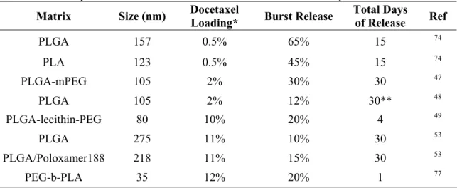
phase typically represents 10-30% of the total encapsulated docetaxel, but can be higher with reports of 65% burst release (Table 1.3). Particles then release cargo at a slower rate over a week to a month.45, 47, 74, 79
Table 1.3 Examples of release from docetaxel loaded PLGA/PLA particles.
Matrix Size (nm) Docetaxel Loading* Burst Release Total Days of Release Ref
PLGA 157 0.5% 65% 15 74
PLA 123 0.5% 45% 15 74
PLGA-mPEG 105 2% 30% 30 47
PLGA 105 2% 12% 30** 48
PLGA-lecithin-PEG 80 10% 20% 4 49
PLGA 275 11% 10% 30 53
PLGA/Poloxamer188 218 11% 15% 30 53
PEG-b-PLA 35 12% 20% 1 77
*See Table 1.2 for encapsulation values **This particle released a maximum of 40%
1.6.3 In vitro cytotoxicity of docetaxel containing PLGA nanoparticles
lines exhibiting similar toxicity (A549).79 Targeting can further enhance docetaxel toxicity, presumably by enhancing internalization. Farokhzad et al. compared the toxicity of docetaxel loaded PLGA-PEG nanoparticles which were untargeted to nanoparticles targeted with a PMSA aptamer on LNCaP cells. Viability decreased from 61% to 42% at 30 minutes and from 48% to 30% at 2 hours with the addition of the targeting ligand.46 Docetaxel can cross the cell membrane on its own so internalization is not necessary for cytotoxicity, but by delivering a higher payload directly into the cell higher toxicity can be achieved.
1.6.4 In vivo efficacy via intratumoral injection
enhancement seen with targeting could be due to reduced clearance or to increased internalization or both.46
1.6.5 In vivo efficacy via intravenous injection
showed tumor regression while vehicle and saline treated mice did not. Furthermore the particle formulations showed enhanced efficacy, with the PEGylated formulations showing the most regression. As a measure of health the mice were also weighed during the course of the study. Mice which received free docetaxel exhibited significant weight loss. Mice which received nanoparticles without PEG also showed some weight loss while mice which received PEGylated nanoparticles did not. This is not addressed by the authors but could be due to increased accumulation in other organs as compared to the PEGylated formulations.47
In addition to increased tumor accumulation, avoidance of cardiotoxicity is a major concern with docetaxel therapy. Esmaeili et al. investigated the biodistribution of docetaxel loaded PLGA nanoparticles (175 nm) sterically stabilized with PVOH compared to Taxotere® in healthy mice. They showed decreased accumulation in the heart, kidneys, and interestingly the liver with nanoparticle formulations.79 This could partly be due to the fact that Taxotere® forms nanoaggregates which would be trapped by the liver. The favorable comparison to Taxotere® makes PLGA nanoparticle systems even more attractive.
1.6.6 Chemotherapeutic PLGA particles in clinical trials
Taxol®, a Cremaphor EL solubilized form similar to Taxotere®. Also approved is Abraxane®, a nanoassembly with albumin. Genexol® did not show enhanced in vitro cytotoxicity, but as previously discussed the anti-tumor efficacy was improved over Taxol® due to the improved tumor delivery. A combination of Genexol® with cisplatin is has proven more effective with less systemic toxicity in patients with advanced non-small cell lung cancer than Taxol® with cisplatin. Genexol® showed a higher response rate in women with metastatic breast cancer over Taxol®. This particular application has progressed to Phase III clinical trials and demonstrates the advances being made with PLGA drug delivery.52
1.7 RNA interference and siRNA delivery
Figure 1.3 The RNAi pathway.
results in the formation of a tumor. Turning these genes off using siRNA therapy could be effective at fighting the disease; however siRNA is easily degraded in vivo and does not readily cross the cell membrane so the development of effective therapies will fully require the development of efficient delivery systems.
1.7.1 Incorporating siRNA in cationic PLGA nanoparticles for effective transfection
The design of PLGA nanoparticles for siRNA delivery is more complex than for the delivery of chemotherapeutics. First siRNA is hydrophilic making it more difficult to encapsulate using traditions two-phase processes that typically rely on water as the anti-solvent. For this reason modifications such as the use of double emulsions (w/o/w) have been employed. Secondly the delivery of chemotherapeutics does not require particle internalization, but the delivery of siRNA does. The siRNA must be introduced into the cytoplasm so both particle internalization and endosomal escape are necessary. The most common method used to achieve efficient delivery is the incorporation of poly(ethylene imine) (PEI) (Figure 1.4).
Figure 1.4 The chemical structure of branched poly(ethylene imine) (PEI).
found that particles with pre-formed PEI/pDNA complexes had the most efficient transfection.82 Cationic complexes are traditionally used for in vitro transfection, but can not be used in vivo due to toxicity and immunogenicity. Cationic complexes are commonly pre-formed for the incorporation in liposomes used as a drug delivery vehicle in vivo. This research demonstrates that though incorporated in a solid polymer particle, having a cationic complex may still provide benefits over a homogeneously mixed system.
Other groups have also demonstrated the enhancement in knockdown provided by a cationic PLGA particle. Andersen et al. compared PLGA particles to PLGA particles with PEI and cetylated PEI. They found that the PLGA particle did not result in gene knockdown while the PLGA/PEI and PLGA/cetylated PEI particles showed 62% and 41% knockdown respectively.83 In addition to PEI, other cationic moieties have been investigated; the second most common being chitosan. Chitosan is a naturally occurring polymer with high primary amine density. Tahara et al. coated PLGA particles containing DOTAP/siRNA complexes in PVOH or a 1:1 mixture of PVOH and chitosan. They only observed gene knockdown in the sample coated with chitosan.84
1.7.2 The effect of N/P ratio on in vitro gene knockdown
1.7.3 In vivo gene knockdown by PLGA nanoparticles
A few groups have taken cationic PLGA siRNA systems in vivo. In 2008 Murata et al. reported anti-tumor efficacy using PLGA/PEI and PLGA/arginine microparticles. They chose an anti-VEGF siRNA as a therapeutic vector against S-180 (sarcoma) xenograft tumors. Vascular endothelial growth factor (VEGF) is responsible for angiogenesis which allows a tumor to grow and spread. By reducing VEGF and therefore the blood supply, many believe a tumor can be contained. They first examined the ability of PEI and arginine to complex with siRNA and produce knockdown. They found, as previously described, a higher N/P ratio resulted in higher knockdown and were able to reach 54% knockdown with arginine complexes and 68% knockdown with PEI complexes. They incorporated these complexes in microspheres that would act as injectable depots using a w/o/w emulsion. The two formulations showed similar release profiles with a burst of 20% and another 20% released over 3 weeks at a steady rate. In vitro these particles exhibited 60% and 43% knockdown with PLGA/arginine and PLGA/PEI respectively (formulation difference not significant; p>0.05). For in vivo evaluation the authors injected microspheres intratumorally and monitored tumor volume. They looked at vehicle controls and compared PLGA/siRNA particles to PLGA/arginine/siRNA particles and PLGA/PEI/siRNA particles. The PLGA/siRNA particles showed some significant growth inhibition (p<0.05), while the PLGA/arginine particles showed a higher level of growth inhibition (p<0.01), and the PLGA/PEI particles were the most efficient formulation with little growth over 21 days.86
for defense against sexually transmitted diseases including human immunodeficiency virus (HIV). Using an N/P ratio of 8:1 they demonstrated PLGA/spermidine/siRNA particles were able to provide more sustained knockdown than commercial transfection agents in vitro, >14 days as compared to <7 days with the commercial agent. They tested particle distribution and found 7 days after in vivo application particles were detected throughout the reproductive tract. An EFGP reporter was used to test in vivo knockdown; 10 days after topical administration 50% knockdown was measured in the uterine horn, 60% knockdown was measured in the cervix, and 60% knockdown was measured in the vaginal tract. They compared these particles to lipid delivery systems which showed equal knockdown, but caused inflammation and epithelial disruption in the vaginal tissue whereas the PLGA nanoparticles did not.87 While PLGA particles do not have a lot of clout as siRNA delivery systems yet, there is a growing body of evidence suggesting these particles can be tailored with varying cationic moieties and show equal knockdown with less toxicity than traditional carriers.
1.8 Future Directions
1.8.1 Combination therapy
Amid PLGA carriers showing promise in both chemotherapy and siRNA, some researchers have started to consider the combination of the two for higher efficacy. Already patients are treated with cocktails of chemotherapeutics which have different mechanisms of toxicity. The combination of a cytotoxin with gene therapy aimed at reducing the cancer’s ability to resist may bring about more rapid and more complete regression. Researchers have demonstrated that the knockdown of certain genes can sensitize cells to chemotherapeutics.88, 89 Combination therapies without carriers have been shown to be more effective against tumor growth than the individual components.90 Saad et al. developed a cationic liposome which incorporated doxorubicin, siRNA against MDR1/MRP1 (drug efflux pump) and siRNA against BCL2 (cellular anti-apoptotic defense) in the same nanoparticle. They achieved efficacy in multi-drug resistant cells which could not be approached by any of the individual components.91
1.8.2 Higher Degree of Control
References
1. E. J. Woodward, C. T., Scheduling of Taxanes: A Review. Current Clinical Pharmacology 2010, Epub ahead of print.
2. A. D. Bangham, M. M. S., J. C. Watkins, Diffusion of univalent ions across the lamellae of swollen phospholipids. Journal of Molecular Biology 1965, 13, (1), 238-252.
3. Gregoriadis, G., The Carrier Potential Of Liposomes In Biology And Medicine Part 2. New England Journal of Medicine 1976, 295, (14), 765-770.
4. Gregoriadis, G., The Carrier Potential Of Liposomes In Biology And Medicine Part 1. New England Journal of Medicine 1976, 295, (13), 704-710.
5. Gregoriadis, G., Engineering liposomes for drug delivery: progress and problems. Trends in Biotechnology 1995, 13, (12), 527-537.
6. K. Kataoka, A. H., Y. Nagasaki, Block copolymer micelles for drug delivery: design, characterization and biological significance. Advanced Drug Delivery Reviews 2001, 47, (1), 113-131.
7. Y. Matsumura, K. K., Preclinical and clinical studies of anticancer agent-incorporating polymer micelles. Cancer Science 2009, 100, (4), 572-579. 8. A. Robert, J. F., Current Status of Nanomedicine and Medical Nanorobotics.
Journal of Computational and Theoretical Nanoscience 2005, 2, 1-25.
9. Chung, T. W.; Huang, Y. Y.; Liu, Y. Z., Effects of the rate of solvent evaporation on the characteristics of drug loaded PLLA and PDLLA microspheres.
International Journal of Pharmaceutics 2001, 212, (2), 161-169.
10. R. T. Liggins, H. M. B., Paclitaxel loaded poly(L-lactic acid) microspheres: properties of microspheres made with low molecular weight polymers. International Journal of Pharmaceutics 2001, 222, 19-33.
11. Lin, S. Y.; Chen, K. S.; Teng, H. H.; Li, M. J., In vitro degradation and
dissolution behaviours of microspheres prepared by three low molecular weight polyesters. Journal of Microencapsulation 2000, 17, (5), 577-586.
13. P. Patel, R. C. M., V. R. Babu, D. Jain, V. Rangaswamy, T. M. Aminabhavi, Microencapsulation of Doxycycline into Poly(lactide-coglycolide) by Spray Drying Technique: Effect of Polymer Molecular Weight on Process Parameters. Journal of Applied Polymer Science 2008, 108, 4038-4046.
14. M. A. Holgado, J. L. A., M. J. C’ozar, J. Alvarez-Fuentes, Synthesis of lidocaine-loaded PLGA microparticles by flow focusing: Effects on drug loading and release properties. International Journal of Pharmaceutics 2008, 358, 27-35. 15. C. E. Astete, C. M. S., Synthesis and characterization of PLGA nanoparticles.
Journal of Biomaterial Science 2006, 17, (3), 247-289.
16. C. Wischke, S. S., Principles of encapsulating hydrophobic drugs in PLA/PLGA microparticles. International Journal of Pharmaceutics 2008, 364, 298-327. 17. Almeida, A. J.; Alpar, H. O.; Brown, M. R. W., Immune-Response to Nasal
Delivery of Antigenically Intact Tetanus Toxoid Associated with Poly(L-Lactic Acid) Microspheres in Rats, Rabbits and Guinea-Pigs. Journal of Pharmacy and Pharmacology 1993, 45, (3), 198-203.
18. Asmatulu, R.; Zalich, M. A.; Claus, R. O.; Riffle, J. S., Synthesis, characterization and targeting of biodegradable magnetic nanocomposite particles by external magnetic fields. Journal of Magnetism and Magnetic Materials 2005, 292, 108-119.
19. Chiou, S. H.; Wu, W. T.; Huang, Y. Y.; Chung, T. W., Effects of the
characteristics of chitosan on controlling drug release of chitosan coated PLLA microspheres. Journal of Microencapsulation 2001, 18, (5), 613-625.
20. Liang, L. S.; Jackson, J.; Min, W. X.; Risovic, V.; Wasan, K. M.; Burt, H. M., Methotrexate loaded poly(L-lactic acid) microspheres for intra-articular delivery of methotrexate to the joint. Journal of Pharmaceutical Sciences 2004, 93, (4), 943-956.
21. Liggins, R. T.; Burt, H. M., Paclitaxel loaded poly(L-lactic acid) microspheres: properties of microspheres made with low molecular weight polymers.
International Journal of Pharmaceutics 2001, 222, (1), 19-33.
22. Mumper, R. J.; Jay, M., Biodegradable Radiotherapeutic Polyester Microspheres - Optimization and Invitro Invivo Evaluation. Journal of Controlled Release 1992, 18, (3), 193-204.
24. Uchida, M.; Jin, Y.; Natsume, H.; Kobayashi, D.; Sugibayashi, K.; Morimoto, Y., Introduction of poly-L-lactic acid microspheres into the skin using supersonic flow: effects of helium gas pressure, particle size and microparticle dose on the amount introduced into hairless rat skin. Journal of Pharmacy and Pharmacology 2002, 54, (6), 781-790.
25. Wake, M. C.; Gerecht, P. D.; Lu, L. C.; Mikos, A. G., Effects of biodegradable polymer particles on rat marrow-derived stromal osteoblasts in vitro. Biomaterials 1998, 19, (14), 1255-1268.
26. Zielhuis, S. W.; Nijsen, J. F. W.; Figueiredo, R.; Feddes, B.; Vredenberg, A. M.; van het Schip, A. D.; Hennink, W. E., Surface characteristics of holmium-loaded poly(L-lactic acid) microspheres. Biomaterials 2005, 26, (8), 925-932.
27. Kim, H. K.; Park, T. G., Comparative study on sustained release of human growth hormone from semi-crystalline poly(L-lactic acid) and amorphous poly(D,L-lactic-co-glycolic acid) microspheres: morphological effect on protein release. Journal of Controlled Release 2004, 98, (1), 115-125.
28. Chung, T. W.; Huang, Y. Y.; Tsai, Y. L.; Liu, Y. Z., Effects of solvent evaporation rate on the properties of protein-loaded PLLA and PDLLA microspheres fabricated by emulsion-solvent evaporation process. Journal of Microencapsulation 2002, 19, (4), 463-471.
29. Han, K.; Lee, K. D.; Gao, Z. G.; Park, J. S., Preparation and evaluation of poly(L-lactic acid) microspheres containing rhEGF for chronic gastric ulcer healing. Journal of Controlled Release 2001, 75, (3), 259-269.
30. Nakaoka, R.; Inoue, Y.; Tabata, Y.; Ikada, Y., Size effect on the antibody production induced by biodegradable microspheres containing antigen. Vaccine 1996, 14, (13), 1251-1256.
31. Zhu, K. J. Z., J. X.; Wang, C.; Yasuda, H.; Ichimaru, A.; Yamamoto, K., Preparation and in vitro release behavior of 5-fluorouracil-loaded microspheres based on poly (L-lactide) and its carbonate copolymers. . Journal of
Microencapsulation 2003, 20, (6), 731-743.
32. Matsuyama, K.; Zhang, D. H.; Urabea, T.; Mishima, K., Formation of L-poly(lactic acid) microspheres by rapid expansion Of CO2 saturated polymer suspensions. Journal of Supercritical Fluids 2005, 33, (3), 277-283.
34. Tom, J. W.; Debenedetti, P. G., Formation of Bioerodible Polymeric
Microspheres and Microparticles by Rapid Expansion of Supercritical Solutions. Biotechnology Progress 1991, 7, (5), 403-411.
35. Falk, R. F.; Randolph, T. W., Process variable implications for residual solvent removal and polymer morphology in the formation of gentamycin-loaded poly(L-lactide) microparticles. Pharmaceutical Research 1998, 15, (8), 1233-1237. 36. Jarmer, D. J.; Lengsfeld, C. S.; Randolph, T. W., Manipulation of particle size
distribution of poly(L-lactic acid) nanoparticles with a jet-swirl nozzle during precipitation with a compressed antisolvent. Journal of Supercritical Fluids 2003, 27, (3), 317-336.
37. Tu, L. S. D., F.; Foster, N. R., Micronization and microencapsulation of pharmaceuticals using a carbon dioxide antisolvent. Powder Technology 2002, 126, (2), 134-149.
38. Choi, S.; Lee, K.; Kim, H., Preparation of poly(L-lactic acid) nano- and micro-particles using supercritical antisolvent. Journal of Chemical Engineering of Japan 2005, 38, (8), 571-577.
39. Kim, M. Y. L., Youn Woo; Byun, Hun-Soo; Lim, Jong Sung., Recrystallization of Poly(L-lactic acid) into Submicrometer Particles in Supercritical Carbon Dioxide. Industrial & Engineering Chemistry Research 2005, ACS ASAP.
40. Patel, M. M.; Zeles, M. G.; Manning, M. C.; Randolph, T. W.; Anchordoquy, T. J., Degradation kinetics of high molecular weight poly (L-lactide) microspheres and release mechanism of lipid : DNA complexes. Journal of Pharmaceutical Sciences 2004, 93, (10), 2573-2584.
41. J. Guan, N. F., L. J. Lee, D. J. Hansford, Fabrication of polymeric microparticles for drug delivery by soft lithography. Biomaterials 2006, 27, 4034-4041.
42. J. Guan, H. H., L. J. Lee, D. J. Hansford, Fabrication of Particulate Reservior-Containing, Capsulelike, and Self-Folding Polymer Microstructures for Drug Delivery. Small 2007, 3, 412-418.
43. J. A. Champion, Y. K. K., S. Mitragotri, Particle Shape: A new design parameter for micro- and nanoscale drug delivery systems. Journal of Controlled Release 2007, 121, 3-9.
44. G. Acharya, C. S. S., M. McDermott, H. Mishra, H. Park, I. C. Kwon, K. Park, The hydrogel template method for the fabrication of homogeneous
45. F. Esmaeili, M. H. G., S. N. Ostad, F. Atyabi, M. Seyedabadi, M. D. Malekshahi, M. Amini, and R. Dinarvnand, Folate-receptor-targeted delivery of docetaxel nanoparticles prepared by PLGA-PEG-folate conjugate. Journal of Drug Targeting 2008, 16, (5), 415-423.
46. O. C. Farokhzad, J. C., B. A. Teply, I. Sherifi, S. Jon, P. W. Kantoff, J. P. Richie, and R. Langer, Targeted nanoparticle-aptamer bioconjugates for cancer
chemotherapy in vivo. PNAS 2006, 103, (16), 6315-6320.
47. M. Senthilkumar, P. M., and N. K. Jain, Long circulating PEGylated poly(D,L-lactide-co-glycolide) nanoparticulate delivery of docetaxel to solid tumors. Journal of Drug Targeting 2008, 16, (5), 424-435.
48. S. Murugesan, S. G., R. K. Averinei, M. Nahar, P. Mishra, N. K. Jain, PEGylated Poly(lactide-co-glycolide) (PLGA) Nanoparticulate Delivery of Docetaxel: Synthesis of Diblock Copolymers, Optimization of Preparation Variables on Formulation Characteristics and In Vitro Release Studies. Journal of Biomedical Nanotechnology 2007, 3, 52-60.
49. J. M. Chan, L. Z., K. P. Yuet, G. Liao, J. Rhee, R. Langer, O. C. Farokhzad, PLGA-lecithin-PEG core-shell nanoparticles for controlled drug delivery. Biomaterials 2009, 30, 1627-1634.
50. Mittal, G. S., D. K.; Bhardwaj, V.; Ravi Kumar, M. N. V., Estradiol loaded PLGA nanoparticles for oral administration: effect of polymer molecular weight and copolymer composition on release behavior in vitro and in vivo. Journal of Controlled Release 2007, 119, 77-85.
51. Lee, S. K., M. S.; Kim, J. S.; Park, H. J.; Woo, J. S.; Lee, B. C.; Hwang, S. J., Controlled delivery of a hydrophilic drug from a biodegradable microsphere system by supercritical anti-solvent precipitation technique. Journal of Microencapsulation 2006, 23, 741-749.
52. G. Gaucher, R. H. M., and J. Leroux, Polyester-based micelles and nanoparticles for the parenteral delivery of taxanes. Journal of Controlled Release 2010, 143, (1), 2-12.
53. Yan, F. Z., C.; Zheng, Y.; Mei, L.; Tang, L.; Song, C.; Sun, H.; Huang, L., The effect of poloxamer 188 on nanoparticle morphology, size, cancer cell uptake, and cytotoxicity. Nanomedicine 2010, 6, 170-178.
55. J. Lu, X. W., C. Marin-Muller, H. Wang, P. H. Lin, Q. Yao, C. Chen, Current advances in research and clinical applications of PLGA-based nanotechnology. Expert Reviews in Molecular Diagnostics 2009, 9, (4), 325-341.
56. F. Alexis, E. P., L. K. Molnar, and O. C. Farokzhad, Factors Affecting the Clearance and Biodistribution of Polymeric Nanoparticles. Molecular Pharmaceutics 2008, 5, (4), 505-515.
57. P. Xu, E. G., L. Tong, C. B. Highley, D. R. Errabelli, T. Hasan, J. Cheng, D. S. Kohane, Y. Yeo, Intracellular Drug Delivery by poly(lactic-co-glycolic acid) Nanoparticles, Revisited. Molecular Pharmaceutics 2008, 6, (1), 190-201.
58. S. E. A. Gratton, P. A. R., P. D. Pohlhaus, J. C. Luft, V. J. Madden, M. E. Napier, J. M. DeSimone, The effect of particle design on cellular internalization
pathways. PNAS 2008, 105, (33), 11613–11618.
59. J. Panyam, V. L., Biodegradable nanoparticles for drug and gene delivery to cells and tissue. Advanced Drug Delivery Reviews 2003, 55, 329-347.
60. J. K. Vasir, V., Labhasetwar, Biodegradable nanoparticles for cytosolic delivery of therapeutics. Advanced Drug Delivery Reviews 2007, 59, 718-728.
61. J. Panyam, W. Z., S. Prabha, S. K. Sahoo, V. Labhasetwar, Rapid endo-lysosomal escape of poly(D,L-lactide-co-glycolide) nanoparticles: implications for drug and gene delivery. The FASEB Journal 2002, 16, 1217-1226.
62. M. S. Cartiera, K. M. J., V. Rajendran, M. J. Caplan, W. M. Saltzman, The uptake and intracellular fate of PLGA nanoparticles in epithelial cells. Biomaterials 2009, 30, 2790-2798.
63. J. A. Champion, S. M., Role of target geometry in phagocytosis. PNAS 2006, 103, (13), 4930-4934.
64. J. J. Yu, H. A. L., J. H. Kim, W. H. Kong, Y. Kim, Z. Y. Cui, K. G. Park, W. S. Kim, H. G. Lee, and S. W. Seo, Biodistribution and anti-tumor efficacy of
PEG/PLA nanoparticles loaded doxorubicin. Journal of Drug Targeting 2007, 15, (4), 279-284.
65. Z. Panagi, A. G., G. Evangelatos, E. Livaniou, D. S. Ithakissios, K. Avgoustakis, Effect of dose on the biodistrbution and pharmacokinetics and PLGA and PLGA-mPEG nanoparticles. International Journal of Pharmaceutics 2001, 221, 143-152. 66. K. Avagoustakis, A. B., Z. Panagi, P, Klepetsanis, E. Livaniou, G. Evangelatos,
67. J. Cheng, B. A. T., I. Sherifi, J. Sung, G. Luther, F. X. Gu, E. Levy-Nissenbaum, A. F. Radovic-Moreno, R. Langer, O. C. Farokhzad, Formulation of
functionalized PLGA-PET nanoparticles for in vivo targeted drug delivery. Biomaterials 2007, 28, 869-876.
68. F. Xu, Y. Y., X. Shan, C. Liu, X. Tao, Y. Sheng, and H. Zhou, Long-circulation of hemoglobin-loaded polymeric nanoparticles as oxygen carriers with modulated surface charges. International Journal of Pharmaceutics 2009, 377, 199-206. 69. Y. Li, Y. P., X. Zhang, Z. Gu, Z. H. Zhou , W. Yuan, J. Zhou, J. Zhu, X. Gao,
PEGylated PLGA nanoparticles as protein carriers: synthesis, preparation and biodistribution in rats. Journal of Controlled Release 2001, 71, 203-211.
70. S. Zhang, X. G., K. Shen, P. Yang, and X. Ju, Evaluation of poly(D,L-lactide-co-glycolide) microspheres for the lung-targeting of yuanhuacine, a novel DNA topoisomerase I inhibitor. Journal of Drug Targeting 2009, 17, (4), 286-293. 71. H. Lee, H. F., B. Hoang, R. M. Reilly, and C. Allen, The Effects of Particle Size
and Molecular Targeting on the Intratumoral and Subcellular Distribtuion of Polymeric Nanoparticles. Molecular Pharmaceutics ASAP.
72. D. A. Christian, S. C., O. B. Garbuzenko, T. Harada, A. L. Zajac, T. Minko, and D. E. Discher, Flexible Filaments for In Vivo Imaging and Delivery: Persistent Circulation of Filomicelles Opens the Dosage Window for Sustained Tumor Shrinkage. Molecular Pharmaceutics 2009, 6, (5), 1343-1352.
73. Lyseng-Williamson, K. A. F., C., Docetaxel: A Review of its Use in Metastatic Breast Cancer. Drugs 2005, 65, 2513-2531.
74. T. Musumeci, C. A. V., I. Giannone, B. Ruozi, L. Montenegro, R. Pignatello, and G. Puglisi, PLA/PLGA nanoparticles for sustained release of docetaxel.
International Journal of Pharmaceutics 2006, 325, 172-179.
75. Garrec, D. L. G., S.; Luo, L.; Lessard, D.; Smith, D. C.; Yessine, M. A.; Ranger, M.; Leroux, J. C., Poly(N-vinylpyrrolidone)-block-poly(D,L-lactide) as a new polymeric solubilizer for hydrophobic anticancer drugs: in vitro and in vivo evaluation. Journal of Controlled Release 2004, 99, 83-101.
76. Karnik, R. G., F.; Basto, P.; Cannizzaro, C.; Dean, L.; Kyei-Manu, W.; Langer, R.; Farokhzad, O.C., Microfluidic platform for conrolled synthesis of polymeric nanoparticles. Nanoletters 2008, 8, 2906-2912.
78. Wang, X. W., Y.; Chen, X.; Wang, J.; Zhang, X.; Zhang, Q., NGR-modified micelles enhance their interaction with CD13-overexpressing tumor and endothelial cells. Journal of Controlled Release 2009, 139, 56-62.
79. F. Esmaeili, R. D., M. H. Ghahremani, S. N. Ostad, H. Esmaily, and F. Atyabi, Cellular cytotoxicity and in-vivo biodistribution of docetaxel poly(lactide-co-glycolide) nanoparticles. Anti-Cancer Drugs 2010, 21, (1), 43-52.
80. C. G. Oster, N. K., L, Grode, L. Barbu-Tudoran, A. K. Schaper, S. H. E. Kaufmann, T. Kissel, Cationic microparticles consisting of poly(lactide-co-glycolide) and polyethylenimine as carrier systems for parental DNA vaccination. Journal of Controlled Release 2005, 104, 359-377.
81. Y. Patil, J. P., Polymeric nanoparticles for siRNA delivery and gene silencing. International Journal of Pharmaceutics 2009, 367, 195-203.
82. X. Zhang, J. I., A. K. Salem, Comparative study of poly(lactic-co-glycolic acid)-polyethyleneimine-plasmid DNA microparticles prepared using double emulsion methods. Journal of Microencapsulation 2008, 25, (1), 1-12.
83. M. Andersen, A. L., A. Arpanaei, S. M. R. Jensen, H. Kaur, D. Oupicky, F. Besenbacher, P. Kingshott, J. Kjems, K. A. Howard, Surface functionalization of PLGA nanoparticles for gene silencing. Biomaterials 2010, 31, 5671-5677. 84. K. Tahara, H. Y., N. Hirashima, Y. Kawashima, Chitosan-modified
poly(D,L-lactide-co-glycolide) nanospheres for improving siRNA delivery and gene-silencing effects. European Journal of Pharmaceutics and Biopharmaceutics 2010, 74, 421-426.
85. H. Katas, E. C., H. O. Alpar, Preparation of polyethyleneimine incorporated poly(D,L-lactide-co-glycolide) nanoparticles by spontaneous emulsion diffusion method for small interfering RNA delivery International Journal of
Pharmaceutics 2009, 329, 144-154.
86. N. Murata, Y. T., K. Toyoshima, M. Yamamoto, H. Okada, Anti-tumor effects of anti-VEGF siRNA encapsulated with PLGA microspheres in mice. Journal of Controlled Release 2008, 126, 246-254.
87. K. A. Woodrow, Y. C., C. J. Booth, J. K. Saucier-Sawyer, M. J. Wood, and W. M. Saltzman Intravaginal gene silencing using biodegradable polymer
88. J. Halder, C. N. L. J., S. K. Lutgendorf, Y. Li, N. B. Jennings, D. Fan, G. M. Nelkin, R. Schmandt, M. D. Schaller, A. K. Sood Focal Adhesion Kinase Silencing Augments Docetaxel-Mediated Apoptosis in Ovarian Cancer Cells. Clinical Cancer Research 2005, 11, (24), 8829-88-36.
89. C. Gill, C. D., A. J. O'Neill, R. W. G. Watson, Effects of cIAP-1, cIAP-2, and XIAP triple knockdown on prostate cancer cell susceptibility to apoptosis, cell survival, and proliferation. Molecular Cancer 2009, 8, (39), 1-12.
90. S. M. Thomas, M. J. O., M. L. Freilino, S. Strychor, D. R. Walsh, W. E. Gooding, J. R. Grandis, W. C. Zamboni, Antitumor Mechanisms of Systemically
Administered Epidermal Growth Factor Receptor Antisense Oligonucleotides in Combination with Docetaxel in Squamous Cell Carcinoma of the Head and Neck. Molecular Pharmacology 2007, 73, (3), 627-638.
CHAPTER 2
2.1 Introduction to the PRINT® Process
of material which connects the cavities, termed the “scum” or “flash” layer, which must then be removed by an additional step such as etching. As feature size decreases this becomes an increasing problem as the layer being etched approximates the size of the feature being molded. Using Fluorocur instead of PDMS eliminates the connecting layer; the cavities of a Fluorocur mold can be filled without wetting the land area between. This versatile technology platform has applications from advanced drug delivery to energy collection and storage.
2.1.1 Control of Size, Shape, and Composition
Figure 2.1 A variety of sizes and shapes generated using the PRINT process (A) 80nm x 360nm cylinders, (B) 200nm x 200nm cylinders, (C) 200nm x 600nm cylinders, (D) 1 µm sphere approximates, (E) 2 µm cubes with ridges on the sides, (F) 3 µm particles with
center fenestrations, (G) 3 µm arrows, (H) 10 µm boomerangs, and (I) 7 µm discs.
ligands can be added to the particles for enhanced uptake specificity or markers of self can be added to the particles to prevent uptake. Control over composition independent of size and shape is unique since in most particle fabrication techniques the composition and size are interrelated. Complete and independent control over size, shape, and composition allows for the rational design of drug delivery vehicles with the PRINT process.
2.1.2 Mold Fabrication
The Fluorocur mold is the keystone in the PRINT process. Fluorocur is a mixture of a perfluorinated polyether oil (Figure 2.2) and a soluble photoinitiator.
Figure 2.2 An example of the perfluorinated polyether oil structure. The chain length (n and m) can be varied as well as the end group functionalization.
wafer that does not have a pattern (Figure 2.3 D-F). This flat substrate is used in solvent evaporation, pressure fill methods as described below.
Figure 2.3 Mold and substrate fabrication (A) Fluorocur is poured onto a template, (B) The Fluorocur wets the template completely and crosslinks upon exposure to UV light, (C) The mold generated can be peeled from the template and now has the inverse pattern
of the template, (D) Fluorocur is poured onto a flat silicon wafer, (E) The Fluorocur crosslinks upon exposure to UV light, (F) The substrate generated can be peeled from the
template and is smooth on the wafer side.
2.2 The solvent evaporation, pressure fill PRINT method
2.2.1 Experimental
2.2.1.1 Materials
Poly(L-lactic acid) (PLLA; 130,000 g/mol), dichloromethane (DCM) and 1,4-dioxane were purchased from Sigma Aldrich. Fluorocur® was synthesized first in lab as previously reported1 and then supplied by Liquidia Technologies.
2.2.1.2 Particle visualization and characterization
For visualization by scanning electron microscopy (SEM) samples were coated with 3 nm gold palladium alloy using a Cressington 108 auto sputter coater. Images were taken at an accelerating voltage of 2 kV using a Hitachi model S-4700 SEM. For thermal characterization, measurements were made on a Seiko 120 differential scanning calorimeter (DSC) using a heating rate of 10oC/min. Electron diffraction measurements were made on a high resolution FEI-Philips Tecnai 12 transmission electron microscope (TEM).
2.2.1.3 Particle fabrication procedure
substrate. The entire apparatus is then placed under vacuum at ambient temperature for 24 hours to thoroughly remove the solvent. When all the solvent evaporates, the polymer solidifies in the cavities of the mold (Figure 2.4 C). The apparatus can then be removed from vacuum, taken apart, and the mold can be peeled from the substrate (Figure 2.4 D). The particles remain in the mold due to the higher surface area contact with the mold compared to the substrate.
Figure 2.4 The solvent evaporation, pressure fill PRINT method (A) a mold and substrate are fabricated from Fluorocur, (B) a solution of polymer in organic solvent is injected between the mold and substrate, (C) pressure is applied to the sandwich which is
placed under vacuum to remove the solvent solidifying the particles, and (D) the mold and substrate are separated revealing isolated particles in the mold.
2.2.2 Results