CELLULAR AND PLASMA PROTEIN CROSSTALK IN ARTERIAL AND VENOUS THROMBOSIS
Maria M. Aleman
A dissertation submitted to the faculty of the University of North Carolina at Chapel Hill in partial fulfillment of the requirements for the degree of Doctor of Philosophy in the
Department of Pathology and Laboratory Medicine.
Chapel Hill 2014
ABSTRACT
Maria M. Aleman: Cellular and Plasma Protein Crosstalk in Arterial and Venous Thrombosis (Under the direction of Dr. Alisa Wolberg)
Coagulation is an enzymatic cascade culminating in the formation of a clot. Intravascular coagulation is termed thrombosis. Studies using platelet-rich plasma, whole blood, and animal models reveal the complex crosstalk between clotting factors and the cellular environment that promote thrombosis. Three distinct, yet related, studies included in this dissertation exemplify this crosstalk.
First, the vascular bed-dependent prothrombotic effects of elevated prothrombin were examined. We found that elevated prothrombin increased venous, but not arterial,
thrombosis in mice. The prothrombotic effects of elevated prothrombin were manifested by increased fibrin deposition but no significant increase in platelet accumulation. These data show that elevated prothrombin would not be expected to contribute to platelet-dominated arterial thrombosis.
Second, the procoagulant properties of microparticles from different cell types were investigated. We found that monocyte-derived microparticles contributed to initiation and propagation of clotting via the extrinsic coagulation pathway, while platelet-derived
microparticles contributed to primarily to propagation of clotting via the intrinsic coagulation pathway. These data suggest monocyte-derived microparticles may contribute to the
Third, the role of fibrinogen and factor XIII (FXIII) in venous thrombosis was studied. We discovered the binding site of FXIII on fibrinogen, the necessity for FXIII activity for red blood cell retention in clots, and that reduction or deficiency of FXIII activity reduces venous thrombus size. These data indicate a novel role for FXIII in the pathogenesis of thrombosis and implicate FXIII as a novel therapeutic target.
ACKNOWLEDGEMENTS
Foremost, I would like to thank Dr. Alisa Wolberg for providing me with exceptional training opportunities, an excellent environment for growth, and her complete and thorough attention on various projects and goals. While a trainee with Alisa, I was afforded
opportunities to mentor undergraduate and graduate students, to attend and present at national and international meetings, and to develop a research project that has subsequently driven the focus of the lab into new and exciting territories. She supported my ambitions and always put my interests first. A better mentor could not be had.
Success in the lab also depends on having great lab mates. Thus, I would also like to thank members of the Wolberg Lab, past and present, especially Christa DeVette, Heyman Peraza, Jian-Guo (Kevin) Wang, James Byrnes, Bethany Walton, Kellie Machlus, and Robert Campbell. The contributions they all made to my studies were tremendous.
I would like to thank my dissertation committee members, Drs. Maureane Hoffman, Bill Coleman, Frank Church, and Nigel Key, for their time, thoughtfulness, and invaluable guidance during my graduate training. It’s been a joy working with each of you. Many thanks, as well, to the UNC McAllister Heart Institute Thrombosis & Hemostasis group, especially Dr. Nigel Mackman. The opportunity to present my work to this excellent group of fellow ‘clotters’ was always rewarding and their critiques provided much needed feedback during the development of my research projects.
Karla, and my dear friends Brianna, Nicole, and Ryan, provided much needed distraction and levity. Likewise, my mother, Susan, and father, Robert, have been great supporters.
Thanks to the American Heart Association (AHA), the National Institute of
Environmental Health Sciences (NIEHS), and the National Heart Lung and Blood Institute (NHLBI) for funding. I was supported by the NIEHS for three years (2009-2012) on a departmental training grant (T32-ES007017). I was later supported briefly on a pre-doctoral fellowship from the AHA (12PRE10420007) before resigning in favor of a Ruth L.
PREFACE
Chapter 2 represents work that was published previous to writing this dissertation with the following citation:
Aleman MM, Walton BL, Byrnes JR, Wang J-G, Heisler M, Machlus KR, Cooley BC, Wolberg AS. Elevated prothrombin promotes venous, but not arterial, thrombosis in mice. Arterioscler Thromb Vasc Biol. 2013;33(8):1829-1836.
Permission to include the article in its entirety in a PhD dissertation was obtained from Lippincott, Williams, & Wilkins (publisher of Arteriosclerosis Thrombosis and Vascular Biology).
Chapter 3 represents work that was published previous to writing this dissertation with the following citation:
Aleman MM, Gardiner C, Harrison P, Wolberg AS. Differential contributions of monocyte- and platelet-derived microparticles towards thrombin generation and fibrin formation and stability. J Thromb Haemost. 2011;9(11):2251-2261.
TABLE OF CONTENTS
LIST OF TABLES ... x
LIST OF FIGURES ... xi
LIST OF ABBREVIATIONS ... xiii
CHAPTER 1: INTRODUCTION ... 1
1.1 Coagulation & Blood Cells ... 1
1.2 Arterial & Venous Thrombosis ... 3
1.3 Hyperprothrombinemia ... 5
1.4 Microparticles & Thrombosis ... 9
1.5 FXIII & RBCs ... 10
1.6 Focus of this Dissertation... 13
1.7 References ... 14
CHAPTER 2: ELEVATED PROTHROMBIN PROMOTES VENOUS, BUT NOT ARTERIAL, THROMBOSIS IN MICE... 19
2.1 Introduction ... 19
2.2 Materials & Methods ... 21
2.3 Results ... 26
2.4 Discussion ... 37
CHAPTER 3: DIFFERENTIAL CONTRIBUTIONS OF MONOCYTE- AND PLATELET-DERIVED MICROPARTICLES TOWARDS THROMBIN
GENERATION AND FIBRIN FORMATION AND STABILITY ... 48
3.1 Introduction ... 48
3.2 Materials & Methods ... 50
3.3 Results ... 55
3.4 Discussion ... 67
3.5 References ... 72
CHAPTER 4: FACTOR XIII ACTIVITY IS REQUIRED FOR RETENTION OF RED BLOOD CELLS IN VENOUS THROMBI ... 76
4.1 Introduction ... 76
4.2 Materials & Methods ... 78
4.3 Results ... 85
4.4 Discussion ... 99
4.5 References ... 107
CHAPTER 5: SUMMARY AND FUTURE DIRECTIONS ... 113
5.1 Summary & Future Directions ... 113
LIST OF TABLES
LIST OF FIGURES
Figure 1.1. Simplified coagulation cascade ...2
Figure 1.2. Arterial thrombosis. ...6
Figure 1.3. Venous thrombosis ...8
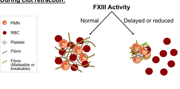
Figure 1.4. Model of FXIII-dependent RBC retention during clot retraction ...11
Figure 2.1. Human prothrombin supports thrombin generation in murine plasma and mouse thrombomodulin reduces thrombin generation in mouse plasma spiked with human prothrombin ...26
Figure 2.2. Human prothrombin circulates in mice 12 hours post-infusion ...28
Figure 2.3. In the absence of vessel injury, elevated prothrombin does not activate coagulation ...30
Figure 2.4. Elevated prothrombin increases the rate and extent of fibrin deposition following electrolytic injury to the femoral vein ...32
Figure 2.5. Elevated prothrombin produces larger venous thrombi by increasing thrombin generation following IVC ligation ...34
Figure 2.6. Elevated prothrombin does not increase thrombin generation or the rate of platelet or fibrin accumulation, and does not shorten the TTO in arterial injury models ...36
Figure 3.1. M-MPs and PMPs have similar size distributions ...56
Figure 3.2. M-MPs, but not PMPs, promote thrombin generation in a TF-dependent manner ...58
Figure 3.3. M-MPs initiate fibrin formation ...60
Figure 3.4. M-MPs, but not PMPs, increase fibrin network density ...62
Figure 3.5. M-MPs increase clot resistance to fibrinolysis ...64
Figure 3.6. PMPs increase thrombin generation and the rate of fibrin formation during TF-initiated clotting ...66
Figure 4.2. Procoagulant activity is normal in Fibγ390-396A mice ...86 Figure 4.3. RBCs are extruded from Fibγ390-396A clots during clot retraction,
resulting in decreased clot weight ...88 Figure 4.4. RBCs adhere to fibrinogen from WT and Fibγ390-396A mice ...90 Figure 4.5. FXIII-A2B2 does not co-precipitate with Fibγ390-396A fibrinogen ...92
Figure 4.6. Compared to WT, plasma from Fibγ390-396A mice exhibits delayed FXIII activation and consequently, delayed fibrin cross-
linking during TF-initiated coagulation ...94 Figure 4.7. FXIII-deficient mice retain fewer RBCs after clot retraction,
and RBC extrusion from clots can be induced in normal blood
with FXIII inhibition ...96 Figure 4.8. FXIII-deficient humans retain fewer RBCs after clot retraction,
and RBC extrusion from clots can be induced in normal human
LIST OF ABBREVIATIONS
α Alpha
A23187 Calcium ionophore ANOVA Analysis of variance APC Allophycocyanin AU Arbitrary unit
β Beta
CTI Corn trypsin inhibitior CVD Cardiovascular disease DSS Dextran sodium sulfate DTT Dithiothreitol
DVT Deep vein thrombosis
EDTA Ethylenediamine tetraacetic acid ELISA Enzyme-linked immunosorption assay EPCR Endothelial protein C receptor
Fgn Fibrinogen FVIII Factor VIII FXIII Factor XIII
FXIIIa Activated factor XIII
g Gram
γ Gamma
HBSS Hank's balanced salt solution HMW Higher molecular weight IgG Immunoglobulin G
II Prothrombin
IIa Thrombin
IVC Inferior vena cava
L Liter
LPS Lipopolysaccharide
M Molar
M-MP Monocyte-derived microparticle MDP Microparticle-depleted plasma Min Minute
mg Milligram
mL Milliliter mM Millimolar MP Microparticle
ng Nanogram
nM Nanomolar
NTA Nanoparticle tracking analysis OD Optical density
PBMC Peripheral blood mononuclear cell PBS Phosphate-buffered saline
PFP Platelet-free plasma
pM Picomolar
PMP Platelet-derived microparticle PPP Platelet-poor plasma
PS Phosphatidylserine RBC Red blood cell SCD Sickle cell disease SD Standard deviation SDS Sodium dodecyl sulfate SEM Standard error of the mean
T1 Magnetic resonance longitudinal relaxation time
TAT Thrombin-antithrombin complex TEM Transmission electron microscopy TF Tissue factor
THP-1 Human monocyte cell line THP-MP THP-1 cell microparticles TM Thrombomodulin
tPA Tissue plasminogen activator TRAP Thrombin receptor agonist peptide TTO Time to occlusion
U Unit
µL Microliter
Va Activated factor V
VTE Venous thromboembolism WBC White blood cell
CHAPTER 1: INTRODUCTION
1.1 Coagulation & Blood Cells
Blood coagulation is a complex enzymatic cascade culminating in the formation of a fibrin protein meshwork that turns liquid blood into a gel-like solid. Two initiating
coagulation pathways—intrinsic and extrinsic—converge on a common pathway that
produces the serine protease thrombin (Figure 1.1). Thrombin activates platelets and cleaves fibrinogen into fibrin monomers that polymerize into fibers forming a net-like structure that is stabilized by the transglutaminase factor XIII (FXIII). Misbalance of this system leads to a failure of hemostasis (bleeding) or excessive clotting (thrombosis).
Clotting can be reduced down to two essential components—thrombin and
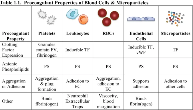
fibrinogen. However, physiologic coagulation involves the entire cascade and, importantly, anionic phospholipid surfaces such as on an activated platelet. Thus, the simple coagulation cascade has evolved into a cell-based model1 that emphasizes the role of blood cells in initiation or propagation of coagulation (Table 1.1). Platelets, when activated, support coagulation by expression of anionic phosphatidylserine (PS) on their plasma membrane, release of granules rich in clotting factors such as factor V and fibrinogen, and by their ability to aggregate and occlude vessels at sites of trauma or atherosclerotic plaque rupture.2 Leukocytes, such as monocytes and neutrophils, contribute to coagulation by various means: a) expression of tissue factor (TF)—the key initiator of extrinsic coagulation—can be
microparticles, especially TF-bearing microparticles6-8, and c) by neutrophil extracellular trap-mediated activation of the intrinsic coagulation pathway9,10. Endothelial cells contribute to clotting by release of von Willebrand Factor which binds platelets11,12, their ability to bind fibrin and modulate its network structure13, and by induced expression of TF14, though their ability to express it in vivo is controversial15. Finally, red blood cells (RBCs) contribute to coagulation by exposure of PS that supports thrombin generation16,17, and RBCs increase blood viscosity18 and marginate platelets toward the endothelium19, placing them in close proximity to sites of vascular trauma. Lastly, recent studies from our laboratory indicate RBCs directly dictate the size of clots (Chapter 4).
1.2 Arterial & Venous Thrombosis
Thrombosis is a pathologic process leading to inappropriate intraluminal blood coagulation that is a key component of myocardial infarction, ischemic stroke, and venous thromboembolism (VTE; deep vein thrombosis (DVT)/pulmonary embolism (PE)). Cardiovascular disease (CVD), which includes myocardial infarction and ischemic stroke, currently afflicts 83.6 million Americans, caused 32.3% of all deaths in the United States in 2009, and costs an estimated $312.6 billion each year.20 One million Americans suffer from VTE each year21, and incidence rates increase with age in both men and women20. The annual health care costs for VTE, including hospital-acquired and recurrent events, are estimated to range from $15.4 to $34.4 billion.22 These pathologies are significant health and economic burdens and the need for improved understanding and treatment is essential.
Table 1.1. Procoagulant Properties of Blood Cells & Microparticles
Procoagulant Property
Platelets Leukocytes RBCs Endothelial Cells Microparticles Clotting Factor Expression Granules contain FV, fibrinogen
Inducible TF Inducible TF, vWF TF
Anionic
Phospholipids PS PS PS PS PS
Aggregation or Adhesion Aggregation & plug formation Adhesion to EC Aggregation, adhesion to EC Supports adhesion Adhesion to other cells
Other fibrin(ogen) Binds
Neutrophil Extracellular Traps Viscocity, blood margination Binds fibrin(ogen)
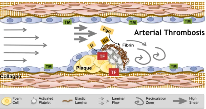
procoagulant sub-endothelial material and cells (Figure 1.2).23 This process occurs under high shear leading to the formation of platelet-rich thrombi—so called “white clots”—and detrimental vascular occlusion. Treatment of arterial thrombosis primarily involves anti-platelet drugs, sometimes with anticoagulation, and/or thrombolytic therapy. Risk factors for arterial thrombosis include CVD, hypertension, diabetes mellitus, obesity, and smoking.24
Venous thrombosis, in contrast, is thought to originate in the hypoxic environment of venous valve pockets (Figure 1.3).25,26 In these areas, zones of blood flow recirculation lead to endothelial activation, leukocyte accumulation and activation, and the exposure of tissue factor and release of neutrophil extracellular traps thereby initiating coagulation and the formation of a thrombus rich in RBCs and fibrin—so called “red clots.” Treatment for acute DVT is heparin, followed by warfarin for 3 to 6 months. Novel anticoagulants to replace warfarin include anti-Xa and anti-thrombin drugs. Like arterial thrombosis, risk factors for venous thrombosis include CVD, hypertension, diabetes mellitus, obesity, and smoking, but additional risk factors include trauma, infection, immobility, and cancer.27 The differences between arterial and venous thrombosis triggering events, cellular compositions, treatments, and risk factors highlight the context-dependent nature of coagulation in vivo.
1.3 Hyperprothrombinemia
What happens to coagulation when a single clotting factor is elevated? The answer depends on which factor is elevated and its functionality in the clotting cascade. Previous studies by our laboratory painstakingly determined the effect of elevated clotting factors on
in vitro thrombin generation and fibrin formation28-31, as well as thrombus formation in
regardless of TF level used to initiate clotting30, elevated factor VIII (FVIII, a cofactor in the intrinsic pathway) only increased fibrin formation with low levels of TF31. Similarly, while elevated fibrinogen shortened the time to occlusion (TTO) in a FeCl3 injury model30,
shortening of the TTO with elevated FVIII was dependent on the extent of injury31. These data show how the effects of elevated factors on coagulation kinetics are context-dependent.
Prothrombin is the inactive zymogen form of thrombin. Previous studies by our laboratory described the in vitro procoagulant effects of elevated prothrombin levels
(hyperprothrombinemia). In both purified and plasma-based systems, elevated prothrombin increases thrombin generation28,29 and significantly increases fibrin formation and network density29. Interestingly, elevated prothrombin has no effect on platelet activation29,
suggesting prothrombotic effects of hyperprothrombinemia are due to effects on fibrin formation.
Risk of thrombosis for hyperprothrombinemia patients is vascular bed-specific. While there is a clear association between venous thrombosis and elevated prothrombin levels32,33, the association is less clear between elevated prothrombin and arterial thrombosis risk.34,35 This suggests that vascular bed-specific mechanisms dictate the prothrombotic effects of elevated prothrombin. To investigate the role of elevated prothrombin in vascular bed-specific thrombosis, we utilized multiple mouse models of venous and arterial
thrombosis in a study described in Chapter 2. Briefly, we determined that acute
1.4 Microparticles & Thrombosis
Cellular microparticles, also known as microvesicles, are sub-micron plasma membrane blebs released from various cell types. In particular, platelets, monocytes, red blood cells, endothelial cells, and tumor cells, have been shown to release microparticles.37-39 These microparticles circulate in the blood and are primarily of platelet origin in healthy individuals.40 However, microparticles of various cellular origins are elevated in diseases including CVD41-43, VTE44, and cancer45,46.
Like their parent cells, microparticles have procoagulant properties that can include the presence of TF and/or PS on the outer leaflet of their plasma membrane (Table 1.1). While PS exposure appears to be a hallmark of microparticles from all cell types, TF expression is restricted to microparticles stemming from TF-expressing parent cells such as monocytes and tumor cells. The presence or absence of TF confers differential procoagulant activity on microparticles. In work detailed in Chapter 3, we show that TF-bearing
microparticles from monocytes or a monocytic cell line support extrinsic thrombin generation and fibrin formation, whereas non-TF-bearing microparticles from platelets support intrinsic thrombin generation and fibrin formation.6 This study suggests TF-bearing microparticles may be causative for VTE, whereas non-TF-bearing microparticles may only contribute to thrombosis after an initiating event.
Cancer patients, particularly pancreatic cancer patients, with VTE have increased microparticle-associated tissue factor activity47, suggesting these cellular blebs may be causative for the development of VTE. To test this concept, we performed a study to determine the role of tumor-derived TF in the activation of coagulation and thrombus
of coagulation only occurred in mice with TF-positive tumors.48 Interestingly, of two TF-positive pancreatic tumor cell lines that activated coagulation, only one provided detectable levels of circulating TF-positive MPs. These data suggested activation of coagulation was due to TF expression by the tumor itself rather than by the MPs. Tumor-bearing mice with elevated levels of TF-positive MPs exhibited increased thrombosis in a saphenous vein model, while mice injected with TF-bearing microparticles showed increased thrombus formation in a concentration-dependent manner in an inferior vena cava stenosis model.48 These data suggest tumors contribute to activation of coagulation and circulating TF that might contribute to thrombosis.
1.5 FXIII & RBCs
FXIII is a 320 kDa pro-transglutaminase that circulates in plasma at ~70 nM and consists of two catalytic subunits (FXIII-A) and two non-catalytic subunits (FXIII-B) in a noncovalent, heterotetramer (FXIII-A2B2). Essentially all FXIII in plasma circulates in
complex with fibrinogen49,50, whereas less than 1% of fibrinogen has FXIII bound. FXIII is activated by thrombin51,52 and calcium53-55. Of note, FXIII activation is accelerated when it is bound to fibrinogen56,57 suggesting localization of FXIII on circulating fibrinogen is an important factor in its activation. However, the residues in fibrinogen that mediate
The most abundant cell in blood, RBCs are flexible, biconcave, anucleate cells derived from bone marrow that circulate at 4-6.x109/mL in humans. Primary RBC function is oxygen transport via its hemoglobin-rich cytoplasm. RBCs are readily identifiable by most macro and microscopic techniques, but are often discarded during blood processing for hematological tests. However, this centrifugal waste may be more important than realized as recent studies suggest RBCs are not just passive bystanders, but play an active role in
coagulation (Table 1.1).
A growing body of evidence suggests that abnormal RBC quantity and quality contribute to clot formation in vivo. Bleeding times shorten as hematocrit rises in anemic, normal, and polycythemic individuals58, and elevated RBC levels are associated with increased risk of venous thrombosis in patients with polycythemia vera or patients on erythropoietin59,60. Patients with sickle cell disease (SCD) have abnormal hemoglobin polymerization resulting in dysfunctional RBCs with a characteristic “sickled” appearance; notably, SCD patients have an increased incidence of large-vessel thrombosis, including pulmonary embolism.61 However, an etiologic role for RBCs in VTE remains unclear.
1.6 Focus of this Dissertation
The pathophysiologic interplay between plasma clotting factors and blood cells is an
important research topic to improve our understanding of mechanisms that tip the hemostatic balance towards thrombosis in both arterial and venous vascular beds. The studies comprised within this dissertation emphasize the importance of complementary in vitro and in vivo
1.7 REFERENCES
1. Hoffman M, Monroe DM, 3rd. A cell-based model of hemostasis. Thromb Haemost. 2001;85(6):958-965.
2. Heemskerk JW, Bevers EM, Lindhout T. Platelet activation and blood coagulation.
Thromb Haemost. 2002;88(2):186-193.
3. Brand K, Fowler BJ, Edgington TS, Mackman N. Tissue factor mRNA in THP-1 monocytic cells is regulated at both transcriptional and posttranscriptional levels in response to lipopolysaccharide. Mol Cell Biol. 1991;11(9):4732-4738.
4. Gregory SA, Morrissey JH, Edgington TS. Regulation of tissue factor gene expression in the monocyte procoagulant response to endotoxin. Mol Cell Biol. 1989;9(6):2752-2755. 5. Darbousset R, Thomas GM, Mezouar S, et al. Tissue factor-positive neutrophils bind to
injured endothelial wall and initiate thrombus formation. Blood. 2012;120(10):2133-2143.
6. Aleman MM, Gardiner C, Harrison P, Wolberg AS. Differential contributions of monocyte- and platelet-derived microparticles towards thrombin generation and fibrin formation and stability. J Thromb Haemost. 2011;9(11):2251-2261.
7. Bernimoulin M, Waters EK, Foy M, et al. Differential stimulation of monocytic cells results in distinct populations of microparticles. J Thromb Haemost. 2009;7(6):1019-1028.
8. Mesri M, Altieri DC. Endothelial cell activation by leukocyte microparticles. J Immunol. 1998;161(8):4382-4387.
9. Fuchs TA, Brill A, Duerschmied D, et al. Extracellular DNA traps promote thrombosis.
Proc Natl Acad Sci U S A. 2010;107(36):15880-15885.
10. von Bruhl ML, Stark K, Steinhart A, et al. Monocytes, neutrophils, and platelets cooperate to initiate and propagate venous thrombosis in mice in vivo. J Exp Med. 2012;209(4):819-835.
11. Andre P, Denis CV, Ware J, et al. Platelets adhere to and translocate on von Willebrand factor presented by endothelium in stimulated veins. Blood. 2000;96(10):3322-3328. 12. Brill A, Fuchs TA, Chauhan AK, et al. von Willebrand factor-mediated platelet adhesion
is critical for deep vein thrombosis in mouse models. Blood. 2011;117(4):1400-1407. 13. Jerome WG, Handt S, Hantgan RR. Endothelial cells organize fibrin clots into structures
14. Campbell RA, Overmyer KA, Selzman CH, Sheridan BC, Wolberg AS. Contributions of extravascular and intravascular cells to fibrin network formation, structure, and stability.
Blood. 2009;114(23):4886-4896.
15. Osterud B, Bjorklid E. Tissue factor in blood cells and endothelial cells. Front Biosci (Elite Ed). 2012;4:289-299.
16. Peyrou V, Lormeau JC, Herault JP, Gaich C, Pfliegger AM, Herbert JM. Contribution of erythrocytes to thrombin generation in whole blood. Thromb Haemost. 1999;81(3):400-406.
17. Whelihan MF, Zachary V, Orfeo T, Mann KG. Prothrombin activation in blood coagulation: the erythrocyte contribution to thrombin generation. Blood.
2012;120(18):3837-3845.
18. Shiga T, Maeda N, Kon K. Erythrocyte rheology. Crit Rev Oncol Hematol. 1990;10(1):9-48.
19. Goldsmith HL, Turitto VT. Rheological aspects of thrombosis and haemostasis: basic principles and applications. ICTH-Report--Subcommittee on Rheology of the
International Committee on Thrombosis and Haemostasis. Thromb Haemost. 1986;55(3):415-435.
20. Go AS, Mozaffarian D, Roger VL, et al. Heart disease and stroke statistics--2013 update: a report from the American Heart Association. Circulation. 2013;127(1):e6-e245.
21. Heit JA. Venous thromboembolism epidemiology: implications for prevention and management. Semin Thromb Hemost. 2002;28 Suppl 2:3-13.
22. Mahan CE, Borrego ME, Woersching AL, et al. Venous thromboembolism: annualised United States models for total, hospital-acquired and preventable costs utilising long-term attack rates. Thromb Haemost. 2012;108(2):291-302.
23. Aird WC. Vascular bed-specific thrombosis. J Thromb Haemost. 2007;5 Suppl 1:283-291.
24. Lowe GD. Common risk factors for both arterial and venous thrombosis. Br J Haematol. 2008;140(5):488-495.
25. Sevitt S. The structure and growth of valve-pocket thrombi in femoral veins. J Clin Pathol. 1974;27(7):517-528.
26. Brooks EG, Trotman W, Wadsworth MP, et al. Valves of the deep venous system: an overlooked risk factor. Blood. 2009;114(6):1276-1279.
28. Machlus KR, Colby EA, Wu JR, Koch GG, Key NS, Wolberg AS. Effects of tissue factor, thrombomodulin and elevated clotting factor levels on thrombin generation in the calibrated automated thrombogram. Thromb Haemost. 2009;102(5):936-944.
29. Wolberg AS, Monroe DM, Roberts HR, Hoffman M. Elevated prothrombin results in clots with an altered fiber structure: a possible mechanism of the increased thrombotic risk. Blood. 2003;101(8):3008-3013.
30. Machlus KR, Cardenas JC, Church FC, Wolberg AS. Causal relationship between hyperfibrinogenemia, thrombosis, and resistance to thrombolysis in mice. Blood. 2011;117(18):4953-4963.
31. Machlus KR, Lin FC, Wolberg AS. Procoagulant activity induced by vascular injury determines contribution of elevated factor VIII to thrombosis and thrombus stability in mice. Blood. 2011;118(14):3960-3968.
32. Makris M, Preston FE, Beauchamp NJ, et al. Co-inheritance of the 20210A allele of the prothrombin gene increases the risk of thrombosis in subjects with familial
thrombophilia. Thromb Haemost. 1997;78(6):1426-1429.
33. Poort SR, Rosendaal FR, Reitsma PH, Bertina RM. A common genetic variation in the 3'-untranslated region of the prothrombin gene is associated with elevated plasma
prothrombin levels and an increase in venous thrombosis. Blood. 1996;88(10):3698-3703. 34. Kim RJ, Becker RC. Association between factor V Leiden, prothrombin G20210A, and
methylenetetrahydrofolate reductase C677T mutations and events of the arterial
circulatory system: a meta-analysis of published studies. Am Heart J. 2003;146(6):948-957.
35. Ye Z, Liu EH, Higgins JP, et al. Seven haemostatic gene polymorphisms in coronary disease: meta-analysis of 66,155 cases and 91,307 controls. Lancet. 2006;367(9511):651-658.
36. Aleman MM, Walton BL, Byrnes JR, et al. Elevated prothrombin promotes venous, but not arterial, thrombosis in mice. Arterioscler Thromb Vasc Biol. 2013;33(8):1829-1836. 37. Burnier L, Fontana P, Kwak BR, Angelillo-Scherrer A. Cell-derived microparticles in
haemostasis and vascular medicine. Thromb Haemost. 2009;101(3):439-451.
38. Davila M, Amirkhosravi A, Coll E, et al. Tissue factor-bearing microparticles derived from tumor cells: impact on coagulation activation. J Thromb Haemost. 2008;6(9):1517-1524.
40. Berckmans RJ, Neiuwland R, Boing AN, Romijn FP, Hack CE, Sturk A. Cell-derived microparticles circulate in healthy humans and support low grade thrombin generation.
Thromb Haemost. 2001;85(4):639-646.
41. Augustine D, Ayers LV, Lima E, et al. Dynamic Release and Clearance of Circulating Microparticles During Cardiac Stress. Circ Res. 2014;114(1):109-113.
42. Canault M, Leroyer AS, Peiretti F, et al. Microparticles of human atherosclerotic plaques enhance the shedding of the tumor necrosis factor-alpha converting enzyme/ADAM17 substrates, tumor necrosis factor and tumor necrosis factor receptor-1. Am J Pathol. 2007;171(5):1713-1723.
43. Preston RA, Jy W, Jimenez JJ, et al. Effects of severe hypertension on endothelial and platelet microparticles. Hypertension. 2003;41(2):211-217.
44. Chirinos JA, Heresi GA, Velasquez H, et al. Elevation of endothelial microparticles, platelets, and leukocyte activation in patients with venous thromboembolism. J Am Coll Cardiol. 2005;45(9):1467-1471.
45. Hron G, Kollars M, Weber H, et al. Tissue factor-positive microparticles: cellular origin and association with coagulation activation in patients with colorectal cancer. Thromb Haemost. 2007;97(1):119-123.
46. Kanazawa S, Nomura S, Kuwana M, Muramatsu M, Yamaguchi K, Fukuhara S.
Monocyte-derived microparticles may be a sign of vascular complication in patients with lung cancer. Lung Cancer. 2003;39(2):145-149.
47. Manly DA, Wang J, Glover SL, et al. Increased microparticle tissue factor activity in cancer patients with Venous Thromboembolism. Thromb Res. 2010;125(6):511-512. 48. Wang JG, Geddings JE, Aleman MM, et al. Tumor-derived tissue factor activates
coagulation and enhances thrombosis in a mouse xenograft model of human pancreatic cancer. Blood. 2012;119(23):5543-5552.
49. Greenberg CS, Shuman MA. The zymogen forms of blood coagulation factor XIII bind specifically to fibrinogen. J Biol Chem. 1982;257(11):6096-6101.
50. Karimi M, Bereczky Z, Cohan N, Muszbek L. Factor XIII Deficiency. Semin Thromb Hemost. 2009;35(4):426-438.
51. Takagi T, Doolittle RF. Amino acid sequence studies on factor XIII and the peptide released during its activation by thrombin. Biochemistry. 1974;13(4):750-756. 52. Schroeder V, Vuissoz JM, Caflisch A, Kohler HP. Factor XIII activation peptide is
53. Lorand L, Gray AJ, Brown K, et al. Dissociation of the subunit structure of fibrin stabilizing factor during activation of the zymogen. Biochem Biophys Res Commun. 1974;56(4):914-922.
54. Curtis CG, Brown KL, Credo RB, et al. Calcium-dependent unmasking of active center cysteine during activation of fibrin stabilizing factor. Biochemistry. 1974;13(18):3774-3780.
55. Hornyak TJ, Shafer JA. Role of calcium ion in the generation of factor XIII activity.
Biochemistry. 1991;30(25):6175-6182.
56. Credo RB, Curtis CG, Lorand L. Alpha-chain domain of fibrinogen controls generation of fibrinoligase (coagulation factor XIIIa). Calcium ion regulatory aspects. Biochemistry. 1981;20(13):3770-3778.
57. Hornyak TJ, Shafer JA. Interactions of factor XIII with fibrin as substrate and cofactor.
Biochemistry. 1992;31(2):423-429.
58. Ho CH. The hemostatic effect of packed red cell transfusion in patients with anemia.
Transfusion. 1998;38(11-12):1011-1014.
59. Dicato M. Venous thromboembolic events and erythropoiesis-stimulating agents: an update. Oncologist. 2008;13 Suppl 3:11-15.
60. Spivak JL. Polycythemia vera: myths, mechanisms, and management. Blood. 2002;100(13):4272-4290.
61. Novelli EM, Huynh C, Gladwin MT, Moore CG, Ragni MV. Pulmonary embolism in sickle cell disease: a case-control study. J Thromb Haemost. 2012;10(5):760-766. 62. Carr ME, Jr., Hardin CL. Fibrin has larger pores when formed in the presence of
erythrocytes. Am J Physiol. 1987;253(5 Pt 2):H1069-1073.
63. van Gelder JM, Nair CH, Dhall DP. The significance of red cell surface area to the permeability of fibrin network. Biorheology. 1994;31(3):259-275.
64. Wohner N, Sotonyi P, Machovich R, et al. Lytic resistance of fibrin containing red blood cells. Arterioscler Thromb Vasc Biol. 2011;31(10):2306-2313.
CHAPTER 2: ELEVATED PROTHROMBIN PROMOTES VENOUS, BUT NOT ARTERIAL, THROMBOSIS IN MICE1
2.1 Introduction
Arterial thrombosis and venous thrombosis/thromboembolism are traditionally regarded as distinct diseases with respect to their epidemiology and treatment strategies (reviewed in2,3). The presence of certain non-overlapping risk factors suggests that distinct features in the arterial and venous environments confer differential pathophysiology. Venous thrombosis is often associated with acquired or inherited plasma hypercoagulability and is thought to be triggered by expression of cell adhesion molecules and procoagulant activity on intact endothelium in low shear. In contrast, arterial thrombosis is typically associated with atherosclerotic plaque rupture and exposure of subendothelial cells and highly procoagulant material (tissue factor [TF] and collagen) to blood in high shear. Consequently, venous thrombi are high in erythrocyte and fibrin content; whereas, arterial thrombi are platelet-rich. Treatment strategies to minimize venous thrombosis/thromboembolism and arterial
thrombosis (anticoagulants and platelet antagonists, respectively) have reduced efficacy vice versa2-4, supporting the premise that unique pathophysiological mechanisms promote
thrombosis in veins and arteries.
general European population8, making it the 2nd most common genetic risk factor for venous thrombosis in Caucasians. In contrast, association of either elevated prothrombin or the G20210A mutation with arterial thrombosis is unclear.9,10 Although the G20210A mutation has been weakly-associated with arterial disease11 including coronary heart disease12, ischemic stroke13, and risk of myocardial infarction in young women14 and men15, other investigations have failed to support these findings in patients with cerebral ischemia16,17, myocardial infarction18, or peripheral arterial events19. Patient heterogeneity may reconcile differences between these studies since arterial risk is increased in patients with additional risk factors (e.g., smoking, hypertension, diabetes and/or obesity)14,15; however, the
independent association between elevated prothrombin and arterial thrombosis is difficult to discern in a human cohort and remains unresolved.
The operant pathological mechanisms of hyperprothrombinemia in either venous or arterial vascular beds are also unknown. Individuals with elevated prothrombin do not have increased circulating prothrombin fragment 1.2 levels7, suggesting thrombosis does not result from constitutive activation of coagulation. However, in vitro studies show that following coagulation activation, high prothrombin levels increase thrombin generation20-23, induce activated protein C resistance24, and promote formation of abnormal fibrin networks20 in clots that resist fibrinolysis24,25. These findings suggest elevated prothrombin levels promote thrombosis; however, this effect has never been directly demonstrated in vivo.
not activate coagulation or increase baseline thrombin generation in vivo in the absence of overt vascular injury. Following vascular injury, elevated prothrombin increased in vivo
thrombin generation, but did not increase the rate of platelet accumulation in either arterial or venous thrombi. Elevated prothrombin increased the rate of fibrin deposition and produced larger thrombi in models of venous thrombosis. In contrast, elevated prothrombin did not increase the rate of fibrin deposition or shorten the time to occlusion (TTO) in models of arterial thrombosis. These data are the first to show that elevated prothrombin levels directly promote venous thrombosis in vivo, and show elevated prothrombin has little to no
independent contribution to arterial thrombosis in the absence of additional risk factors.
2.2 Materials & Methods
Proteins and materials. Human and murine prothrombin and human thrombin were from Haematologic Technologies, Inc (Essex Junction, VT). Murine thrombin was from Enzyme Research Laboratories (South Bend, IN). Human prothrombin was treated with
Marschall Runge [University of North Carolina (UNC)] and Charles Esmon (Oklahoma College of Medicine).
Murine platelet-poor plasma (PPP) was prepared from blood drawn from the inferior vena cava (IVC) of 49 female C57BL/6 mice into 3.2% sodium citrate and processed by centrifugation (4,000xg, 20 minutes).
Thrombosis models. Procedures were approved by the UNC and Medical College of Wisconsin Institutional Animal Care and Use Committees. Mice (6-8 week old male C57BL/6) were purchased from Charles River Laboratories (Raleigh, NC) or Harlan Laboratories (Indianapolis, IN). The left saphenous vein or jugular vein was exposed and catheterized as described. 27-29 Prothrombin or vehicle (HBS) was administered through the catheter on a per weight basis [blood volume (mL) is 7% of body weight (g), plasma is 50% of blood volume] 5 minutes before injury.
intervals were analyzed for relative fluorophore intensity (ImageJ 1.45s) within the thrombus zone and normalized for inter-animal comparisons. Rates of platelet and fibrin accumulation were determined by linear fit to 3 or more points exhibiting the maximal rate of increase.
The FeCl3/carotid artery thrombosis model was performed as described.28,29 Briefly,
mice were anesthetized with 1.5-2% isoflurane in oxygen and infused with prothrombin (to 200%, final) or vehicle (HBS) via left saphenous vein catheter 5 minutes before thrombus induction. The right common carotid artery was dissected and placed on parafilm. FeCl3
(7.5%) was applied to the artery for 2 minutes. The artery was washed with warm saline and blood flow was monitored via Doppler transonic flow probe (Indus Instruments, Webster, TX). The TTO was defined as the time between FeCl3 administration and lack of flow for 60
consecutive seconds.
Detection of circulating human prothrombin antigen. PPP (1:10 dilution) was separated on Novex 10% Tris-Glycine gels and transferred to polyvinylidene difluoride membranes (Invitrogen). Membranes were blocked for 1 hour at room temperature with Odyssey blocking buffer (LI-COR Biosciences, Lincoln, NE). Primary antibodies were incubated
overnight at 4°C. After washing three times with 10 mM phosphate (pH 7.4), 150 mM NaCl
containing 0.1% Tween-20, membranes were incubated with fluorescently-labeled secondary antibodies (1:15,000 dilution) for 1 hour at room temperature. Membranes were then washed 3 times and scanned using an Odyssey® Infrared Imaging System (LI-COR Biosciences). Band intensity was quantified by densitometry.
Detection of circulating human prothrombin activity. The final level of circulating prothrombin was confirmed by clotting assay in separate, uninjured mice as described.29
Briefly, blood was drawn from the IVC 5 minutes after infusion into 3.2% sodium citrate and processed to PPP by centrifugation (5,000xg, 10 minutes). Murine PPP was mixed with human prothrombin-deficient PPP (5% murine PPP, 95% prothrombin-deficient PPP), and clotting was initiated with Kontact aPTT reagent (40%, final) and calcium chloride (10 mM, final). Clot formation was followed in a SpectraMax Plus 340 plate reader (Molecular Devices, Sunnyvale, CA)34 and compared to a standard curve. The prothrombin level of mice infused to 300% prothrombin (final, endogenous murine plus infused human) was as expected (323±12% of normal activity, n=2).
was measured as described.35 Briefly, murine PPP (10 µL) was added to 30 µL HBS, and 10
µL of PPP Reagent Low or Calibrator Reagent. After 10 minutes at 37 °C, reactions were initiated by addition of 10 µL fluorogenic substrate/CaCl2. Thrombin generation was
detected on a Fluoroskan Ascent fluorometer (Thermo Labsystems) and evaluated using Thrombinoscope softwarev3.0.0.29.
Measurement of circulating thrombin-antithrombin (TAT) complexes. TAT levels were measured by ELISA (Enzygnost TAT microELISA, Siemens Healthcare Diagnostics Inc., Deerfield, IL) using citrated PPP prepared from IVC blood draws. This kit detects mouse antithrombin in complex with either mouse or human thrombin, but is slightly (1.3-fold) more sensitive to human/mouse chimeric TAT complexes than mouse/mouse TAT complexes (data not shown).
Statistical Methods. Descriptive statistics (mean and standard deviation [SD] or standard error of the mean [SEM], as indicated) were calculated for each experiment. Analysis of variance (ANOVA) was used to identify significance in experiments with more than two dosing groups, with posthoc Student-Newman-Keuls tests for between-groups comparisons. Datasets with only two comparison groups (control and elevated prothrombin) were
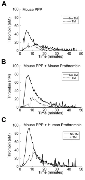
Figure 2.1. Human prothrombin supports thrombin generation in murine plasma and mouse thrombomodulin reduces thrombin generation in mouse plasma spiked with human prothrombin. Thrombin generation was measured in the absence and presence of 200 nM thrombomodulin in A) murine PPP spiked with vehicle, B) murine PPP spiked to 200% with murine prothrombin, and C) murine PPP spiked to 200% with human
2.3 Results
Human prothrombin is active in murine plasma. The in vivo hyperprothrombinemia model was developed by infusing mice with human prothrombin. Human prothrombin and the thrombin B chain have 81.4% and 88.8% amino acid identity with murine prothrombin and thrombin, respectively, and highly-conserved substitutions in non-identical residues.36 Human and mouse thrombin bind and cleave human and mouse fibrinogen37, activate platelets to form aggregates with pseudopodia38, bind murine thrombomodulin, and support activated protein C generation39,40. To assess the ability of human (pro)thrombin to support thrombin generation in murine plasma, murine plasma was spiked with vehicle or murine or human prothrombin to 200% (final, endogenous plus spiked human prothrombin) and thrombin generation was measured by calibrated automated thrombography in the absence and presence of 200 nM murine thrombomodulin. Thrombin generation peaks were 49.4±7.2, 82.4±13.8 and 90.4±5.2 nM (mean±SD, n=2-3) for plasma plus vehicle, plasma plus murine prothrombin, or plasma plus human prothrombin, respectively, and addition of murine thrombomodulin reduced the thrombin peaks by 73, 74, and 64%, respectively (Figure 2.1). These data demonstrate that human (pro)thrombin is compatible with the murine procoagulant and anticoagulant systems.
In the absence of vessel injury, elevated prothrombin does not activate coagulation. Platelet-poor plasma (PPP) from patients with elevated prothrombin demonstrates increased thrombin generation ex vivo7,23; however, these patients do not have higher circulating prothrombin fragment 1.2 compared to age-matched controls.7 We hypothesized that elevated
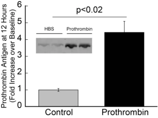
Figure 2.2. Human prothrombin circulates in mice 12 hours post-infusion. Twelve hours after infusion, blood was drawn from the IVC into 3.2% sodium citrate and processed to PPP. PPP samples were separated by 4-12% SDS-PAGE, transferred to PVDF
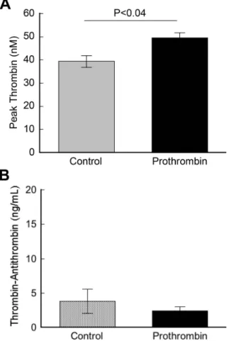
injury, but increases thrombin generation following TF exposure. To test this hypothesis, we infused prothrombin (to 300% of normal levels; total prothrombin equals endogenous murine prothrombin plus infused human prothrombin) or vehicle [20 mM HEPES (pH 7.4)/150 mM NaCl (HBS), Control] into uninjured mice and measured prothrombin antigen by western blotting, thrombin generation ex vivo by calibrated automated thrombography, and thrombin-antithrombin complex (TAT) levels by ELISA 12 hours after infusion. Human prothrombin still circulated in mice 12 hours post-infusion and was strongly detected by the rabbit anti-human prothrombin antibody (Figure 2.2). Consistent with anti-humans, thrombin generation was significantly elevated in PPP from prothrombin-infused mice compared to controls (49.5±2.1 versus 39.3±2.5 peak thrombin, respectively, mean±SEM, P<0.04) (Figure 2.3A) following initiation of coagulation ex vivo. However, also consistent with that seen in humans, circulating TAT levels in mice with elevated prothrombin, even at this high level, were not elevated compared to controls (2.4±1.4 versus 3.8±4.0 ng/mL, respectively, mean±SEM, P=0.26) (Figure 2.3B). These data reconcile outwardly discordant findings regarding thrombin generation in hyperprothrombinemic individuals measured ex vivo and in vitro by showing that elevated prothrombin does not increase baseline hemostatic “idling” in the absence of vascular injury, but augments thrombin generation following a procoagulant trigger.
Elevated prothrombin accelerates fibrin deposition and produces larger thrombi in venous thrombosis models. To characterize the effect of elevated prothrombin on venous thrombosis
Figure 2.3. In the absence of vessel injury, elevated prothrombin does not activate coagulation. HBS (Control) or human prothrombin was infused into mice via tail vein injection to 300%, final (mouse plus human). Twelve hours after infusion, blood was drawn from the IVC into 3.2% sodium citrate and processed to PPP. A) PPP from mice infused with prothrombin (to 300%) or HBS (Control) was diluted 1:3 and coagulation was triggered by addition of TF/lipid. Thrombin generation was measured by calibrated automated
elevated prothrombin to thrombus formation. The electrolytic injury model induces mural thrombus formation via iron-mediated injury that causes early platelet accumulation followed by fibrin accumulation30 (Figure 2.4A, Supplemental Videos I and II, available online1). Thrombus formation in this model is reduced by heparin, consistent with the sensitivity of venous thrombosis to thrombin generation.41 We tested 2 levels of prothrombin: 130% and 200% (final); these levels were chosen to approximate the mean and upper end of the pathophysiologic range. Neither prothrombin concentration significantly increased the rate or total amount of platelet accumulation in femoral vein thrombi (Figures 2.4B, D).
However, the plasma prothrombin level showed a dose-dependent effect on fibrin
accumulation in the vein. At 60 minutes, fibrin accumulation in mice infused to 130% and 200% prothrombin was 1.7- (P<0.06) and 3.5- (P<0.002) fold higher, respectively, than control mice (Figure 2.4C), and mice infused with 200% prothrombin exhibited a
significantly (2.3-fold, P=0.006) increased fibrin accumulation rate than control mice (Figure 2.4D). Furthermore, in contrast to control thrombi that remained relatively localized to the thrombus induction site, thrombi in prothrombin-infused mice showed considerable
downstream elongation of a mass containing both fibrin and platelets (Supplemental Videos I, II).
Figure 2.4. Elevated prothrombin increases the rate and extent of fibrin deposition following electrolytic injury to the femoral vein. Mice were infused with vehicle (control) or prothrombin to 200% of normal. Thrombosis was induced by electrolytic injury as
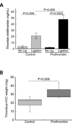
300% so that levels remained elevated for the duration of thrombus formation (12 hours). Following IVC ligation, TATs were more than 2-fold higher in prothrombin-infused mice compared to control mice (47.9±6.5 versus 21.2±4.5 ng/mL, respectively, mean±SEM,
P<0.009, Figure 2.5A), suggesting elevated prothrombin augmented thrombin generation during venous thrombogenesis. Following IVC ligation, all prothrombin-infused and control mice developed thrombi within 12 hours. Similar to that seen in the electrolytic injury model, thrombi in prothrombin-infused mice were significantly larger than thrombi in control mice [27.6 (26.1-43.6) versus 22.6 (9.5-27.7) mg, respectively, median (range), P=0.01, Figure 2.5B], and in some mice extended into the iliac branches. Together, these data show that elevated prothrombin augments venous thrombus formation in vivo by increasing thrombin generation and intravascular fibrin deposition.
Elevated prothrombin has little to no effect on the TTO or rate of platelet accumulation in arterial thrombosis models. We then used the electrolytic injury and real-time fluorescence detection method to measure the kinetics of platelet and fibrin accumulation in control and prothrombin-infused mice during arterial thrombosis (Figures 2.6A-D, Supplemental Videos III and IV, available online1). Platelet accumulation in the artery was 3.2-fold faster
and 200% of normal (final). Neither prothrombin concentration significantly increased either the rate or total amount of platelet accumulation, or the rate of fibrin accumulation in carotid artery thrombi (Figures 2.6B-D). Elevated prothrombin increased fibrin deposition at 60 minutes (~2-fold, Figure 2.6C), but this difference did not reach statistical significance (P<0.085).
We then tested the effect of elevated prothrombin in a second model of arterial thrombosis. The FeCl3 application/carotid artery model triggers arterial thrombosis via
generation of reactive oxygen species and exposure of collagen, resulting in a platelet-rich thrombus.43-45 Since neither prothrombin concentration significantly altered arterial thrombus formation in the electrolytic model, we only tested the higher concentration of prothrombin (infusing to 200%, final) in the FeCl3/carotid artery model. Prothrombin
infusion transiently elevated TAT levels (12.6±3.2 ng/mL), likely reflecting trace (<0.004%) thrombin contamination in the prothrombin concentrate that was immediately inhibited by endogenous antithrombin (present in 1000-fold excess, 2.5 µM plasma level) with no physiologic effects (data not shown and 46,47). For both control and prothrombin-infused mice, circulating TATs were significantly higher in mice that underwent FeCl3 injury than in
mice that did not (Figure 2.6E), demonstrating activation of coagulation following FeCl3
injury. Following FeCl3 injury, TAT levels were significantly different between control and
prothrombin-infused mice (13.9±1.7 versus 23.5±2.6, for control and prothrombin-infused mice, respectively, P<0.007, Figure 2.6E). Interestingly, the absolute increase in TAT levels following arterial injury was similar in both control and prothrombin-infused mice,
Figure 2.6. Elevated prothrombin does not increase thrombin generation or the rate of platelet or fibrin accumulation, and does not shorten the TTO in arterial injury models. A) Mice were infused with vehicle (Control) or prothrombin to 130 or 200% of normal. Thrombosis was induced by electrolytic injury as described in Methods. Representative images of platelet and fibrin accumulation 60 minutes after induction of carotid artery thrombosis via electrolytic injury are shown. Flow is from left to right; field dimensions are 3.20 x 0.85 mm (width x height). B-D) Relative fluorescence intensity of platelet (B) and fibrin (C) accumulation following electrolytic injury to the carotid artery (mean±SEM, n=7-8/group). Symbols are: control (open circles) and prothrombin-infused (closed triangles, 130%; closed circles, 200%). D) Maximum rate of platelet and fibrin accumulation during carotid artery thrombosis following electrolytic injury (mean±SEM, n=7-8/group). E) Mice were infused with vehicle (Control) or prothrombin to 200% of normal. Thrombosis was induced by 7.5% FeCl3 application to the carotid artery for 2 minutes. Following thrombus
formation, blood was collected from the IVC into citrate and processed to PPP. TAT levels were measured in FeCl3-treated (n=7-10/group) and uninjured mice (n=4/group) in parallel.
thrombi, and the time to vessel occlusion in prothrombin-infused mice was not different from controls (9.5 [4.3-40.0] versus 6.4 [4.5-8.5] minutes, respectively, median [range], Figure 2.6F). Thus, elevated prothrombin did not significantly increase platelet or fibrin
accumulation during arterial thrombus formation or shorten the time to artery occlusion. Together, these data show that although elevated prothrombin promotes venous thrombus formation, it does not significantly augment arterial (platelet-dependent) thrombosis.
2.4 Discussion
Elevated prothrombin is a well-established risk factor for venous thrombosis, but its relationship to arterial thrombosis is unclear. Using state-of-the-art in vivo models of venous thrombosis and arterial thrombosis, we show that elevated prothrombin did not increase baseline prothrombotic markers in unchallenged mice, but did increase thrombin generation following venous injury. The presence of elevated prothrombin did not accelerate
intravascular platelet accumulation following either venous or arterial injury. In venous thrombosis models, mice with elevated prothrombin exhibited an increased rate and amount of fibrin accumulation, thrombus extension and formation of thrombi with increased mass. However, in arterial thrombosis models, elevated prothrombin slightly (non-significantly) increased the total amount of fibrin deposited, but did not increase the rate of fibrin
accumulation or shorten the TTO. These findings suggest elevated prothrombin has little to no independent contribution to arterial thrombosis, and are the first to show that elevated prothrombin levels directly promote venous thrombosis in vivo.
arterial and venous thrombogenic processes.48 An important strength of our study was the use of complementary arterial and venous models to delineate both kinetic processes and their consequences for thrombus composition. Complementary, integrated information from the two arterial models and two venous models reveals both common and vascular bed-specific processes operant in these vessels that are consistent with the histological appearance of arterial and venous thrombi isolated from humans. Arterial injury produced rapid platelet accumulation; whereas, venous injury resulted in slower thrombus formation with fibrin accumulation. We used models that produced both occlusive (FeCl3/carotid and IVC) and
non-occlusive (electrolytic injury) thrombi. Findings were consistent within vessels, but differed between arteries and veins, suggesting these models are sensitive to the unique physical and biochemical environments within the different vessels.
We detected both common and vascular bed-specific effects of elevated prothrombin on arterial and venous thrombosis. Elevated prothrombin significantly augmented
endogenous thrombin generation in the venous model, but only slightly (non-significantly) in the arterial model (Figures 2.5A, 2.6E). Consistent with our findings, a recent study showed that ApoE-/- mice expressing half of the prothrombin level of wild type mice (FII-/+) are not protected from arterial thrombosis, supporting the conclusion that variation in the
generation significantly increased fibrin deposition and therefore, venous thrombosis, a fibrin-dominated process. We previously showed that elevated prothrombin also promotes the thrombin concentration-dependent formation of plasma clots with an abnormally dense fibrin network.20 Although fibrin network structure is difficult to assess in thrombi in the presence of cells, combined, these results suggest that in veins, elevated prothrombin
promotes thrombi with fibrin networks that have both increased mass and increased network density. Both properties are associated with increased clot stability in vitro and in vivo, and have been correlated with increased thrombosis risk.50
It is interesting that while elevated prothrombin did not accelerate arterial occlusion after FeCl3 injury, elevated plasma factor VIII does51-53. Both elevated prothrombin and
elevated factor VIII increase thrombin generation in vitro20-22,53,54 and in vivo (Figure A and
51). However, elevated factor VIII significantly shortens the lag time to platelet aggregation
in vitro51 and trends towards increased platelet accumulation in vivo53; whereas, elevated prothrombin did not significantly change the rate of platelet aggregation in vitro20 or in vivo
(Figures 2.4D, 2.6D). These data suggest elevated factor VIII modulates an early
(amplification) phase of coagulation when thrombin levels are relatively low and platelet activation is taking place. Consequently, platelet-dependent arterial thrombosis models are sensitive to the effects of elevated factor VIII, but not to elevated prothrombin. This
observation is consistent with the premise that procoagulant factors have complementary, but distinct, roles in different phases of coagulation.55
Previous studies on the association between elevated prothrombin and arterial
only in specific groups. Of interest are observations that relative risk increases when another identifiable cardiovascular risk factor is also present and appears higher than from either risk factor alone, suggesting an additive or synergistic interaction.14,15 A strength of our murine model in which prothrombin levels were acutely elevated in healthy wild type mice is the clear absence of other risk factors. Nonetheless, the co-existence of additional known or unidentified risk factors may augment the positive associations detected in prior studies with human cohorts. For example, on an atherosclerosis-prone background (ApoE-/-), chronic plasma hypercoagulability (TMPro/Pro) increases atherogenesis and plaque formation, both of which are associated with atherothrombosis, and reduced prothrombin levels attenuate atherosclerotic lesion formation.49
This study has potential limitations. First, we used human prothrombin to increase circulating levels in the mouse. However, published studies36-40 as well as our data show human prothrombin is stable in murine circulation and participates in murine pro- and anti-coagulant pathways. Moreover, the infusion strategy enabled us to precisely control the level of circulating prothrombin. Second, the infusion model used in these experiments does not reflect pathologic effects that chronic exposure to elevated prothrombin could have on the vasculature. Atherosclerotic disease reflects chronic vascular injury with occurrences of acute injury (plaque disruption and TF exposure). However, a major strength of the infusion model is that it enabled us to isolate and investigate the immediate, direct effects of elevated prothrombin on thrombus formation. These data on acute effects will be critical for
interpreting findings from mice with genetically-induced chronic plasma hypercoagulability (e.g., factor V Leiden mice); comparison of short-term and long-term exposure to
individuals to thrombosis. Third, the thrombosis models we used differed in methodologic aspects, including anesthesia and analgesia protocols. However, the observation that elevated prothrombin exhibited consistent effects in each of the venous and each of the arterial models suggests the observed effects were not due to the methodologies, but to the prothrombin level, itself. Finally, the arterial and venous models used in this study were sensitive to thrombus formation, but did not reflect additional effects elevated prothrombin may have on thrombus stability. For example, although groups have demonstrated increased activation of the thrombin activatable fibrinolysis inhibitor in plasma with increased
prothrombin, we did not evaluate the long-term resistance of thrombi to fibrinolysis. In summary, our findings demonstrate that elevated prothrombin does not trigger endogenous thrombin generation in the absence of vascular injury, suggesting that in lieu of a signal that initiates coagulation, plasma hypercoagulability is not independently
prothrombotic. These data suggest that increased coagulation biomarkers (e.g., fragment 1.2 or TATs) indicate vascular dysfunction that, when coupled to additional plasma
prothrombotic potential, promote thrombosis. Our findings further show that elevated prothrombin increases thrombin generation following vascular injury. Elevated prothrombin does not accelerate platelet activation in either the artery or the vein, but significantly
2.5 REFERENCES
1. Aleman MM, Walton BL, Byrnes JR, et al. Elevated prothrombin promotes venous, but not arterial, thrombosis in mice. Arterioscler Thromb Vasc Biol. 2013;33(8):1829-1836. 2. Lijfering WM, Flinterman LE, Vandenbroucke JP, Rosendaal FR, Cannegieter SC.
Relationship between venous and arterial thrombosis: a review of the literature from a causal perspective. Semin Thromb Hemost. 2011;37(8):885-896.
3. Turpie AG, Esmon C. Venous and arterial thrombosis--pathogenesis and the rationale for anticoagulation. Thromb Haemost. 2011;105(4):586-596.
4. Brighton TA, Eikelboom JW, Mann K, et al. Low-dose aspirin for preventing recurrent venous thromboembolism. N Engl J Med. 2012;367(21):1979-1987.
5. Poort SR, Rosendaal FR, Reitsma PH, Bertina RM. A common genetic variation in the 3'-untranslated region of the prothrombin gene is associated with elevated plasma prothrombin levels and an increase in venous thrombosis. Blood. 1996;88(10):3698-3703.
6. Makris M, Preston FE, Beauchamp NJ, et al. Co-inheritance of the 20210A allele of the prothrombin gene increases the risk of thrombosis in subjects with familial
thrombophilia. Thromb Haemost. 1997;78(6):1426-1429.
7. Kyrle PA, Mannhalter C, Beguin S, et al. Clinical studies and thrombin generation in patients homozygous or heterozygous for the G20210A mutation in the prothrombin gene. Arterio Thromb Vasc Biol. 1998;18(8):1287-1291.
8. Rosendaal FR, Doggen CJ, Zivelin A, et al. Geographic distribution of the 20210 G to A prothrombin variant. Thromb Haemost. 1998;79(4):706-708.
9. Kim RJ, Becker RC. Association between factor V Leiden, prothrombin G20210A, and methylenetetrahydrofolate reductase C677T mutations and events of the arterial
circulatory system: a meta-analysis of published studies. Am Heart J. 2003;146(6):948-957.
10. Ye Z, Liu EH, Higgins JP, et al. Seven haemostatic gene polymorphisms in coronary disease: meta-analysis of 66,155 cases and 91,307 controls. Lancet.
2006;367(9511):651-658.
11. Arruda VR, Annichino-Bizzacchi JM, Goncalves MS, Costa FF. Prevalence of the prothrombin gene variant (nt20210A) in venous thrombosis and arterial disease. Thromb Haemost. 1997;78(6):1430-1433.
12. Watzke HH, Schuttrumpf J, Graf S, Huber K, Panzer S. Increased prevalence of a
13. De Stefano V, Chiusolo P, Paciaroni K, et al. Prothrombin G20210A mutant genotype is a risk factor for cerebrovascular ischemic disease in young patients. Blood.
1998;91(10):3562-3565.
14. Rosendaal FR, Siscovick DS, Schwartz SM, Psaty BM, Raghunathan TE, Vos HL. A common prothrombin variant (20210 G to A) increases the risk of myocardial infarction in young women. Blood. 1997;90(5):1747-1750.
15. Doggen CJ, Cats VM, Bertina RM, Rosendaal FR. Interaction of coagulation defects and cardiovascular risk factors: increased risk of myocardial infarction associated with factor V Leiden or prothrombin 20210A. Circulation. 1998;97(11):1037-1041.
16. Martinelli I, Franchi F, Akwan S, Bettini P, Merati G, Mannucci PM. The transition G to A at position 20210 in the 3'-untranslated region of the prothrombin gene is not
associated with cerebral ischemia. Blood. 1997;90(9):3806.
17. Lalouschek W, Aull S, Series W, Zeiler K, Mannhalter C. The prothrombin G20210A mutation and factor V Leiden mutation in patients with cerebrovascular disease. Blood. 1998;92(2):704-705.
18. Coulet F, Godard V, Verdy E, Soubrier F. Lack of association of the prothrombin gene variant G20210A with myocardial infarction in Caucasian males. Thromb Haemost. 2000;83(5):796-797.
19. Ferraresi P, Marchetti G, Legnani C, et al. The heterozygous 20210 G/A prothrombin genotype is associated with early venous thrombosis in inherited thrombophilias and is not increased in frequency in artery disease. Arterioscler Thromb Vasc Biol.
1997;17(11):2418-2422.
20. Wolberg AS, Monroe DM, Roberts HR, Hoffman M. Elevated prothrombin results in clots with an altered fiber structure: a possible mechanism of the increased thrombotic risk. Blood. 2003;101(8):3008-3013.
21. Allen GA, Wolberg AS, Oliver JA, Hoffman M, Roberts HR, Monroe DM. Impact of procoagulant concentration on rate, peak and total thrombin generation in a model system. J Thromb Haemost. 2004;2(3):402-413.
22. Butenas S, van't Veer C, Mann KG. "Normal" thrombin generation. Blood. 1999;94(7):2169-2178.
23. Castoldi E, Simioni P, Tormene D, et al. Differential effects of high prothrombin levels on thrombin generation depending on the cause of the hyperprothrombinemia. J Thromb Haemost. 2007;5(5):971-979.
24. Binetti BM, Rotunno C, Tripodi A, et al. Hyperprothrombinaemia-induced APC
25. Colucci M, Binetti BM, Tripodi A, Chantarangkul V, Semeraro N.
Hyperprothrombinemia associated with prothrombin G20210A mutation inhibits plasma fibrinolysis through a TAFI-mediated mechanism. Blood. 2004;103(6):2157-2161. 26. Wolberg AS, Monroe DM, Roberts HR, Hoffman M. Elevated prothrombin results in
clots with an altered fiber structure: a possible mechanism of the increased thrombotic risk. Blood. 2003;101(8):3008-3013.
27. Buyue Y, Whinna HC, Sheehan JP. The heparin-binding exosite of factor IXa is a critical regulator of plasma thrombin generation and venous thrombosis. Blood. 2008;112(8):3234-3241.
28. Machlus KR, Cardenas JC, Church FC, Wolberg AS. Causal relationship between hyperfibrinogenemia, thrombosis, and resistance to thrombolysis in mice. Blood. 2011;117(18):4953-4963.
29. Machlus KR, Lin FC, Wolberg AS. Procoagulant activity induced by vascular injury determines contribution of elevated factor VIII to thrombosis and thrombus stability in mice. Blood. 2011;118(14):3960-3968.
30. Cooley BC. In vivo fluorescence imaging of large-vessel thrombosis in mice.
Arterioscler Thromb Vasc Biol. 2011;31(6):1351-1356.
31. Maroney SA, Cooley BC, Ferrel JP, Bonesho CE, Mast AE. Murine hematopoietic cell tissue factor pathway inhibitor limits thrombus growth. Arterioscler Thromb Vasc Biol. 2011;31(4):821-826.
32. Henke PK, Varga A, De S, et al. Deep vein thrombosis resolution is modulated by monocyte CXCR2-mediated activity in a mouse model. Arterioscler Thromb Vasc Biol. 2004;24(6):1130-1137.
33. Wrobleski SK, Farris DM, Diaz JA, Myers DD, Jr., Wakefield TW. Mouse complete stasis model of inferior vena cava thrombosis. J Vis Exp. 2011(52):e2738.
34. Pratt CW, Monroe DM. Microplate coagulation assays. Biotechniques. 1992;13(3):430-433.
35. Dargaud Y, Spronk HM, Leenders P, Hemker HC, Ten Cate H. Monitoring platelet dependent thrombin generation in mice. Thromb Res. 2010;126(5):436-441.
36. Degen SJ, Schaefer LA, Jamison CS, et al. Characterization of the cDNA coding for mouse prothrombin and localization of the gene on mouse chromosome 2. DNA Cell Biol. 1990;9(7):487-498.