CASE SERIES
Arachnoid cysts: the role of the BLADE technique
Mavroidis P
1,2, Roka V
3, Kostopoulos S
4, Batsikas G
5, Lavdas E
6,71Department of Radiation Oncology, University of North Carolina, Chapel Hill, NC, USA
2Department of Medical Physics, Karolinska Institutet & Stockholm University, Stockholm, Sweden 3Health Center of Farkadona, Trikala, Greece
4Department of Medical Instruments Technology, Technological Education Institute of Athens, Athens, Greece 5Department of Medical Imaging, IASO Thessalias Hospital, Larissa, Greece
6Department of Medical Radiological Technologists, Technological Education Institute of Athens, Athens, Greece 7Department of Medical Imaging, Animus kyanoys Larissa Hospital, Larissa, Larissa, Greece
Abstract
Background: This study aims at demonstrating the ability of BLADE sequences to reduce or even eliminate all the
im-age artifacts as well as verifying the significance of using this technique in certain pathological conditions.
Material and Methods: This study involved fourteen consecutive patients (5 females, 9 males), who routinely
under-went magnetic resonance imaging (MRI) brain examination, between 2010-2014. The applied routine protocol for brain MRI examination included the following sequences: i) T2-weighted (W) fluid-attenuated inversion recovery (FLAIR) axial; ii) T2-W turbo spin echo (TSE) axial; iii) T2*-W axial, iv) T1-W TSE sagittal; v) Diffusion-weighted (DWI) axial; vi) T1-W TSE axial; vii) T1-W TSE axial+contrast. Additionally, the T2-W FLAIR BLADE sequence was added to the protocol in cases of cystic tumors. Two radiologists independently evaluated all the images at two separate settings, which were performed 3 weeks apart. The presence of image artifacts such as motion, flow, chemical shift and Gibbs ringing artifacts, were also evaluated by the radiologists. In the measurements of the cysts, the extent of the divergence by the two MRI techniques (conventional and BLADE) was used by the two radiologists to evaluate the accuracy of the two techniques to determine the size of the cysts.
Results: BLADE sequences were found to be more reliable than the conventional onesregarding the estimation of the
cyst size. The qualitative analysis showed that the T2 FLAIR BLADE sequences were superior to the conventional T2 FLAIR with statistical significance (p <0.001) in the following fields: i) overall image quality, ii) cerebrospinal fluid (CSF) nulling; iii) contrast between pathology and its surrounding; iv) borders of the pathology; v) motion artifacts; vi) flow artifacts; vii) chemical shift artifacts and viii) Gibbs ringing artifacts.
Conclusions: BLADE sequence was found to decrease both flow artifacts in the temporal lobes and motion artifacts
from the orbits. Additionally, it was shown to improve flow artifacts and image quality in cystic pathologies such as arachnoid cysts. Hippokratia 2016, 20(3): 244-248
Key words: Arachnoid cysts, magnetic resonance imaging, BLADE sequence, flow artifacts, motion artifacts
Corresponding author: Panayiotis Mavroidis, Associate Professor, Department of Radiation Oncology, University of North Carolina, 101 Manning Dr. Chapel Hill, NC 27599-7512, tel: +19849748438, fax: +19198439127, e-mail: [email protected]
Introduction
With the increasing use of magnetic resonance imag -ing (MRI) and computed tomography (CT) in brain ex-aminations, a corresponding increase has been reported in the number of arachnoid cysts that are incidentally
diagnosed1-5. Arachnoid cysts are benign, congenital, intra-arachnoidal space-occupying lesions that are filled with clear cerebrospinal fluid (CSF) not
communicat-ing with the ventricular system6,7. The typical arachnoid cyst has no identifiable internal architecture and does not enhance. The cyst has the same signal intensity as CSF at all sequences including the fluid-attenuated inversion
recovery (FLAIR). This characteristic is in contrast to
other lesions which may give a variety of signals8. Oc-casionally, however, hemorrhage, high protein content, or lack of flow within the cyst may complicate the MR appearance6,9-10.
An epidermoid cyst is the most difficult lesion to dis-tinguish from the arachnoid cyst. On MR images, epi-dermoid cysts appear isointense to CSF. Arachnoid cysts displace adjacent arteries and cranial nerves rather than engulf them, as epidermoid cysts often do. Also, chronic subdural hematoma and a porencephalic cyst can be
often clinically “silent”, a variety of symptoms may de-velop, depending on the location and size of the cyst.
Although asymptomatic patients whose arachnoid cyst is an incidental finding, are not considered to be candidates for surgery, according to some groups, it is advisable to surgically treat them similarly to
symptom-atic arachnoid cysts, which warrant surgical treatment 12-18. It is therefore very important to be able to see the exact structures and signals within the cyst without artifacts. The purpose of this study was firstly to determine the ex-tent by which BLADE sequences can reduce the previ-ously mentioned image artifacts and secondly to examine the impact of this technique in certain pathological condi-tions.
Material and Methods Patients
In this retrospective study, we included fourteen con-secutive patients (5 females, 9 males) with mean age 32 years (range: 8 months - 45 years). These patients rou-tinely underwent MRI brain examinations from February 2010 to February 2014. Written informed consent was obtained from all the subjects participating in the study protocol. Since this was a retrospective study where the patients received standard care as per institutional guide-lines, with all the data being anonymized, an ethical com-mittee approval was deemed unnecessary.
MR imaging techniques
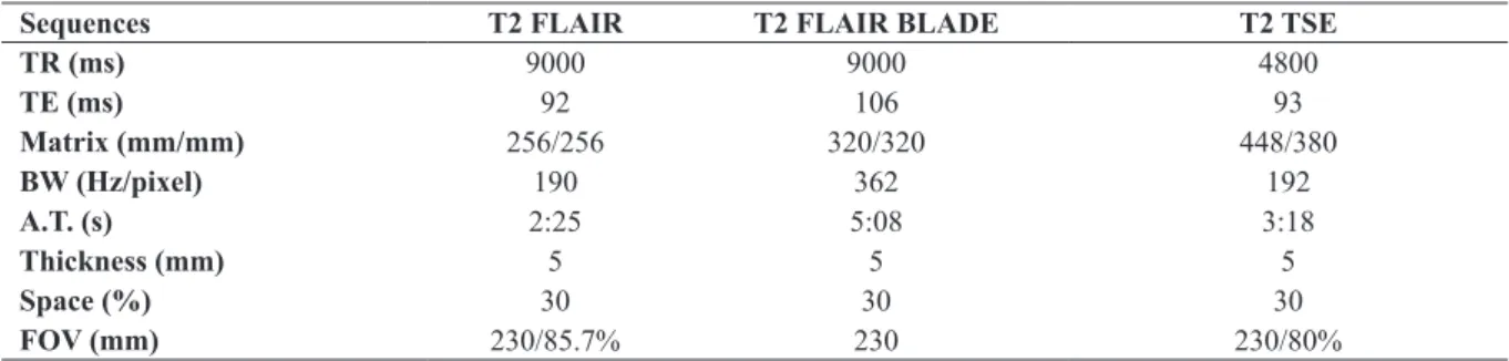
A brain examination was performed on all the pa-tients using a 1.5-T scanner (Magnetom Avanto, Siemens Medical Systems, Erlangen, Germany) with the Siemens 12-channel head matrix coil. The parameters of the dif-ferent sequences are presented in Table 1.
The routine protocol of brain MR examination includ-ed the following sequences: i) T2-weightinclud-ed (W) FLAIR axial; ii) T2-W turbo spin echo (TSE) axial; iii) T2*-W axial; iv) T1-W TSE sagittal; v) Diffusion-weighted (DWI) axial; vi) T1-W TSE axial; and vii) T1-W TSE axial+contrast. The T2-W FLAIR BLADE sequence was added to the protocol in cases of cystic tumors. In few cases, the T1-W FLAIR BLADE sequence was also ap-plied.
The average time to acquire a T2-W FLAIR sequence was 2.25 min, whereas for the T2-W FLAIR BLADE it
was 5.08 min. The decision for the additional imaging was made by the physician and it had been incorporated into the routine imaging protocol.
The term BLADE is the product name (used by Sie-mens Medical System, Erlagen, Germany) for a TSE se-quence that uses the ‘Periodically Rotated Overlapping Parallel Lines with Enhanced Reconstruction’ (PROPEL-LER) k-space trajectory. The BLADE method acquires a number of blades that are rotated around the center of the k-space. Each blade consists of a number of lowest phase encoding lines of a conventional rectilinear k-space
tra-jectory that are acquired after a single radiofrequency
excitation. This technique can potentially eliminate pul-sation in MR images when that pulpul-sation is caused by the unwanted modulation of k-space data.
Qualitative analysis
All the acquired images were evaluated independent-ly at two separate sittings with three weeks interval by
two radiologists, who did not have access to any other
image information when they made their assessments. The radiologists graded the images on a 5-point scale (0: non-visualization, 1: poor, 2: average, 3: good, 4: ex-cellent) for each of the following image characteristics: i) overall image quality, ii) CSF nulling, iii) contrast at the pathology and its surrounding, iv) size of pathology, and vi) limits of pathology.
Also, the presence of image artifacts (motion, flow, chemical shift, Gibbs ringing) was evaluated by the two radiologists, using a separate scoring scale (4: minimum, 3: slight, 2: moderate, 1: severe, 0: maximum). The
statis-tical significance of the qualitative data was determined
by the Kruskal-Wallis nonparametric test.
Quantitative analysis
The radiologists compared the techniques used (con-ventional and BLADE) in order to evaluate the size of the cyst, by assessing the extent by which the measure-ments of the cysts diverge. The T2-W TSE axial sequence was used as a reference because it is not prone to flow artifacts and arachnoid cysts are depicted with enhanced signal-to-noise ratio (SNR) resulting in better contrast from their surroundings. The quantitative evaluation was performed by means of the Kolmogorov-Smirnov non-parametric test.
Table 1: Summary of the sequences that were applied in brain MR examination in the current case series.
Sequences T2 FLAIR T2 FLAIR BLADE T2 TSE
TR (ms) 9000 9000 4800
TE (ms) 92 106 93
Matrix (mm/mm) 256/256 320/320 448/380
BW (Hz/pixel) 190 362 192
A.T. (s) 2:25 5:08 3:18
Thickness (mm) 5 5 5
Space (%) 30 30 30
FOV (mm) 230/85.7% 230 230/80%
Results
The findings of the analysis indicate that the BLADE sequences were superior to the conventional ones regard-ing the overall image quality, CSF nullregard-ing, contrast of the pathology and its surrounding, size of pathology, borders of the pathology, motion artifacts, flow artifacts, chemi-cal shift artifacts, and Gibbs ringing artifacts.
The qualitative measurements show that T2 FLAIR BLADE sequences are significantly superior to the
con-Figure 1: Axial T2 fluid attenuated inversion recovery (FLAIR) (left), Axial T2 FLAIR BLADE (right) images of the brain. The signal within the arachnoid cyst in the con-ventional image cannot be assessed accurately due to the image flow artifacts. In T2 FLAIR BLADE, these artifacts are reduced resulting in very good depiction of the pathol-ogy, which provides the possibility for more accurate mea-surements so that a potential pressure on the medial rectus muscle or internal carotid can be estimated.
Figure 2: Axial T2 fluid attenuated inversion recovery (FLAIR) (upper left), Axial T2 FLAIR BLADE (upper right), Axial T1 tur-bo spin echo (TSE) (lower left), Axial T1 FLAIR BLADE (lower right) images of the brain. It is shown that in the T2 FLAIR im-age more flow artifacts are depicted and worse CSF nulling is achieved compared to the conventional T1 TSE image. In both corresponding BLADE images all the artifacts are reduced and excellent cerebrospinal fluid (CSF) nulling is achieved resulting
in correct delineation of the arachnoid cyst and more accurate
depiction of the ischemic lesion in the temporal lobes.
Figure 3: Axial T2 fluid attenuated inversion recovery (FLAIR) (upper left), Axial T2 FLAIR BLADE (upper right) and T2 turbo spin echo (TSE) (lower) images of the brain. It is very important to depict the exact limits of an arachnoid cyst in order to be able to determine any possible enlarge-ment of the cyst. Here, it is shown that chemical shift arti-facts are reduced in T2 FLAIR BLADE image and better ce-rebrospinal fluid (CSF) nulling is achieved, which results in a more accurate depiction of the pathology. BLADE images provide the possibility for performing more precise measure-ments compared to those obtained from T2 TSE images.
ventional T2 FLAIR with statistically significantly differ-ences in terms of: i) overall image quality (p =0.003) (Fig-ure 1), ii) CSF nulling (p <0.001) (Fig(Fig-ures 2 and 3), iii) contrast of the pathology and its surrounding (p <0.001) (Figure 3), iv) borders of the pathology (p <0.001) (Fig-ure 3), v) motion artifacts (p <0.001) (Fig(Fig-ure 4), vi) flow artifacts (p <0.001) (Figure 1, Figure 2, Figure 4), vii) other artifacts (p <0.001) [chemical shift artifacts (Figure 3) and Gibbs ringing artifacts].
Similarly, the quantitative results about the extent of the cyst size indicate that the BLADE measurements were found to be much more reliable than the conven-tional ones with the differences being statistically signifi-cant (p <0.001).
Discussion
The number of incidentally diagnosed arachnoid cysts has increased with the technological progress of diagnostic brain imaging. The optimum diagnostic case scenario is a sharply demarcated extra-axial cyst that can displace or deform adjacent brain tissue. Also, an often seen sign is the scalloping of the adjacent calvarium. The
size of arachnoid cysts varies, from small and incidental
to large space-occupying lesions12. Arachnoid cysts are generally stable over time, although cases of sudden or progressive enlargement, as well as spontaneous resolu-tion, have been reported.
Practice has proven that image artifacts like motion
artifacts, flow artifacts and other, are very often seen in
MR images and their presence complicates diagnosis. Many studies have been conducted showing that BLADE
sequences significantly reduce those artifacts19-21. That is of particular significance not only for the surgical op-erations but also for better patient management, which requires the depiction of arachnoid cysts with a clear sig-nal, high contrast, and clearly demarcated borders. While these lesions are often clinically silent, a variety of symp-toms may develop, depending on the location and size of the cyst. Although they generally have a congenital origin, they may appear after head trauma or infection. Some of them appear to be due to a ball-valve commu-nication with the subarachnoid space, and may progres-sively enlarge and become symptomatic, and then they
require surgery22. Since the initial diagnosis of an arach-noid cyst, the follow-up evaluation of its size has become essential, especially in the cases where the cyst displaces the adjacent arteries and cranial nerves. This may trig-ger several symptoms depending on the exact location and size of the cyst. Unfortunately, due to image artifacts (motion, flow chemical shift), it is difficult to evaluate the borders and therefore the exact size of the cyst. Most authors agree that symptomatic arachnoid cysts warrant
surgical treatment12,23,24.
The basic protocols for the study of the brain include T1-W TSE, T2-W TSE, and T2-W FLAIR sequences. Normal CSF has long T1 and long T2 times that manifest themselves as a dark signal on T1-weighted images and a bright signal on T2-weighted images. FLAIR imaging re-sults in nulling and dark CSF signal25. According to some authors, operative strategies can be more easily worked out on the FLAIR images. Furthermore, the differences between the arachnoid and epidermoid cysts may be de-picted on Τ2 FLAIR images11. It has been observed that on FLAIR images, flow artifacts are more prominent
and severe25-28. However, recent studies have shown that FLAIR images produce better results when combined with the BLADE technique25,28,29 since they improve CSF
nulling and further reduce flow-related artifacts. Thus, with BLADE sequences, the extent of the cyst size was calculated more accurately (see Figure 3). Chemical shift artifacts are reduced in T2 FLAIR BLADE image, and better CSF nulling is achieved. The measurements of the size of the cyst were compared with those obtained from T2-W TSE images.
Von Kalle T et al30, who studied pediatric patients, found pulsation artifacts to be more prominent than move-ment artifacts, and significantly less often in BLADE im-ages. Pulsation artifacts were more frequent and severe in conventional FLAIR images, but sometimes these artifacts were also observed in FLAIR BLADE images. Nyberg E et al19, found that T2 FLAIR BLADE was su-perior to conventional T2 FLAIR in reducing motion re-lated artifacts. Notwithstanding, his research focused on
the reduction of motion artifacts and the two techniques
were not compared for other artifacts.
The present study indicates that image artifacts and especially the flow artifacts are more prominent in cystic lesions; thereby the use of BLADE was even more ef-fective in decreasing them and improving the contrast of the pathology compared to the conventional sequences. In Figure 1, the flow artifacts are so severe that we cannot estimate the type of signal within the arachnoid cyst or where are the exact limits of the cyst. BLADE sequence decreases both flow artifacts in the temporal lobes and motion artifacts in the orbits (Figure 2). It is shown that in the conventional T2 FLAIR image more flow artifacts are depicted in comparison to the conventional T1 TSE, and worse CSF nulling is achieved. As it can be seen, the corresponding BLADE sequences reduced the flow arti-facts, and excellent CSF nulling was achieved resulting in correct delineation of the arachnoid cyst. The ischemic lesions in the temporal lobes are shown better in the T2 FLAIR BLADE than in the T2 FLAIR image. Moreover, a brain with cystic lesions tend to produce more pulsa-tion related artifacts (ghosts) than a normal brain and that makes the effects of the BLADE technique even more important.
In Figure 4, as it can be seen, the flow artifacts that are present in the arachnoid cyst in the conventional T2 FLAIR were reduced in the T2 FLAIR BLADE and bet-ter CSF nulling was achieved, which leads to a more ac-curate estimation of the content of the arachnoid cyst. An equally important benefit of BLADE is that the better CSF nulling that it achieved led to better contrast between the pathology and its surrounding and better delineation of the limits of the pathology (Figure 3). BLADE images were found superior to the conventional ones in all the examined cases. It is therefore very important to perform more studies, like the present one, in order to evaluate whether the use of BLADE is indispensable for the depic-tion of specific pathologies.
Conflict of interest
Acknowledgment
Results of this work were presented in the 57th AAPM Annual Meeting, Anaheim, CA, USA, July 12-16, 2015 (abstract is included in Med Phys. 2015; 42: 3257).
References
1. Katzman GL, Dagher AP, Patronas NJ. Incidental findings on brain magnetic resonance imaging from 1000 asymptomatic vol-unteers. JAMA. 1999; 282: 36-39.
2. Kim BS, Illes J, Kaplan RT, Reiss A, Atlas SW. Incidental find-ings on pediatric MR images of the brain. AJNR Am J Neurora-diol. 2002; 23: 1674-1677.
3. Vernooij MW, Ikram MA, Tanghe HL, Vincent AJ, Hofman A, Krestin GP, et al. Incidental findings on brain MRI in the general population. N Engl J Med. 2007; 357: 1821-1828.
4. Weber F, Knopf H. Incidental findings in magnetic resonance imaging of the brains of healthy young men. J Neurol Sci. 2006; 240: 81-84.
5. Zada G, Krieger MD, McNatt SA, Bowen I, McComb JG. Patho-genesis and treatment of intracranial arachnoid cysts in pediatric patients younger than 2 years of age. Neurosurg Focus. 2007; 22: E1.
6. Osborn AG. Miscellaneous tumors, cysts, and metastases. Os-born AG (Ed). Diagnostic neuroradiology. Mosby, St Louis, 1994, 631-649.
7. Dutt SN, Mirza S, Chavda SV, Irving RM. Radiologic differ-entiation of intracranial epidermoids from arachnoid cysts. Otol Neurotol. 2002; 23: 84-92.
8. Oprişan A, Popescu BO. Intracranial cysts: an imagery diagnos-tic challenge. ScientificWorldJournal. 2013; 2013: 172154. 9. Burger PC, Scheithauer BW, Vogel FS. Intracranial meninges.
Burger PC, Scheithauer BW, Vogel FS (Eds). Surgical pathology of the brain and its coverings. 4th edition, Churchill Livingstone,
Philadelphia, 2002, 89-93.
10. Osborn AG. Arachnoid cyst. Diagnostic imaging: brain. Amir-sys, Salt Lake City, 2004, I-7-I-26.
11. Osborn AG, Preece MT. Intracranial cysts: radiologic-patholog-ic correlation and imaging approach. Radiology. 2006; 239: 650-664.
12. Fernández Molina G. Neuroendoscopic management of middle fossa arachnoid cysts. World Neurosurg. 2013; 79: S19.e19-e23.
13. Martínez-Lage JF, Valentí JA, Piqueras C, Ruiz-Espejo AM, Román F, Nuño de la Rosa JA. Functional assessment of intrac-ranial arachnoid cysts with TC99 m-HMPAO SPECT: a prelimi-nary report. Childs Nerv Syst. 2006; 22: 1091-1097.
14. Parsch CS, Krauss J, Hofmann E, Meixensberger J, Roosen K. Arachnoid Cysts Associated with Subdural Hematomas and Hy-gromas: Analysis of 16 Cases, Long-term Follow-up, and Re-view of the Literature. Neurosurgery. 1997; 40: 483-490. 15. Sgouros S, Chapman S. Congenital middle fossa arachnoid cysts
may cause global brain ischaemia: a study with 99Tc-hexame-thylpropyleneamineoxime single photon emission computerised
tomography scans. Pediatr Neurosurg. 2001; 35: 188-194. 16. Wester K, Helland CA. How often do chronic extra-cerebral
hae-matomas occur in patients with intracranial arachnoid cysts? J Neurol Neurosurg Psychiatry. 2008; 79: 72-75.
17. Wester K, Hugdahl K. Verbal laterality and handedness in pa-tients with intracranial arachnoid cysts. J Neurol. 2003; 250: 36-41.
18. Kang JK, Lee KS, Lee IW, Jeun SS, Son BC, Jung CK, et al. Shunt-independent surgical treatment of middle cranial fossa arachnoid cysts in children. Childs Nerv Syst. 2000; 16: 111-116.
19. Nyberg E, Sandhu GS, Jesberger J, Blackham KA, Hsu DP, Gris-wold MA, et al. Comparison of brain MR images at 1.5T using BLADE and rectilinear techniques for patients who move during data acquisition. AJNR Am J Neuroradiol. 2012; 33: 77-82. 20. Pipe JG. Motion correction with PROPELLER MRI: application
to head motion and free-breathing cardiac imaging. Magn Reson Med. 1999; 42: 963-969.
21. Lavdas E, Mavroidis P, Hatzigeorgiou V, Roka V, Arikidis N, Oikonomou G, et al. Elimination of motion and pulsation ar-tifacts using BLADE sequences in knee MR imaging. Magn Reson Imag. 2012; 30: 1099-1110.
22. Choong CT, Lee SH. Subdural hygroma in association with middle fossa arachnoid cysts: acetazolamide therapy. Brain Dev. 1998; 20: 319-322.
23. Lee EJ, Ra YS. Clinical and neuroimaging outcomes of surgical-ly treated intracranial cysts in 110 children. J Korean Neurosurg Soc. 2012; 52: 325-333.
24. Gui S, Zong X, Li C, Zhang Y. Endoscopic treatment of convex-ity arachnoid cysts. Childs Nerv Syst. 2013; 29: 505-508. 25. Lisanti C, Carlin C, Banks KP, Wang D. Normal MRI
appear-ance and motion-related phenomena of CSF. AJR Am J Roent-genol. 2007; 188: 716-725.
26. Bakshi R, Caruthers SD, Janardhan V, Wasay M. Intraventricu-lar CSF pulsation artifact on fast fluid-attenuated inversion-re-covery MR images: analysis of 100 consecutive normal studies. AJNR Am J Neuroradiol. 2000; 2: 503-508.
27. Bailey WM. Fast Fluid Attenuated Inversion Recovery (FLAIR)
imaging and associated artefacts in Magnetic Resonance Imag
-ing (MRI). Radiography. 2007; 13: 283-290.
28. Lavdas E, Mavroidis P, Kostopoulos S, Glotsos D, Roka V, To-palzikis T, et al. Improvement of image quality using BLADE sequences in brain MR imaging. Magn Reson Imag. 2013; 31: 189-200.
29. Lavdas E, Mavroidis P, Kostopoulos S, Ninos C, Strikou AD, Glotsos D, et al. Reduction of motion, truncation and flow ar-tifacts using BLADE sequences in cervical spine MR imaging. Magn Reson Imag. 2015; 33: 194-200.

![Aqua[{[2 (2 hydroxyphenyl)ethylidene]amino}acetato]copper(II) monohydrate](data:image/gif;base64,R0lGODlhAQABAIAAAP///wAAACH5BAEAAAAALAAAAAABAAEAAAICRAEAOw==)