A Thesis Submitted for the Degree of PhD at the University of Warwick
http://go.warwick.ac.uk/wrap/871
This thesis is made available online and is protected by original copyright. Please scroll down to view the document itself.
development.
Stéphanie Tételin
A thesis submitted to the University of Warwick
for the degree of Doctor of Philosophy.
Genes and Development,
Department of Biological Sciences,
University of Warwick,
Coventry,
CV4 7AL.
Contents
Page number
Contents ii
List of figures. xiii
List of tables. xviii
Acknowledgements. xxi
Declaration. xxii
Summary. xxiii
Abbreviations. xxiv
Chapter 1 Introduction. 1
1.1 Overview, aims of the project. 1
1.2 Why use X. laevis embryos in developmental studies? 1
1.3 The kidney as a model organ to study organogenesis. 4
1.3.1 The three kidney forms, function, organisation and similarities. 4
1.3.1.1 The mesonephros in mammals. 6
1.3.1.2 The metanephros in mammals. 8
1.3.2 The pronephros in frog. 10
1.3.2.1 Pronephros formation. 10
1.3.2.2 Pronephros anatomy. 14
1.3.2.3 The pronephros function. 14
1.4 Molecular markers of early kidney development, used in this thesis. 17
1.4.1 Markers of pronephros specification. 17
1.4.1.1 lhx1, the first marker of pronephros specification. 17 1.4.1.2 pax8, a second marker of pronephros specification. 20 1.4.1.3 wt1, a marker of medial patterning during pronephros specification.
20
1.4.1.4 wnt4, a source of dorsoventral patterning. 21 1.4.1.5 pax2 is associated with the onset of the pronephros
morphogenesis.
1.4.1.6 Functional synergism between lhx1, pax8 and pax2. 21 1.4.2 Markers of pronephros morphogenesis. 23
1.4.2.1 The notch signalling pathway is involved in pronephric tubule differentiation.
23
1.4.2.2 irx3 directs nephron segment identity. 23 1.4.2.3 hnf1, marker of pronephros morphogenesis. 24 1.4.2.4 sall1, marker of anterior tubule epithelium. 25 1.4.2.5 tcf21, marker of the pronephric glomus. 25 1.4.3 Markers of pronephros maturation. 26
1.4.3.1 Antibodies 3G8 and 4A6, markers of the proximal tubules and intermediate/distal tubules, respectively.
26
1.4.3.2 slc5a1.1, an highly specific molecular marker for pronephric proximal tubule epithelia undergoing maturation and terminal differentiation.
27
1.4.3.3 clc-k, specific marker for the Xenopus pronephric intermediate/distal and connective tubule.
28
1.4.3.4 grem1, a marker of differentiated intermediate/distal tubule. 28
1.5 The wnt gene family. 29
1.5.1 Introducing the wnt molecules. 29 1.5.2 The wnt signalling transduction pathways. 30 1.6 wnts molecules used in this study. 34 1.6.1 The canonical wnt molecules used in this study. 34
1.6.1.1 wnt6, a somite epithelialisation factor from the ectoderm of chick embryos, plays a role in mouse kidney formation, and is expressed in various organs as they form during Xenopus
embryogenesis.
34
1.6.1.2 wnt7b induces neural crest markers in Xenopus embryos and tubulogenesis in cell lines.
38
1.6.1.3 wnt8, a wnt1-related gene, is responsive to mesoderm-inducing growth factors and plays a role in dorsoventral patterning of Xenopus embryos.
39
1.6.1.4 wnt9 genes have a role to play during Xenopus and mouse kidney development.
1.6.1.4.1 Mouse Wnt9b is required for mammalian urogenital system formation.
40
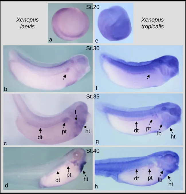
1.6.1.4.2 The Xenopus paralogues wnt9a and wnt9b have a different expression pattern during early Xenopus
development.
41
1.6.2 The non-canonical wnt molecules used in this study. 42
1.6.2.1 wnt4 is critically required for pronephros and metanephros tubulogenesis.
42
1.6.2.2 Non-canonical wnt5a is involved in morphogenetic movements during early embryonic development.
45
1.6.2.3 wnt11 (formerly Xenopus wnt11-related), is involved in various organ formation.
46
1.6.2.4 Maternal wnt11b (formerly Xenopus wnt11) is required for dorso-ventral axis formation, for convergent extension movements during gastrulation, in heart formation and in neural crest cells migration.
49
Chapter 2 Materials and Methods. 53
2.1 Materials. 53
2.2 Media and stock solutions. 53
2.3 Escherichia coli bacterial strains. 53
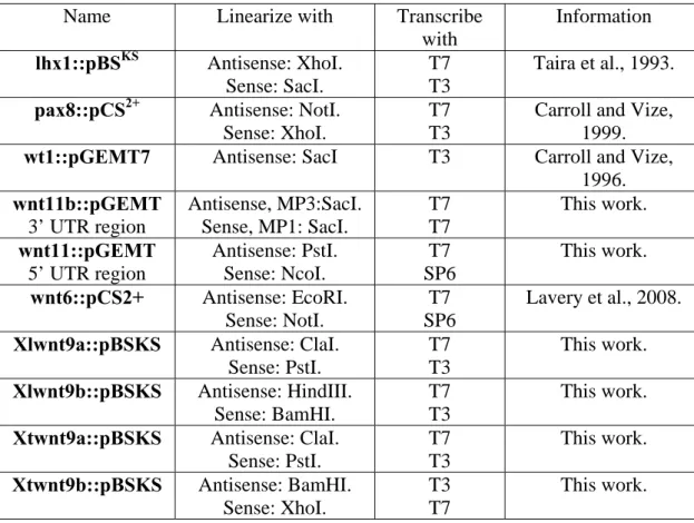
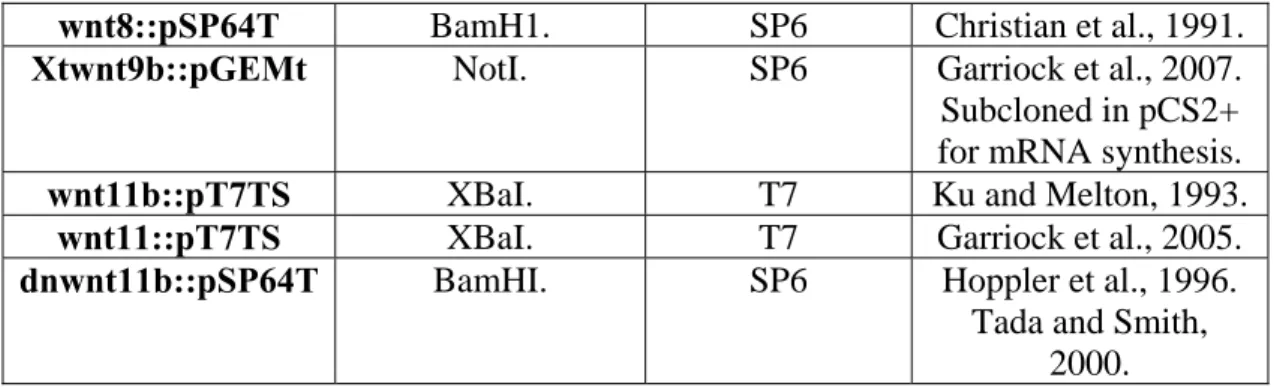
2.4 Plasmid template DNA. 53
2.4.1 Plasmid template DNA used to clone PCR product. 53 2.4.2 Plasmid DNA used as template in situ hybridization. 54 2.4.3 Plasmid template DNA used for capped mRNA synthesis for microinjection.
54
2.5 Antisense oligo morpholinos. 55
2.6 DNA techniques. 55
2.6.1 Agarose gel electrophoresis. 55
2.6.2 Restriction enzyme digestion. 55
2.6.3 DNA minipreps. 56
2.6.5 Transformation of plasmid DNA into competent Escherichia coli. 56
2.6.6 Primer design. 56
2.6.7 Automated DNA sequencing. 57
2.7 RNA techniques. 57
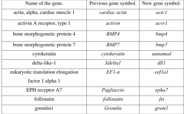
2.7.1 RNA extraction from embryos and animal caps. 57 2.7.2 New Xenopus gene names and symbols. 58
2.7.3 Reverse transcription PCR. 60
2.7.4 RACE PCR. 62
2.7.5 Preparation of in vitro transcription of mRNA. 63 2.7.6 Wholemount in situ hybridisation. 63
2.8 Protein techniques. 63
2.8.1 Protein expression in oocytes. 63
2.8.2 Protein extraction from oocytes. 63
2.9 Wholemount immunohistochemistry. 64
2.10 Embryo manipulations. 65
2.10.1 In vitro fertilisation of Xenopus eggs. 65 2.10.2 Microinjection of embryos and oocytes. 65
2.10.3 Growth factor treatments. 66
2.10.4 Dissections of animal caps and embryonic material. 66
2.10.5 Holtfreter sandwich cultures. 66
Chapter 3 Identification of Xenopus wnt9a and wnt9b genes potentially
involved in pronephros formation.
67
3.1 Introduction. 67
3.2 Identification of Xlwnt9b. 68
3.2.1 Sequence analysis of the genomic database identifies X. tropicalis wnt9b in scaffold 43.
68
3.2.2 Strategy for amplification of Xlwnt9b. 70 3.2.3 Verification of Xlwnt9b sequence amplified by RT-PCR. 70 3.3 Temporal and spatial expression of wnt9a and wnt9b in X. laevis and X. tropicalis embryos.
3.3.1 Generation of X. tropicalis and X. laevis wnt9a and wnt9b in situ
probes.
78
3.3.1.1 Design of wnt9a and wnt9b primers to amplify gene for species specific probes.
79
3.3.1.2 Verification of sequences amplified by PCR and generation of an in situ probe clone.
79
3.3.2 Temporal and spatial expression of wnt9a and wnt9b in X. laevis
and X. tropicalis embryos.
81
3.3.2.1 Temporal and spatial embryonic expression of Xtwnt9a and
Xlwnt9a.
81
3.3.2.2 Temporal and spatial embryonic expression of Xtwnt9b and
Xlwnt9b.
83
3.3.3 Expression profile of Xlwnt9a and Xlwnt9b in early embryonic development.
85
3.3.4 Expression of wnt9a and wnt9b in adult X. laevis and X. tropicalis
organs.
85
3.4 Over-expression of Xtwnt9b leads to abnormal kidney formation. 87
3.4.1 Xtwnt9b belongs to the canonical wnt class of molecules. 90 3.4.2 Over-expression of Xtwnt9b into the V2 blastomere results in
abnormal pronephros formation.
93
3.4.3 Over-expression of Xtwnt9b in epidermis results in abnormal pronephros formation.
98
3.5 Inhibition of function of Xlwnt9a and Xlwnt9b in X. laevis embryos
using antisense morpholinos.
103
3.5.1 Design of Xlwnt9a and Xlwnt9b antisense morpholinos. 103
3.5.2 Inhibition of Xlwnt9a and Xlwnt9b results in abnormal pronephros formation.
109
3.6 Discussion. 110
3.6.1 Identification of Xlwnt9a and Xlwnt9b genes. 110 3.6.2 Expression of wnt9a and wnt9b in X. laevis and tropicalis embryos and adult organs.
111
3.6.4 Xtwnt9b plays a role in pronephros formation. 112
Chapter 4 Identification of the wnt signalling molecules potentially involved in X. laevis pronephros formation.
114
4.1 Introduction. 114
4.2 Temporal expression of known developmental signalling molecules
screened in isolated Xenopus pronephric anlagen.
114
4.2.1 Localisation and dissection of pronephric anlagen during early stages of X. laevis development.
114
4.2.2 Temporal expression of wnt signalling molecules screened in isolated Xenopus pronephric anlagen.
116
4.2.3 Temporal expression of other signalling molecules screened in isolated X.laevis pronephric anlagen.
119
4.3 Expression of signalling molecules in X. laevis somites. 121
4.3.1 Dissection of anterior and posterior somites in X. laevis stage 17 embryos.
121
4.3.2 Expression of wnt signalling molecules in X. laevis isolated anterior and posterior somites.
123
4.3.3 Expression of other signalling molecules in X. laevis isolated anterior and posterior somites.
125
4.4 Discussion. 127
4.4.1 Control of somite tissue maturity prior analysis of gene expression. 127 4.4.2 The different phases of kidney development are characterised by waves of signalling molecules expression.
127
4.4.3 Identification of the wnts signalling molecules in the pronephric anlagen at the right time and place to have a role to play in pronephric development.
129
Chapter 5 A direct assay of pronephrogenesis using the Holtfreter
sandwich cultures.
131
5.2 Setting up an in vitro model for pronephrogenesis. 131
5.2.1 Assembly of Holtfreter sandwich cultures. 131 5.2.2 Animal caps over-expressing wnt molecules analysed by RT-PCR for the presence of pronephric and other genes.
133
5.2.2.1 Overall morphology of animal caps expressing wnt
signalling molecules.
133
5.2.2.2 Animal caps over-expressing wnt molecules do not induce nervous system, kidney or muscle markers.
135
5.2.3 The general morphology of Holtfreter sandwich cultures before immuno-assay.
138
5.2.4 Somites can induce pronephros from intermediate mesoderm in explants cultures.
141
5.3 Holtfreter sandwich cultures to test the wnt molecules in forming
pronephros.
145
5.3.1 Both non-canonical wnt11b and wnt11 can induce pronephric tubule formation in explants cultures.
145
5.3.2 Somites over-expressing the dnwnt11b cultured with intermediate mesoderm inside normal animal caps surprisingly form pronephric tubules.
147
5.3.3 Non-canonical wnt4 cannot induce pronephric tubules in explant cultures.
148
5.3.4 Non-canonical wnt5a cannot induce pronephric tubules in explant cultures.
148
5.3.5 Canonical wnt6 and wnt8 can induce pronephric tubules in explant cultures.
149
5.3.6 Canonical wnt7b cannot induce pronephric tubules in explant cultures.
151
5.3.7 Statistical analysis shows that some but not all wnt molecules can significantly induce pronephric tubules in in vitro Holtfreter sandwich cultures.
152
5.4 wnt Holtfreter sandwich cultures assayed for somite formation. 158 5.5 Sandwich cultures over-expressing wnt11b cultured with intermediate
mesoderm induce specific pronephric markers.
5.6 Discussion. 166
5.6.1 Holtfreter sandwich cultures, an explant model to study pronephrogenesis, advantages and limitations.
166
5.6.2 Somites over-expressing the dnwnt11b cultured with intermediate mesoderm inside normal animal caps still form pronephric components.
167
5.6.3 Animal caps over-expressing wnt molecules and tissue formation. 168 5.6.4 wnt11b and sall1 might share the same pathway in pronephros formation.
168
5.6.5 Direct and indirect formation of pronephric tubules in Holtfreter sandwich cultures over-expressing wnt molecules.
169
Chapter 6 In vivo function of non-canonical wnt11b and wnt11 molecules in X. laevis pronephros development.
172
6.1 Introduction. 172
6.2 wnt11b and wnt11, two closely related genes, have distinct spatial
expression patterns in Xenopus embryos.
172
6.2.1 Generation of X. laeviswnt11 and wnt11b in situ probes. 174 6.2.2 Temporal and spatial expression of wnt11 and wnt11b in Xenopus
embryo.
174
6.3 In vivo function of wnt11b in X. laevis pronephros formation. 176 6.3.1 Over-expression of wnt11b results in formation of abnormal
pronephros.
176
6.3.1.1 Morphological phenotypes of Xenopus embryos over-expressing wnt11b.
176
6.3.1.2 Over-expression of wnt11b in V2 blastomere affects normal pronephros morphogenesis.
178
6.3.1.3. Over-expression of wnt11b in the epidermis leads to abnormal kidney formation.
184
6.3.2 Inhibition of wnt11b using a dominant-negative construct (dnwnt11b) leads to abnormal kidney development in X. laevis
embryos.
185
6.3.2.2 Morphological phenotypes of X. laevis embryos over-expressing dnwnt11b.
188
6.3.2.3 Inhibition of wnt11 using the dnwnt11b affects X. laevis
pronephros morphogenesis during terminal differentiation.
188
6.3.2.4. Inhibition of wnt11b using the dnwnt11b have an effect on
X. laevis pronephros morphogenesis during early differentiation.
192
6.3.2.5 Inhibition of wnt11b using the dnwnt11b have an effect on pronephric glomus formation.
195
6.3.3 Inhibition of both wnt11b and wnt11 using morpholinos result
in a more severely abnormal pronephros phenotype.
198
6.3.3.1 wnt11b and wnt11 morpholinos targeted in V2 blastomere give rise to abnormal kidney formation.
198
6.4 Discussion. 204
6.4.1 wnt11b and wnt11, two closely related genes, have distinct spatial expression patterns in X. laevis embryos.
204
6.4.2 Identification of a possible role for wnt11b in pronephros formation.
205
Chapter 7 The role of the canonical wnt/β-catenin pathway in Xenopus pronephros formation.
206
7.1 Introduction. 206
7.2 The over-expression of the dntcf3 to inhibit all canonical wnt
signalling.
206
7.2.1 The dntcf3 molecular construct and its functional mode of action in the cell.
206
7.2.2 The dntcf3 translates in vivo in X. laevis oocytes. 208 7.2.3 Demonstration of the effectiveness of the dominant-negative construct to inhibit canonical wnt8 signalling in animal caps explants.
208
7.3 Over-expression of dntcf3 causes severe morphological abnormalities in developing X. laevis embryos.
211
7.4 The role of canonical wnt signalling in X. laevis kidney development. 212
laevis pronephros terminal differentiation.
7.4.2 Inhibition of the canonical wnt signalling using dntcf3 over-expression disturbs the normal development of the pronephric glomus.
220
7.5 Spatial and temporal expression of wnt6 in X. laevis and X. tropicalis
embryos.
223
7.6 Over-expression of wnt6 in the epidermis leads to abnormal
pronephros formation.
227
7.7Discussion. 230
7.7.1 Inhibition of the canonical wnt pathway using the dntcf3 leads to abnormal embryos morphology and abnormal pronephros formation.
230
7.7.2 Spatial and temporal expression of wnt6 in X. laevis and X. tropicalis embryos.
232
7.7.3 wnt6 plays a role of in X. laevis pronephros formation. 233
Chapter 8 General Discussion. 234
8.1 wnt4, required for nephrostome and glomus formation. 234
8.2 wnt8, indirect pronephric inducer. 235
8.3 wnt6, a somite epithelialisation factor from the ectoderm, acts as an indirect
pronephric inducer.
235
8.4 wnt11b, direct pronephric inducer, regulates morphogenetic movements with wnt11 during pronephros formation.
236
8.5 wnt5a, modulator of morphogenetic movements during pronephros development.
238
8.6. wnt9a and wnt9b, general organizing molecules of the pronephros. 238 8.7 wnt7b, possibly required for pronephros differentiation. 240
8.8 Conclusion. 240
List of figures
Figure Page number
1.1 Why use Xenopus embryos in developmental studies? 2
1.2 Organisation of nephrons into kidneys. 5
1.3 The first steps of the metanephros development. 9 1.4 Patterning events that subdivide the Xenopus pronephric anlagen. 12 1.5 The anatomy of the Xenopus pronephros at stage 38. 15 1.6 Pronephric compartments and key expression time of some
molecular markers.
18
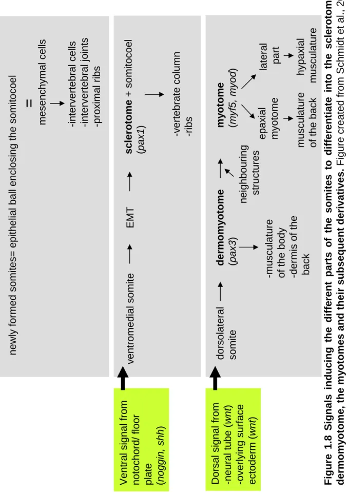
1.7 Simplified schematic representation of the wnt signalling pathways. 31-32 1.8 Signals inducing the different parts of the somites to differentiate
into the sclerotome, the dermomyotome, the myotomes and their subsequent derivatives.
36
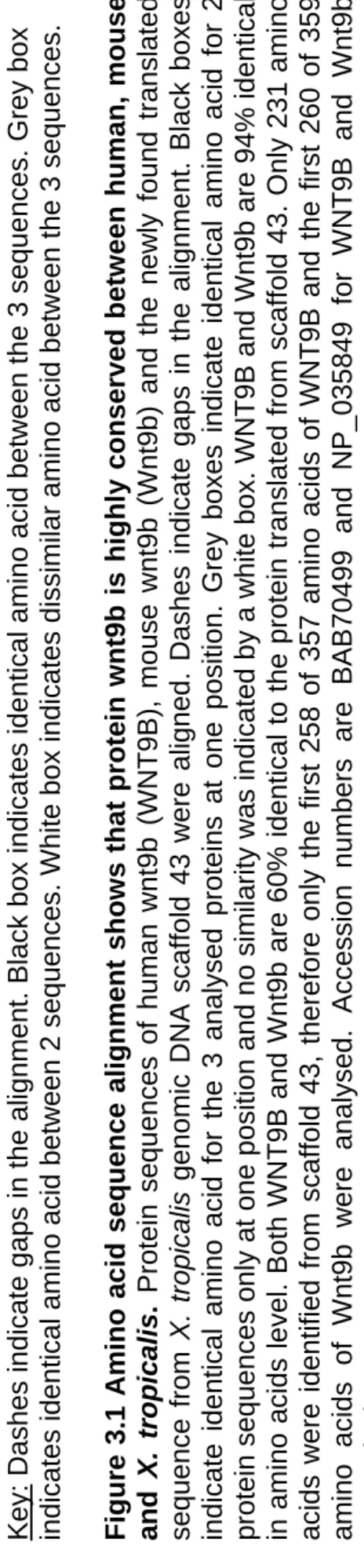
3.1 Amino acid sequence alignment shows that protein wnt9b is highly
conserved between human, mouse and X. tropicalis.
69
3.2 Design of specific Xenopus wnt9b primers based on the complete nucleotide sequences alignment of human and mouse wnt9b with scaffold 43.
71-72
3.3 Design of specific Xenopus wnt9b primers based on the complete nucleotide sequences alignment of X. laevis wnt genes with scaffold 43.
73-76
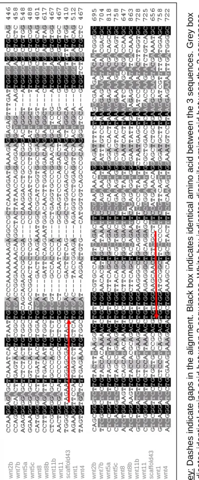
3.4 Partial nucleotide sequence alignments of X. laevis wnt genes with scaffold 43 around the position of primers 3 and 6.
77a
3.5 Nucleotide sequence alignment of PCR product amplified from
Xlwnt9b with X. tropicalis scaffold 43 coding sequence.
77b
3.6 Spatial and temporal embryonic expression of XlWnt9a and
XtWnt9a.
82
XtWnt9b.
3.8 Temporal expression of Xlwnt9b and Xlwnt9a during X. laevis
early development.
86
3.9 Expression of wnt9a and wnt9b in X. tropicalis and X. laevis adult organs.
88-89
3.10 Injection of targeted GFP mRNA injection in 8-cell stage embryos and followed later on in development.
91
3.11 Xtwnt9b is capable of inducing a second axis when over-expressed in the ventral blastomere of 4-cell and 8-cell stage embryos.
92
3.12 Xenopus embryos previously injected into the V2 blastomere with
Xtwnt9b mRNA develop an abnormal pronephros.
94
3.13 Numerical and graphical representation of the pronephros phenotype in X. laevis embryos over-expressing 1 to 2ng of
Xtwnt9b mRNA.
96-97
3.14 Embryos over-expressing Xtwnt9b in the epidermis develop an abnormal pronephros.
99
3.15 Numerical and graphical representation of the pronephros phenotype in X. laevis embryos over-expressing 0.25 to 1ng of
Xtwnt9b mRNA.
101-102
3.16 Novel Xlwnt9b partial DNA and protein sequences. 105 3.17 Novel Xlwnt9a partial DNA and protein sequences. 106 3.18 Amino acid sequences alignment shows that the newly identified
Xlwnt9b protein is highly similar to its Xtwnt9b paralog.
107
3.19 Amino acid sequences alignment shows that the newly identified Xlwnt9a protein is highly similar to its Xtwnt9a paralog.
108
4.1 Diagram representing the pronephros anlagen dissections in X. laevis embryos from stage 12.5 to stage 35.
115
4.2 Identification of wnt signalling molecules expressed in isolated X. laevis pronephric anlagen.
118
4.3 Analysis of some non-wnt signalling molecules expressed in X. laevis pronephric anlagen.
4.4 Diagram illustrating the somite dissections in X. laevis embryos at stage 17.
122
4.5 Identification of wnt signalling molecules expressed in X. laevis
anterior and posterior somites.
124
4.6 Analysis of some non-wnt signalling molecules expressed in X. laevis somites.
126
5.1 Dissection of explants and assembly of Holtfreter sandwich cultures.
132
5.2 Morphological analysis of X. laevis embryos over-expressing wnt
molecules.
134
5.3 Animal caps over-expressing wnt molecules do not induce nervous system, kidney or muscle markers.
136-137
5.4 The general morphology of Holtfreter sandwich cultures before immuno-assay.
139-140
5.5 Somites can induce pronephros in explant cultures. 142 5.6 wnt11b and wnt11 can induce pronephros formation when cultured
with unspecified intermediate mesoderm alone.
146
5.7 Canonical wnt6 and wnt8 over-expressed in animal caps and cultured with unspecified intermediate mesoderm alone can in vitro
induce pronephros formation.
150
5.8 Statistical analyses show that some but not all wnt molecules can significantly induce proximal tubule formation in Holtfreter sandwich cultures.
154
5.9 Statistical analyses show that some but not all wnt molecules can significantly induce intermediate/distal tubules formation in Holtfreter sandwich cultures.
156
5.10 Pronephric markers are expressed in sandwich cultures over-expressing wnt11b.
163-164
6.1 The temporal and spatial expression of wnt11b and wnt11 during X. laevis development.
6.2 Morphological defects caused by wnt11b over-expression in X. laevis embryos.
177
6.3 Embryos over-expressing wnt11b in one V2 blastomere develop abnormal pronephros.
179
6.4 Embryos over-expressing wnt11b in the epidermis develop an abnormal pronephros.
181
6.5 Numerical and graphical representation of the pronephros phenotype in X. laevis embryos over-expressing 1ng of wnt11b
mRNA.
182-183
6.6 Construction of dominant-negative wnt11b (dnwnt11b), according to Smith at al., 2000.
186
6.7 dnwnt11b can inhibit elongation in animal caps treated with activin.
187
6.8 Morphology of X. laevis embryos resulting from dnwnt11b over-expression.
189
6.9 Inhibition of wnt11b using the dnwnt11b affects normal Xenopus
pronephros formation.
190
6.10 Numerical and graphical representation of the pronephros
phenotype in X. laevis embryos over-expressing 1 to 1.5ng of
dnwnt11b mRNA.
191
6.11 Numerical and graphical representation of the pronephros phenotype in X. laevis embryos over-expressing 1.5ng of dnwnt11b
mRNA and analysed by in situ hybridization.
194
6.12 Embryos over-expressing the dnwnt11b show defects in glomus formation.
196
6.13 Numerical and graphical representation of the pronephros glomus phenotype in X. laevis embryos over-expressing 1ng of the
dnwnt11b mRNA and analysed by in situ hybridization using the glomus marker wt1.
197
6.14 Diagram explaining the design of antisense oligonucleotide morpholinos.
199
perturb pronephros formation.
6.16 Numerical and graphical representation of the pronephros phenotype in X. laevis embryos injected with 15ng of wnt11b and
wnt11 antisensemorpholinos.
202
7.1 The dntcf3 construct used in microinjection and its functional mode of action in the cell.
207
7.2 In vivo translation of dntcf3 mRNA in X. laevis oocytes. 209 7.3 dntcf3 inhibits wnt/ß-catenin signalling in animal caps. 210 7.4 Morphological defects caused by over-expression of dntcf3 in X.
laevis embryos.
213
7.5 Inhibition of canonical wnt signalling using the dntcf3 construct affects the X. laevis pronephros development in a dose-dependent manner.
215
7.6 Numerical and graphical representation of the pronephros phenotype and embryo morphology in X. laevis embryos injected with various concentrations of the dntcf3.
216-218
7.7 Inhibition of the canonical wnt signalling using dntcf3 disturbs pronephric glomus formation in X. laevis embryos.
221
7.8 Numerical and graphical representation of the glomus morphology in X. laevis embryos injected with various concentrations of the
dntcf3.
222
7.9 The temporal and spatial expression of wnt6 during X.laevis and X. tropicalis development.
224-225
7.10 Embryos over-expressing wnt6 in the epidermis develop an abnormal pronephros.
228
7.11 Numerical and graphical representation of the pronephros phenotype in X. laevis embryos over-expressing 1ng of wnt6
mRNA in the epidermis.
229
8.1 Simple view of the role of the wnt signalling molecules during early Xenopus pronephros formation.
List of tables
Table Page number
1.1 Some kidney markers and their expression within the three kidneys forms and their phenotypic consequences of targeted mutagenesis in developing mice.
19
2.1 Plasmid DNA used as template in situ hybridization. 54 2.2 Plasmid template DNA used for capped mRNA synthesis for
microinjection.
54
2.3 The oligo morpholino sequences used in this thesis. 55 2.4 Name of genes, a synonym of the previous gene symbol and the new
gene symbol.
58
2.5 Primer sequences and PCR condition used in RT-PCR analysis. 61
3.1 Comparison between the four nucleotide sequences of XlWnt9a, XlWnt9b,XtWnt9a and XtWnt9b generated by PCR amplification.
80
3.13.1 Morphology of embryos injected with 1 to 2ng of Xtwnt9b mRNA into one V2 blastomere at the 8-cell stage and analysed for pronephros phenotype.
96
3.13.2 Number and percentage of embryos showing pronephros phenotype in embryos injected in V2 blastomere of 8-cell stage with 1 to 2ng of
Xtwnt9b mRNA.
97
3.15.1 Morphology of embryos over-expressing 0.25 to 1ng of Xtwnt9b
mRNA in the epidermis and analysed for pronephros phenotype.
101
3.15.2 Number and percentage of embryos showing pronephros phenotype in embryos injected in the animal pole of 2-cell stage with 0.25 to 1ng of
Xtwnt9b mRNA.
102
3.5.1 Gene specific primers (GSP) designed from Xlwnt9a and Xlwnt9b
sequences previously identified from generating in situ hybridization probes designed to amplify by 5’ RACE PCR the 5’UTR of XlWnt9a
and XlWnt9b.
5.1 Number of controls and wnt sandwich cultures assayed for presence of pronephric tubules using the specific antibodies 3G8/4A6.
144
5.2 Statistical analyses show that some wnt molecules but not all can significantly induce proximal tubule formation in Holtfreter sandwich cultures.
153
5.3 Statistical analyses show that some wnt molecules but not all can significantly induce intermediate/distal tubules formation in Holtfreter sandwich cultures.
155
5.4 Number of controls and wnt sandwich cultures assayed for presence of somites using the specific antibody 12/101.
159
5.5 Statistical analyses show that some wnt molecules but not all can significantly induce somites formation in Holtfreter sandwich cultures.
160
6.1 Percentage of DNA sequence identity between the PCR amplified
wnt11b 3’ in situ probe, the PCR amplified wnt11 5’ in situ probeand the respective whole gene sequences deposited in GenBank.
173
6.5.1 Morphology of embryos over-expressing 1ng of wnt11b mRNA in the epidermis and analysed for pronephros phenotype.
182
6.5.2 Number and percentage of embryos showing pronephros phenotype in embryos injected in the animal pole of one cell at 2-cell stage with 1ng of wnt11b mRNA.
183
6.10.1 Number and percentage of embryos showing pronephros phenotype in embryos injected in one V2 ventral blastomere at the 8-cell stage with1 to 1.5ng of dnwnt11b mRNA.
191
6.11.1 Number and percentage of embryos showing pronephros phenotype in embryos injected in one V2 ventral blastomere at the 8-cell stage with 1.5ng of dnwnt11b mRNA and analysed by in situ hybridization with the pronephric markers lhx1 and pax8.
194
6.13.1 Number and percentage of embryos showing glomus phenotype in embryos injected in one V2 ventral blastomere at the 8-cell stage with 1ng of dnwnt11b mRNA and analysed by in situ hybridization with the pronephric markers wt1.
6.16.1 Number and percentage of embryos showing pronephros phenotype in embryos injected in one V2 ventral blastomere at the 8-cell stage with15ng of wnt11b and wnt11 antisense morpholinos.
202
7.6.1 Morphology of embryos injected with 100pg to 1.6ng of dntcf3 in one ventral blastomere at the 4-cell stage and analysed for pronephros phenotype.
216
7.6.2 Number and percentage of embryos showing on pronephros phenotype when injected into one ventral blastomere at the 4-cell stage with 100pg to 1.6ng of dntcf3 mRNA.
217
7.8.1 Number and percentage of embryos showing glomus phenotype when injected in one ventral blastomere at the 4-cell stage with 100pg to 400pg of dntcf3 mRNA.
222
7.11.1 Number and percentage of embryos showing pronephros phenotype in embryos injected in the animal pole of one cell at the 2-cell stage with 1ng of wnt6 mRNA.
Acknowledgements
I would like to thank my supervisor Professor Elizabeth Oliver-Jones for her advice, encouragement and support to complete this work and for giving me the unique opportunity to carry on my studies here at the University of Warwick.
I would also like to thank the members of Liz’s lab, Dr Karine Massé, Dr Junichi Kyuno, Richard Naylor and Surinder Bhamra for the cooperative team work, and in particular Karine for teaching me Bioinformatics and Molecular Biology.
But I would not have been part of this adventure without the unconditional support of my very good friends, Dr Laura Howard, Craig Tucker, Nader Amin, Jean-Christophe and Mella Bommé and Didier Borget.
Declaration
The results presented in this thesis are the work of the author unless specified.
Microinjections and dissections were carried out together with Professor Elizabeth Oliver-Jones.
Hormone injections to X. laevis females were given by Mr Robert Taylor and Mr Paul Jarrett.
Sources of information have been acknowledges by reference.
Summary
The aim of the project was to characterise known wnt signalling molecules during the early pronephros patterning using novel and effective bioassays in Xenopus embryos. Anterior somites have been shown previously to have unique biological activity to induce pronephros formation in Xenopus embryos (Seufert et al., 1999).
A molecular approach consisted of analysing the expression of canonical wnts (wnt6,
wnt7b, wnt8, wnt9a and wnt9b) and the non-canonical wnts (wnt4, wnt5a, wnt11 and
wnt11b) genes in isolated pronephric anlagen and pronephros from stage 12.5 to stage 35 and in anterior and posterior somites. This allowed the identification of potential candidate wnt genes which could act as pronephric inducers. Their potential to induce pronephros was tested in vitro using the Holtfreter sandwich culture pronephrogenesis assay, which consists of unspecified intermediate mesoderm cultured inside two animal caps over-expressing the wnt molecule of interest. Results suggest that the canonical wnt molecules (wnt6 and wnt8) are capable of modifying the intermediate mesoderm leading to formation of somite and neural tissue that in turn, can secondarily induce pronephric tubule formation. By contrast, signals of the non-canonical wnt11 and wnt11b are sufficient to directly specify the intermediate mesoderm to become kidney.
In vivo, the role of the canonical wnt6, non-canonical wnt11b and the closely related
wnt11 gene was investigated by gain and loss-of-function experiments. Results suggest that mis-expression of these genes disturb the normal formation of the pronephric tubules and suggest that both canonical and non-canonical wnt molecules are required for formation of functional pronephros.
This thesis also reports the identification of the novel Xlwnt9a and Xlwnt9b genes,
Abbreviations
AP alkaline phosphatase
ATP adensonsine triphosphate
bFGF basic fibroblast growth factor
bp base pairs
BSA bovine serum albumin
cDNA complementary deoxyribonucleic acid C-terminal carboxyl-terminal
dATP deoxyadenosine triphosphate
dCTP deoxycytidine triphosphate
dGTP deoxyguanosine triphosphate
dH2O distilled water
DIG digoxygenin
DNA deoxyribonucleic acid
DNase deoxyribonuclease
dNTPs deoxyribonucleoside triphosphates
dpc days postcoitum
dTTP deoxythymidine triphosphate
E embryonic day
E.coli Escherichia coli
EDTA ethylene diamine tetra acid
EST expressed sequence tag
FGF fibroblast growth factor g gram GFP green fluorescent protein
HCl hydrochloric acid
H2O water
H2O2 hydrogen peroxide
l litre
LBroth Luria Broth
kD kilo Dalton M molar
MBT mid-blastula transition
mg milligram ml millilitre mM millimolar MMLV moloney murine leukaemia virus
MOPS 3-[N-morpholino] propane sulphonic acid
MO morpholino oligonucleotide
mRNA messenger ribonucleic acid
NaCl sodium chloride
ng nanogram nl nanolitre nM nanomolar nt nucleotides N-terminal amino-terminal
PAGE polyacrylamide gel electrophoresis PBS phosphate buffered saline
PBT phosphate buffered tris
PBST phosphate buffered tris PCR polymerase chain reaction pM picomolar pmol picomoles
RA retinoic acid
RNA ribonucleic acid
RNase ribonuclease
rpm revolutions per minute
RT-PCR reverse transcription PCR
SDS sodium dodecylsulphate
TBE tris buffered EDTA TBS tris buffered saline
TdT Terminal deoxynucleotidyl transferase TGFβ transforming growth factor β
Tris-Cl tris (hydroxymethyl) aminomethane, pH adjusted with HCl
UTR untranslated region
V volts
v/v volume/volume w/v weight/volume U units
Chapter 1 Introduction
1.1 Overview, aims of the project.
The aim of the project was to characterise known wnt signalling molecules during the early pronephros patterning using novel and effective bioassays in Xenopus embryos. Anterior somites have been shown previously to have unique biological activity to signal to unspecified intermediate mesoderm to induce pronephros formation in
Xenopus embryos (Seufert et al., 1999). A molecular approach consisting of analysing the canonical and non-canonical wnt genes expression in isolated pronephric anlagen and pronephros from stage 12.5 to stage 35 and in anterior and posterior somites will allow the identification of potential candidate wnt genes and will be directly tested in pronephrogenesis assay. Gain and loss-of-function experiments of the newly identified pronephric inductive wnt genes will investigate the full role of these genes in pronephric induction and patterning.
1.2 Why use X. laevis embryos in developmental studies?
Panel B Panel A • Animals h o u sed i n t h e lab o ratory. •
1000 plus eggs co
llected w
ith HCG injectio
n.
•
Embryo
g
e
nesis occurs o
u
tsid
e the moth
er. • Defined stages of embry onic development obta ine d by in v it ro fer tilization. •
Embryos are large allow
ing mic rom a n ipula tions • Organ o g
enesis easily mo
nitored. • Transgen ic techni q u e s. • No feedi n
g required up t
o t h e s w imming tadpole. Pic tu re fro m the Geno me N e w s Ne two rk , 2002 . Fi g u
re 1.1 Wh
y use
Xenopus
embry
o
s in develop
mental studies?
Panel A shows the adult African
claw
ed frog
X. laevis,
on the l
e
ft and its cl
ose rel
a
tive, the smaller
X. tropi c alis, o n th e rig h
t. Panel B
summarises the advantages of using
X.
laev
is
embryos in devel
opmental studi
es.
hormone (hCG) and X. tropicalis female adults can lay many hundreds of eggs. Then, eggs can be collected in a Petri dish and are subsequently fertilised in vitro by simply brushing over the male frog testis, thus releasing sperm and providing a synchronously fertilised group of embryos. As embryogenesis occurs outside the mother, development and subsequent organogenesis can be easily monitored. Defined stages of embryonic development can be easily obtained and correctly identified following the Normal Table of Nieuwkoop and Faber, 1994.
Embryos are large, approximately 2mm diameter, and relatively sturdy allowing micromanipulation. These micromanipulations are one of the greatest advantages of using X. laevis embryos in developmental studies as injection of DNA, RNA or protein molecules allow the introduction of defined gene products. Injection of mRNA results in the over-expression of protein and injection of morpholino antisense oligonucleotide results in inhibition of translation of a defined gene product. Morpholino oligos are short chains of 25 morpholino subunits. Each subunit is comprised of a nucleic acid base, a morpholine ring and a non-ionic phosphorodiamidate intersubunit linkage. Morpholinos act via an RNase H- independent steric blocking mechanism. With their high mRNA binding affinity and specificity, they result in knocking down gene expression by blocking translation initiation in the cytosol (by targeting the 5’UTR through the first 25 bases of coding sequence) (Heasman et al., 2000). Both techniques, over-expression or inhibition of specific gene product, allow the alteration of the gene function and therefore give indication about their role during development. Injection can be performed into the fertilized egg or into one blastomere of the early cleaving stage, allowing targeting to specific regions of the embryo (Dale and Slack, 1987 and Moody, 1987). In addition, injection can be restricted to one side of the embryo, allowing the uninjected side to be used as a contralateral control within the same embryo. Additional micromanipulations such as fine dissections, grafting experiments and embryo explant culture can also be carried out in simple saline solution.
During the last decade, Xenopus has also been developed as a genetic system. A number of reports have identified successful strategies for generating both transgenic
extrachromosomal transgenic DNA into genomic DNA. The use of inducible constructs allow the investigation to control the time of the expression of the transgene (Wheeler et al., 2000, Waldner et al., 2006). Moreover, the recently opened European Resource Centre for Xenopus provides a means of obtaining a supply of genetically altered animals along with wild type. To augment these genetic studies, in August 2005, the whole genome of the diploid X. tropicalis was sequenced and assembled by the Joint Genome Institute (JGI). This genomic resource constitutes a great tool for genomic and genetic research in amphibia.
The advantages stated above make Xenopus an excellent model organism to study both early development and organogenesis. In this study, we have exploited this model system to study the formation of the embryonic kidney, the pronephros.
1.3 The kidney as a model organ to study organogenesis.
1.3.1 The three kidney forms, function, organisation and similarities.
The function of the kidney is to filter blood thus retaining useful molecules inside the organism and disposing of metabolic waste outside the organism.
During vertebrate life, the kidney, which derives from the intermediate mesoderm, undergoes spatial and size rearrangement but its basic organisation and function remain identical. The first embryonic kidney to form is the pronephros which is functional in lower vertebrates such as amphibian and fish. Later in development the pronephros is replaced by the mesonephros that constitutes the adult kidney form of the amphibians and fish but acts as the functional embryonic kidney form for the higher vertebrates. In mammals, the adult kidney form is the metanephros.
Fi
gure 1.2
Organisa
tion of
nephrons
in
to
ki
dney
s.
The pronephros has one nephron, the
mesonephros
is
constituted of 10-50 nephrons
a
n
d
the m
o
st compl
e
x
form of kidn
ey, the
metanephros
has one million nephrons
. Figure from Vize
et al., 1997.
into the collecting tubule system. The pronephros is one single nephron, while the mesonephros is composed of 10 to 50 nephrons and the most complicated form of the kidney, the metanephros is formed of about a million nephrons.
1.3.1.1 The mesonephros in mammals.
All three kidney forms of the developing urinary system derive from the intermediate mesoderm. In vertebrates, the second embryonic kidney to form, the mesonephros, follows the primitive kidney, the pronephros and precedes the development of the permanent kidney, the metanephros. Thus, it is also called the intermediate kidney. In adult lower vertebrates, amphibians and teleost fish, the mesonephros serves as the adult excretory organ. The mesonephros undergoes epithelial differentiation, cell migration, developmental changes, apoptosis and is a source of stem cell differentiation and hormonal determination of gene activity.
In mammalian mesonephrogenesis, the caudally descending nephric duct, the pronephric duct, also called the Wolffian or mesonephric duct at later stages, forms in the anterior region from the pronephros (Smith and MacKay, 1991). The duct is permanent, but the pronephric tubules regress and are eventually replaced by a new type of embryonic kidney, the mesonephros. The nephric duct is essential as both the common urinary drain and the terminal inducer of mesonephric tubules. No mesonephric nephrons differentiate if the inductive interaction between the duct and the blastema is disrupted (Waddington, 1938).
The mesonephric kidney regresses prenatally in mammals. Degradation is completed in mouse by day E-15 (Saxén, 1987). Murine regression seems to start from the caudal mesonephric tubules and spread cranially (Sainio et al., 1997a). In females, degradation is complete, but in males, the remaining mesonephric tubules form part of the gonadal ducts (Moore, 1977). The molecular mechanism regulating the mesonephric degradation is not known, but apoptosis has been described by morphological criteria. Mesonephric tubule differentiation and regression are controlled by tissue surrounding the mesonephric tubules.
Some key regulatory molecules in the mesonephros are the same as those in the primitive and permanent kidney forms. For example, hox genes are essential for normal specification and differentiation of the urogenital tissue (Patterson et al., 2001); pax2 is necessary for the development of the excretory system and is associated with the initial steps in the differentiation of the intermediate mesoderm (Torres et al., 1995); bmp4 is a candidate molecule to regulate early steps in the differentiation of the Wolffian duct and budding of the ureter (Miyazaki et al., 2000);
gdnf is a second factor crucial for ureteric budding (Sainio et al., 1997b). wt1
regulates mesonephric tubule differentiation (Kreidberg et al., 1993) and the homeobox gene emx2 regulates at least the Wolffian duct maintenance (Yoshida et al., 1997).
The mesonephros contributes to the development of other organ systems and is a source of several cell lineages. The mesonephros is important for the somatic cell differentiation of both the testis and the ovary (McLaren, 2000), is a source of hematopoietic cells (Medvinsky et al., 1996) and a source of adrenal cortex cells (Worbel and Süß, 2000).
1.3.1.2 The metanephros in mammals.
The mammalian metanephros kidney constitutes the third and final member of the kidney form developing from the intermediate mesoderm. The metanephros is stable throughout adult life.
Figure 1.3 shows the first steps of metanephros formation. Renal development starts when an epithelial ureter bud forms from the nephric duct and invades the metanephric mesenchyme. Continuous interaction between the epithelium of this ureter bud and the metanephric mesenchyme is required for the development of the metanephric kidney. This interaction is referred to as a reciprocal epithelium-mesenchymal interaction. In response to signals from the mesenchyme, the ureter grows, branches and eventually forms the collecting duct system of the mature kidney. In turn, in response to signals from the developing ureter, the mesenchymal cells, condense, aggregate and undergo epithelialisation. Once the ureteric bud has invaded the blastema and adjacent metanephric mesenchyme cells have condensed around the tip, the bud grows and bifurcates. As each tip extends, it remains surrounded by a cap of dense cells, but leaves behind a small condensation of cells, the future nephrons. Once the tip reaches the periphery of the kidney, the condensed cells spread out and form a population of blastema cells that, is the source of all future nephrons. These blastema cells have a rapid growth rate ensuring that most kidney enlargement is at the periphery (Saxén, 1987).
Fi
g
u
re 1.3 T
h
e first steps
of the metanephros
development.
At stage E10.25-10.75, an
epitheli
al ureter
forms from the nephri
c
duct and invades the metanephric
m esenchyme. At stage E11.5 the epitheli u
m of thi
ureter
bud and the metanephric
m esench y m e recip ro
cally inter
a
ct to form the m
e ta ne phr ic kid n e y . In respo to sig n a
ls from the
mese nchyme , the ureter grows, b ra n ch es a n d eve n tu
ally forms the coll
ectin
g
du
ct syste
of the mature kidney. In turn, i
n response to si gn al
s from the devel
opi
n
g ureter, the mesenchymal
condense, aggregate and unde
rgo epithel ial is ation (E 12.5), until they fi n a lly form a functi on al ne ph ron. Figure
adapted from Davi
es and Bard, 1998.
Similar genes that are involved in mesonephros formation, are also found to regulate the metanephros formation. A number of specific genes, such as wt1, pax2 and gdnf
are already expressed prior to induction and mark the metanephric anlagen (Brophy et al., 2001). pax2 and wt1 are required to enable the mesenchyme to respond to inductive signals, they may also be necessary for the proliferation and differentiation of tubular epithelium after induction occurs. Many of the properties associated with the inductive signals emanating from the uteric bud to induce the metanephric mesenchyme are common to the wnt molecules (Stark et al., 1994).
All the three kidney forms have essential developmental and functional similarities. First of all, the three kidney forms undergo the central event of conversion from mesenchymal to epithelial tissue. Secondly, almost all of the cell types found in the metanephros are also found in the pronephros. Finally, many of the genes responsible for the induction and patterning of the pronephros have been shown to be expressed in the mesonephric and metanephric kidneys in higher vertebrates. Additionally, the development of the mesonephros and the metanephros is dependent on the previous kidney form from which they are derived. However, the metanephros remains a complex system to study with many distinct developmental events required to produce a working organ. Therefore using the Xenopus pronephros as a simple model for understanding the function, the organisation, the cell patterning and identification of the genes involved in the early development of the kidney will have predictive value for the study of nephrogenesis in more complex kidney forms such as the metanephros in higher vertebrates.
1.3.2 The pronephros in frog.
1.3.2.1 Pronephros formation.
mesoderm requires inductive signals during early neurulation to specify the pronephric anlagen and then undergo morphogenesis to give rise to a functional renal unit, the nephron. Specification of all three components of the pronephros occurs between stages 12.5 and 14 (Brennan et al., 1998, 1999).
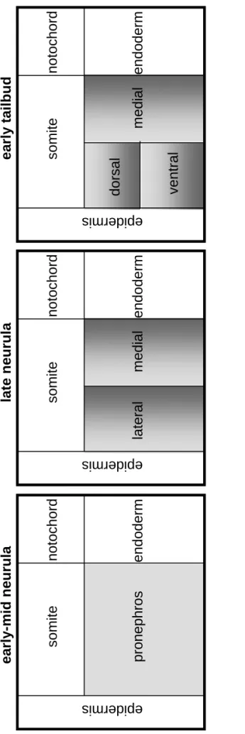
The induction of the pronephric anlagen, in which cells of the intermediate mesoderm are instructed to change their future development by another cell type, starts with a signal coming from the anterior somites (Seufert et al., 1999, Mauch et al., 2000, Mitchell and al., 2007). It has been shown that the anterior somites can induce pronephric tubules when cultured in combination with unspecified intermediate mesoderm. The molecular mechanism responsible for giving such inductive signals has not been yet identified. This constitutes the main research interest of this thesis. However, some genes have been shown to be crucially involved in the patterning of the pronephric mesoderm. Such genes are pax8 (Carroll and Vize, 1999), lhx1 (previously called lim1, Taira et al., 1994) and wt1 (Carroll and Vize 1996). Together pax8, lhx1 and wt1 give the switch to the specified intermediate mesoderm for mediolateral patterning. Figure 1.4 shows the patterning events that subdivide the Xenopus pronephric anlagen. wt1 is only expressed in the medial portion of the developing pronephric mesoderm at stage 18. Shortly after wt1
activation, genes that were initially expressed throughout the pronephric mesoderm, such as pax8 and lhx1, become restricted to the lateral pronephros. From this point, the original pronephric mesoderm is subdivided into the medial region that will form the glomus, the dorsolateral region that will give rise to the proximal tubules and the ventrolateral region that will establish the intermediate/distal tubules. The glomus and the tubules are specified at the same time, stage 12.5 but the glomus can develop after removal of the pronephric tubules primordial, confirming that these structures come from independent origins (Carroll and Vize 1996). Complementary to wt1, epidermal bmp4 constitutes a source for mediolateral patterning (Majumdar et al., 2000).
somite
pr
onephros
notochord endoderm
epidermis
somite
notochord endoderm
epidermis
lateral
m
edial
ea
rly
-mid neurula
late neurul
a
somite
notoc endoderm
epidermis
dorsal
m
e
dial
ventral
e
a
rly
ta
ilbud
Fi
g
u
re 1.4 Patternin
g
events that sub
d
ivi
d
e the
X
e
nopus
pronephric anl
a
gen.
By
lat
e
neur
u
la (
s
tage 20)
, t
he pr
onephr
ic
anlagen
get
s
ubdiv
ided
int
o
th
e lat
e
ral
and medi
al
domai
ns. By late
neurula
(stage
26), the lateral domain is once mor
e
divi
ded into dorsal and ventral domai
ns.
Figure adapted from
Vize
et al., 2003.
mesoderm, the presumptive notochord. Some markers of early dorsoventral patterning include the signalling molecule wnt4 and members of the notch family. At stage 18, wnt4 is the first gene to be activated, followed by delta1 at stage 19, notch1
at stage 20 and serrate1 at stage 22. It has been shown that over-expression of the
notch pathway blocks the intermediate/distal tubules formation, whereas blocking
notch signalling enhances it (McLaughlin et al., 2000). These results suggested that this system plays a role in defining pronephric compartment boundaries.
Once the pronephric mesoderm has been specified, it undergoes morphogenesis, ultimately resulting in a functional organ. The pronephric nephron forms by a process of rearrangement or segregation. The first sign of pronephric morphogenesis is a change in cell shape in the somatic intermediate mesoderm at early tailbud stage. Pronephric cells reorganize to form a compact structure just ventral to the somites. The cell mass that forms is then called the pronephric primordium or anlagen. At stage 25, once the pronephric anlage has formed, the intermediate/distal/ connective tubule that derives from a rudiment, begins its caudal migration which is completed at stage 37 when it fuses with the rostrally migrating rectal diverticulum (Nieuwkoop and Faber, 1994). Following segregation, the pronephric primordium begins to be shaped both dorsally and ventroposteriorly. On the dorsal side, the anlagen change from a round shape into a ‘τ’ shape. On the ventroposterior margin, the future distal tubule is pushed anteriorly so that it lies below the most anterior of the forming dorsal branches. Finally, the proximal and distal tubule grows and extends and the pronephric sinus becomed vascularised.
1.3.2.2 Pronephros anatomy.
The best stage to describe X. laevis pronephros anatomy is at stage 38, when the pronephric components have fully differentiated and are functional. The blood is filtered by the pronephric glomus or glomerulus, which is vascularised by capillaries branching from the dorsal aorta and which projects into the coelom (Fig 1.5, figure adapted from Reggiani et al., 2007). Fluids are then driven from the coelom into the pronephric tubules by the nephrostomes that are thin epithelial ciliated funnels. Nephrostomes have no resorptive or excretory activity (Fig 1.5, arrows pointing three nephrostomes). The nephrostomes are linked to the proximal tubules by short branches. The surface of the proximal tubules is covered with microvilli, called brush borders, which function to reabsorb molecules by enhancing surface area (Fig 1.5 PT1 and PT2). Fluids moving through the proximal tubules drain into a single common tubule that has a greater diameter (Fig 1.5 PT3). Useful molecules that have been reabsorbed by the proximal tubules from the glomerular filtrate, return to the blood circulation through a network of veins that surrounds the proximal tubules known as the pronephric sinus. The sinus receives blood from the glomus, the anterior/posterior cardinal veins, the branchial veins and the external jugular veins (Fig 1.5 red arrow). Urine and molecules that were not reabsorbed by the proximal tubules are moved through the intermediate/distal tubules, which forms an “S” shape (Fig 1.5 IT1, IT2, DT1 and DT2). The intermediate/distal tubules are constituted of two types of cells. Cells that stain with the 4A6 antibody which probably correspond to principal cells and those which do not stain with 4A6 which probably correspond to intercalating cells observed histologically. These latter cells are characterised by large number of mitochondria. Finally, urine is eliminated from the organism by the connecting tubule that connects with the cloaca (Fig 1.5 CT).
1.3.2.3 The pronephros function.
PT1
PT2
PT1
PT1
NS
NS
NS
gl
omus
coelom
The anatomy of the
Xenopus
pronephros. Figure adapted from Vi
ze et al., 1997,
Reggiani
2007 and Raciti
et al
., 2008.
Ke
y
:
Nephrost
o
mes,
NS.
Proxima
l tu
bule, PT1,
PT
2, PT3.
I
tubule,
IT1,
I
T2. Distal
tu
bule DT1, DT2. Connect
ing tubule,
CT.
Fi
g
u
re 1.5 T
h
e anato
m
y
of t
h
e
Xenopus
pro
n
ep
hro
s
at stage 38.
Bloo
d i
s
filtered b
then, useful mol
e
cul
e
s are reabsorb
ed by the pronephric
proximal
tubul
e
s and finally, the wastes are
driven outsi
de the organism by
the connecti
ng tubul
e.
need of minimizing ion loss implicate the pronephros in effective resorptive processes. Molecules that must be effectively recovered from the kidney filtrate are organic and inorganic ions, amino acids, bicarbonates, carbohydrates, lipids and water itself (Dantzler et al., 1988). A key player in active transport of small molecules and ions across the kidney epithelial membranes is the sodium potassium ATPase. This transporter uses the energy from hydrolysis of one molecule of ATP to extrude three cytoplasmic Na+ into the body fluids for two extracellular K+. These, in turn, power cotransporters that utilize the resulting electrochemical gradient to reabsorb the useful molecules from the kidney filtrate (Drummond et al., 1998).
The single pronephric nephron, organised into discrete functional segments, is composed of distinct renal epithelial cell types. Each cell type carries out specific functions to regulate fluid balance, osmolarity and metabolic waste excretion. The segments do not operate independently but rely on the correct spatial organisation along the nephron to insure normal excretory functions. The segmental organisation of the pronephric nephron also demonstrates regionalised expression of transporters and ion channel genes along the proximodistal axis (Eid, et al., 2002). For example,
scl5a11, slc5a2 and slc7a13 mark the proximal tubule whereas, clcnk, slc12a1 and
slc12a3 mark the intermediate/distal and connecting tubules (Reggiani et al., 2007 and Raciti et al., 2008).
1.4 Molecular markers of early kidney development, used in this thesis.
Figure 1.6 shows the three pronephric compartments and the key times of expression of some known molecular markers that have been used in this work. Figure 1.6 does not show the full expression pattern of these genes, it only represents the expression of pronephric genes during the key events of pronephric specification, morphogenesis or maturation. lhx1, pax8 and wt1 will be used to perform in situ
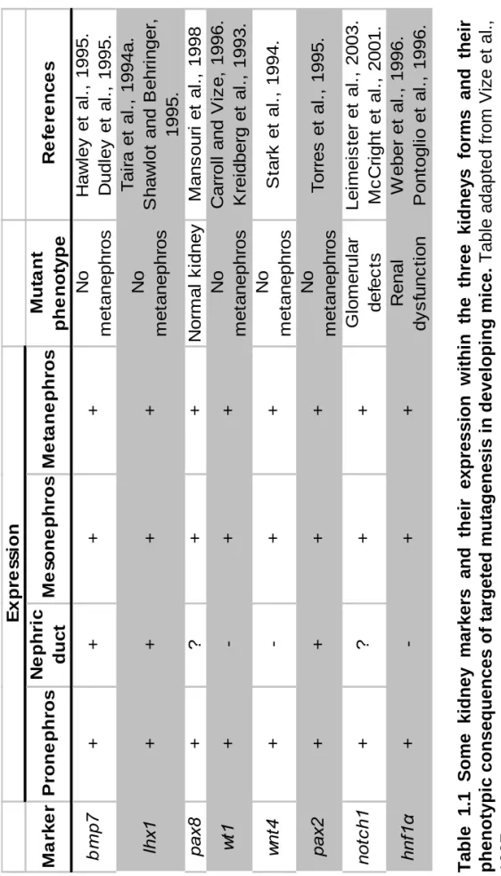
hybridization in whole Xenopus embryos as markers of specification. The antibodies 3G8 and 4A6, recognising specifically the pronephric proximal and intermediate/distal tubules respectively will be used to perform antibody staining in whole mount Xenopus embryos or in embryonic explants to identify these differentiated components. Finally, the remaining pronephric markers will be used in RT-PCR analysis to confirm the presence of pronephric tissue in embryonic explants and whole embryos. Where these molecules have been used in gene function studies in another organism, mouse, they are shown in table 1.1.
1.4.1 Markers of pronephros specification.
1.4.1.1 lhx1, the first marker of pronephros specification.
lhx1 encodes a DNA binding homeodomain and constitutes the earliest marker of nephrogenesis (Taira et al., 1994). lhx1 is activated in the pre-pronephric region immediately following gastrulation. It expression extends from the ventral border of the somites to the ventral mesoderm in a vertical strap-like shape. By stage 22, lhx1
Stage
12.5-14
18
30-38
Pronephric proximal tubules Pronephric inter
m edia te/ dis ta l tubule s Pronephric gl omus
lhx1 pax8 lhx1 pax8
pax2 hnf1 sall1 wt1 tcf21
notc h famil y irx 3 hnf1 4A6 clc grem1 c-ret
Specification of the pronephric primordi
a Morph o ge n e sis Function ? 3G8 slc5a1.1 Fi g u
re 1.6 Pro
n
ep
hric compartments and key
expre s s ion ti me of some molec u lar Dur ing t he pr onephros s p ec if icat ion, m o rphogenesis and func ti on, v a ri ou s se t of genes ar e t specifically marking
one of the three
pronephric compartment
s, proximal tubules,
intermediate/distal
tubules and the glomus.
wt1 tcf21 F igu re ada pted fro m R y 26
pax2 wnt4 pax2 wt1
M a rk e r P rone phr o s N e phr ic duc t M e s o ne phr os M e ta ne phr o s Mu ta n t phe not y pe Re fe re n c e s bm p 7 ++ + + No m e tanephr os H a w ley et al ., 1995. D udl ey et al ., 1995. lh x1 ++ + + No m e tanephr os T a ir a et al ., 1994a. S haw lo
t and B
e hr inger 1995. pax 8 + ? + + N o rm al k idney M ans our i et al ., 1998 wt 1 +-+ + No m e tanephr os C a rr ol
l and V
iz e , 1996. K rei dber g et al ., 1993. wn t4 +-+ + No m e tanephr os S tar k et al ., 1994. pax 2 ++ + + No m e tanephr os T o rr es et al ., 1995. not c h 1 +? + + G lom er ul ar def ec ts Lei m e is te r et al ., 2003. M c C ri ght et al ., 2001. hnf 1 α +-+ + R enal d ysf u n ct io n W eber et al ., 1996. P ont ogl io et al ., 1996. E x pr e s s ion Tab
le 1.1 Some kid
n
ey
markers and t
h eir expre ssion w ithi n the th ree kidney s f o rms and their p h en ot ypi c conseq u e
nces of targeted mutagenesis in develo
p
in
g
mice.
Table adapted from Vize
et al.,
1997.
the posterior portion. This again supports the idea that lhx1 is defining the growth zone of the forming intermediate/distal tubule (Carroll et al., 1999).
1.4.1.2 pax8, a second marker of pronephros specification.
pax8, belongs to the pax gene family of transcription factors playing fundamental roles during organogenesis. This family is characterised by the presence of a 128 amino acid paired domain encoding a unique DNA-binding motif. pax8 is expressed in the prospective otic placode and in the intermediate mesoderm, indicating that
pax8 plays a central role in auditory and excretory system development (Heller and Brändli, 1999).
At the time at which the pronephric anlagen is specified, stage 12.5, pax8 is actively expressed in a patch directly behind the head and ventral to the anterior somites (Carroll and Vize, 1999). It is believed that pax8 plays a role in the specification of the pronephric mesoderm and constitutes a direct response to the anterior somite-derived inductive signal. After neurulation, pax8 expression can be seen in both the pronephric proximal and intermediate/distal tubules. During pronephric kidney morphogenesis, pax8 is found in the proximal tubule and in the elongating distal tubule. By stage 31, expression in the distal tubule progressively ceases, while transcription in the differentiating proximal tubule remains at high levels until stage 36-37, when the pronephric kidney becomes functional (Heller and Brändli, 1999). To summarise, pax8 is one of the initial responses to specification of the pronephric primordia between stage 12.5 and 14. its expression responding to the somitic inducing signal.
1.4.1.3 wt1, a marker of medial patterning during pronephros specification.
The zinc-finger transcription factor encoded by the Wilm’s tumor suppresser gene
stage 20 onwards, wt1 is a marker of the pronephric glomus, but not after stage 35, where its expression is also found in the heart.
1.4.1.4 wnt4, a source of dorsoventral patterning.
At stage 18, just prior to the first morphological signs of pronephric differentiation,
wnt4 is weakly expressed throughout the pronephric mesoderm. As the embryo develops, wnt4 expression increases in the pronephric proximal tubules and proximal intermediate/distal tubule anlagen. During the tailbud stage, wnt4 expression is quite strong in the developing proximal tubule but starts declining in the developing intermediate/distal tubule. By the early tadpole stage, wnt4 ceases to be expressed in the pronephric intermediate/distal tubule and is restricted to the anterodorsal portion of the proximal tubule anlagen corresponding to the dorsal tips of the growing tubule (Carroll et al., 1999). The role of wnt4 in pronephros formation has been shown to be essential for tubulogenesis (Saulnier et al., 2002). This will be discussed in section 1.4.2.1.
1.4.1.5 pax2 is associated with the onset of the pronephros morphogenesis.
pax2, another member of the the pax gene family of transcription factors, is expressed at very low level in the pronephric mesoderm at stage 22. However, its expression strengthens progressively throughout the tailbud stage. By late tailbud stage, pax2 is strongly expressed in both the proximal and intermediate/distal tubule anlagen, suggesting a role in the maintenance of the pronephric fate (Heller and Brändli, 1997). As the tubules develop further, pax2 is relatively stronger in the nephrostomes of the proximal tubule and in cell migrating caudally from the rectal diverticulum to joint the distal tubule.
1.4.1.6 Functional synergism between lhx1, pax8 and pax2.
In Xenopus embryos, targeted over-expression of pax8 mRNA results in the development of large and ectopic pronephroi. Moreover, coinjection of pax8 and