University of Warwick institutional repository: http://go.warwick.ac.uk/wrap
This paper is made available online in accordance with
publisher policies. Please scroll down to view the document
itself. Please refer to the repository record for this item and our
policy information available from the repository home page for
further information.
To see the final version of this paper please visit the publisher’s website.
Access to the published version may require a subscription.
Author(s): Umakhanth Venkatraman Girija, Christopher Furze, Julia
Toth, Wilhelm J. Schwaeble, Daniel A. Mitchell, Anthony H. Keeble and
Russell Wallis
Article Title: Engineering Novel Complement Activity into a Pulmonary
Surfactant Protein
Year of publication: 2010
Link to published version:
http://dx.doi.org/10.1074/jbc.M109.097493
1
Engineering Complement Activity into a Pulmonary-surfactant
Protein
1Umakhanth Venkatraman Girija
*§, Christopher Furze
*§, Julia Toth
*, Wilhelm J.
Schwaeble
*, Daniel A. Mitchell, Anthony H. Keeble
*and Russell Wallis
*‡§
These authors contributed equally to the work
Running title: Engineering complement activity into SP-A
From the Departments of Infection, Immunity and Inflammation* and Biochemistry
‡,
University of Leicester, Leicester, United Kingdom
Address correspondence to:
Dr Russell Wallis RCUK Academic Fellow, Department of
Infection, Immunity and Inflammation, Maurice Shock Building, University of Leicester PO
Box 138, Leicester, LE1 9HN UK; Tel.: +44 (0)116 252 5089; Fax: +44 (0)116 252 5030;
E-mail:
[email protected]
Complement neutralises invading pathogens, stimulates inflammatory and adaptive immune responses and targets non- or altered-self structures for clearance. In the classical and lectin activation pathways it is initiated when complexes composed of separate recognition and activation subcomponents bind to a pathogen surface. Despite its apparent complexity, recognition-mediated activation has evolved independently in three separate protein families, C1q, mannose-binding lectins (MBLs) and serum ficolins. Although unrelated, all have bouquet-like architectures and associate with complement-specific serine proteases: MBLs and ficolins with MBL-associated serine protease-2 (MASP-2) and C1q with C1r and C1s. To examine the structural requirements for complement activation, we have created a number of novel MBLs in which the position and orientation of the MASP-binding sites have been changed. We have also engineered MASP-binding into a pulmonary-surfactant protein (SP-A), which has the same domain structure and architecture as MBL, but lacks any intrinsic complement activity. The data reveal that complement activity is remarkably tolerant to changes in the size and orientation of the collagenous stalks of MBL, implying considerable rotational and conformational flexibility in unbound MBL. Furthermore, novel complement activity is introduced concurrently with MASP binding in SP-A, but is uncontrolled and occurs even in the absence of a carbohydrate target. Thus, the active state is default and must be inhibited in circulating MBLs through additional regions
(not present in SP-A) until facilitated by pathogen recognition.
Complement is a central part of the immune system that neutralises pathogens via antibody-dependent and -independent mechanisms and stimulates a variety of protective responses including phagocytosis, inflammation and adaptive immunity (1). It is triggered via three routes called the classical, lectin and alternative pathways. Although the alternative pathway is initially non-specific and depends on host regulatory proteins to avert activation on self structures, the classical and lectin pathways both selectively target pathogen-associated molecular patterns via circulating complexes composed of recognition and zymogen-protease subcomponents (2). Upon binding to a target, conformational changes in the recognition subcomponent trigger activation of the protease subcomponent, which in turn activates the downstream complement cascade. In the classical pathway, C1q binds to microorganisms, immune complexes, apoptotic and necrotic cells as well as amyloids to initiate the stepwise activationof C1r and C1s. In the lectin pathway, mannose-binding lectin (MBL) and serum ficolins bind to terminal mannose-like epitopes or N-acetyl groups on pathogens to activate MBL-associated serine protease-2 (MASP-2).
2 During biosynthesis, polypeptides associate into
trimeric subunits (4), which assemble to form larger oligomers resembling bouquets (5, 6). In C1q, the N-terminal portions of the collagenous stalks associate with each other and splay apart at a short interruption within the repeating Gly-Xaa-Yaa sequence, called the kink. Although MBL and some ficolins also possesses a kink-like region, recent measurements have revealed that the stalks probably diverge nearer the N-terminus, at the junction with the cysteine-containing domain, and thus form spider-like structures rather than the classical bouquets of C1q (7). Recognition subcomponents possess different numbers of subunits. For example, human C1q is a hexamer assembled from three different polypeptide chains (8) and rat MBLs are homooligomers comprising dimers, trimers and tetramers of subunits (9).
The protease subcomponents, C1r and C1s of the classical pathway and MASP-2 of the lectin pathway are homologous, and bind to the collagenous domains of their respective recognition subcomponent through Ca2+ -dependent interactions (10-13). They each comprise two Ca2+-binding CUB domains (domain found in complement component C1r/C1s, Uegf, and bone morphogenic protein 1), separated by a Ca2+-binding epidermal growth factor-like domain (EGF), followed by two complement-control protein (CCP) modules and a serine protease (SP) domain (14, 15). C1r and C1s form heterotetramers (C1s-C1r-C1r-C1s) and MASPs are homodimers. Although the stoichiometries of the resulting C1 and MBL-MASP complexes differ, interactions between subcomponents are analogous, with equivalent contacts between the CUB domains of the proteases and the collagenous domains of the recognition subcomponents (2). Overall, each C1rs tetramer presents a total of six binding sites, one for each of the collagenous domains of C1q (one site on each CUB1 of C1r and C1s and each CUB2 of C1r). Each MASP-2 dimer presents four binding sites to MBL and ficolins (one site on each CUB domain).
A number of other proteins have bouquet-like architectures, such as adiponectin (16), emilins (17) and some collectins (18), including pulmonary surfactant protein-A (SP-A). Nevertheless, only MBLs, ficolins and C1qs are able to activate complement. As yet, the only established difference between these proteins and their structural analogues is their ability to bind
to MASPs or C1rs tetramers. The MASP-binding site in MBLs and ficolins is characterised by a distinct motif within the collagenous domain: Hyp-Gly-Lys-Xaa-Gly-Pro, where Hyp is hydroxyproline and Xaa is generally an aliphatic residue. Point mutations to this sequence disrupt MASP binding (10, 13). C1q also possesses a similar motif and we have proposed that this region forms the binding sites for C1r and C1s (2).
In this manuscript, we have further probed the functions of the recognition subcomponents by examining the structural requirements for complement activation. Initially, we modified the collagenous domain of MBL to change the position and relative orientations of the MASP-binding site on the collagenous stalks. The data reveal that MASP binding and activation are surprisingly tolerant to such changes, implying considerable rotational and conformational flexibility. To take these studies further, we have engineered the MASP-binding motif into SP-A, which also has a C-type lectin domain and a similar architecture to MBL, but cannot bind MASPs or activate complement. We find that MASP binding comparable to MBL can be engineered through just three amino acid substitutions to the collagenous domain of SP-A. The resulting proteins also activate the MASP, but lack the control mechanisms necessary to target carbohydrate surfaces selectively.
EXPERIMENTAL PROCEDURES
Protein Components Recombinant rat MBL and modified forms of rat MASP-2 were produced in a Chinese hamster ovary cell-expression system and purified as described previously by affinity chromatography on mannose-Sepharose and nickel-Sepharose columns, respectively (9, 19). MASP-2A is a catalytically inactive form of rat MASP-2 in which the active site serine at position 613 has been replaced by an alanine. MASP-2K is a
catalytically active form, in which the arginine residue at the cleavage site for zymogen activation (Arg424) has been changed to a lysine residue to slow down the rate of spontaneous autoactivation during biosynthesis. Mutant forms of MBL were created by PCR and expressed in the same way as the wild-type protein.
SP-3 A isoform A1 was cloned into the polylinker of
the eukaryotic expression vector pED-4 (20), which contains the dihydrofolate reductase cDNA as a selectable marker. Proteins were produced by expression in the Chinese hamster ovary cell line DXB11 following amplification using the dihydrofolate reductase inhibitor, methotrexate, to a final concentration of 25 M. Culture media was harvested using a protocol that has been described previously for production of recombinant MBL (9, 21). Protein was purified by affinity chromatography on mannose-Sepharose columns (1 ml column for 250 mls of media). Following washes with high salt (50 mM Tris pH 7.5, containing 150 mM NaCl and 2.5 mM CaCl2) and low salt buffers (10 mM Tris
pH 7.5, containing 10 mM NaCl and 2.5 mM CaCl2) protein was eluted in 0.5 ml fractions of
10 mM Tris pH 7.5, containing 10 mM NaCl and 2.5 mM EDTA. Fractions containing recombinant protein were identified by SDS-polyacrylamide gel electrophoresis. Mutations were introduced into to the cDNA of SP-A by PCR and the encoded proteins were produced in the same way as the wild type protein. Although SP-A tolerated relatively high salt concentrations when bound to the Sepharose matrix, concentrations >10 mM caused significant aggregation in solution.
Surface plasmon resonance Measurements were performed using a BIAcore 2000 instrument (GE Healthcare) or a BioRad ProteOn XPR36 biosensor. Protein ligands were diluted into 10 mM sodium acetate (pH 4.5 for MBL or pH 5.0 for SP-A), and immobilized on to the carboxymethylated dextran surface of a CM5 sensor chip (GE Healthcare) or a GLM chip (BioRad), using amine coupling chemistry. Binding was measuredin 10 mM Tris-HCl (pH 7.4) containing 140 mM NaCl, 2 mM CaCl2 and
0.005% surfactant SP40, at a flow rate of 25 µl/min and at 25°C. After injectionof ligand, the protein surface was regenerated by injection of 10 µl of 10 mM Tris-HCl buffer (pH 7.4) containing1 M NaCl and 5 mM EDTA. Data were analyzed by fitting association and dissociation curves to Langmuir binding models for several protein concentrations simultaneously, using BIAevaluation 4.1 software (GE Healthcare). Increasingly complex models were testeduntil a satisfactory fit to the data was achieved. Apparent equilibrium
dissociation constants (KD) were calculated from the ratio of the dissociation and association rate constants(koff/kon). MBL was immobilized on the
chip surface rather than used as a soluble ligand, because it bound to the chip thereby masking analysis of the protein-protein interactions. SP-A was also immobilized because it tended to precipitate on the chip surface.
MASP-2 activation assays Activation was measured by following MASP autolysis using a modified version of the protocol described previously (22). Briefly, MASP-2K was mixed with wild-type or mutant MBL or SP-A and added to a suspension of fucose-Sepharose (5 µlof a 1:1 v/v suspension in a total volume of 30 µl), in50 mM Tris-HCl (pH 7.5) containing 150 mM NaCl and 5 mM CaCl2,at
37°C with mixing. A 1.2-fold molar excess of MBL or SP-A was used to ensure that all the MASP-2K was bound to the recognition molecule. The mixture was incubated at 37 °C with shaking and aliquots of the suspension were removed from the reactionmix at varioustimes and immediately frozen on dry ice to quench the reaction. Proteins were separated by SDS-polyacrylamide gel electrophoresis under reducing conditions and the amount of MASP cleaved was quantified by densitometry. Data are the mean ± S.E. from at least two separate experiments using different protein preparations, unless otherwise stated.
RESULTS
Gly-Xaa-4 Yaa triplet would change both the position of
each binding site and its relative orientation. For example, deletion of a triplet would effectively rotate each collagenous stalk by ~180° as well as shift the position of the binding site further towards the N-terminus (Fig. 1). Deletion of a second triplet would restore the original alignment but move the binding sites even further towards the N-terminus. To accommodate all of these changes, the collagenous stalks would not only have to splay further apart (for a deletion) or move closer together (for an insertion), but also rotate independently of each other. Thus, analysis of MASP binding and activation by the modified MBLs provides a useful probe of the conformational and rotational flexibility of MBL. Two deletion mutants were created in which either one or two Gly-Xaa-Yaa triplets were removed from the collagenous domain, thus moving the MASP-binding motif closer to the N-terminus of MBL (Fig. 1). In addition, a single Gly-Pro-Hyp was inserted near the kink-like region to move the MASP binding motif further towards the C-terminus. We also made an MBL in which the kink-like region itself was removed by insertion of a proline residue to restore the Gly-Xaa-Yaa tandem repeats. A previous study has demonstrated that the kink-like region is not essential for complement activation by MBL (24). However, the kinetics of activation were not investigated in this work, so more subtle changes might have been missed. Analysis of the purified, recombinant MBLs by SDS-polyacrylamide gel electrophoresis under non-reducing conditions, shown previously to be a sensitive indicator of MBL assembly (25), demonstrated that all assembled correctly during biosynthesis (Fig. 2).
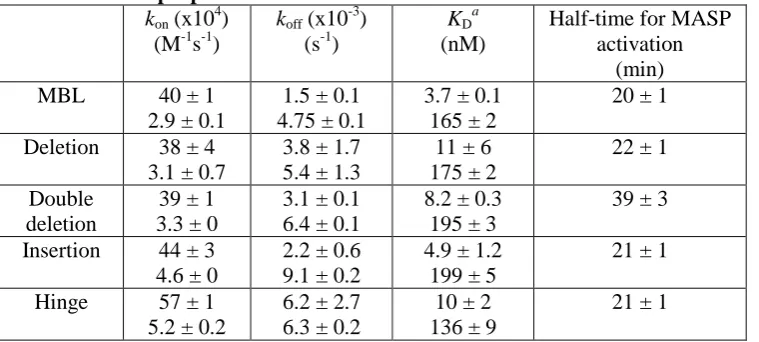
Initially MBLs were tested for their abilities to bind to MASP-2A, using surface plasmon resonance. As expected, wild-type bound with complex kinetics compatible with the formation of 1:1 complexes, together with binding of a second MASP at relatively high protein concentrations (19). The KDs were 3.7
and 164.5 nM consistent with previous measurements (2). Surprisingly, the entire set of mutant MBLs bound to MASP-2A with similar affinities and kinetics (Table I), indicating that none of the changes perturbed MASP binding (Fig. 2). We next examined their complement activities by monitoring MBL-dependent MASP-2K autoactivation with mannose-Sepharose as an
activating target (Fig. 3). In the presence of the target, all mutant MBLs activated the MASP with comparable rates as wild-type (t1/2 ~20
mins). The greatest difference observed was for the double-deletion mutant, which activated the MASP just two-fold more slowly (t1/2 ~40 mins),
despite removal of six amino acid residues from the collagenous domain. In the absence of the target, activation was relatively slow and similar to autoactivation by MASP-2 alone (>1000 min), indicating that all MBLs only activate on a carbohydrate target. Thus, neither insertions, deletions nor removal of the kink-like region disrupt MASP-2 binding or activation, despite large changes to the position and relative orientations of the MASP-binding sites. The tolerance of MBL to these changes must reflect significant adjustment and realignment of the collagenous stalks. Given that collagenous domains themselves are relatively rigid and inflexible, changes must occur at the N-terminus where the stalks converge, probably at the junction between the cysteine-rich domain and the collagenous domain.
5 collagenous domain, thus becoming more similar
to MBL and ficolins (28).
Three new recombinant constructs were created in which all or part of the MASP-binding motif was introduced into the collagenous domain of SP-A (Fig. 4). Changes were made five Gly-Xaa-Yaa triplets towards the C-terminal side of the kink, to ensure that there would be enough space for the MASP to bind within the SP-A bouquet. In SPA-KL, Glu63 and Cys64 were replaced by lysine and leucine residues, respectively. Lysine in the Xaa position is known to be essential for MASP binding and complement activation in both MBLs and ficolins. The adjacent cysteine residue was also replaced to avoid any potential disulfide bond formation which might interfere with MASP binding. In SPA-KLP a proline residue was introduced in place of the glutamate at position 66 to further optimize the MASP-binding motif: Hyp-Gly-Lys-Leu-Gly-Pro. One further change was made in SPA-KLPO, replacing Lys67 by a hydroxyproline residue. Lysine residues in the Yaa position of collagen are often hydroxylated and glycosylated during biosynthesis, so this change was designed to prevent any potential steric inhibition of MASP binding by the sugar residues (23). We created two further variants of SPA-KLPO, in which the kink was removed or modified to change the point at which the stalks splay apart (28), and thus more resemble MBL and ficolins. In one mutant, SPA-KLPOdel the kink sequence (PCPP) was removed completely, and in the other, called SPA-KLPOS, the cysteine residue was replaced by a serine residue to remove the potential for disulfide bond formation, which is believed to tether the separate stalks together in native SP-A (28).
Following production in chinese hamster ovary cells, all proteins bound to mannose- or fucose-Sepharose columns, demonstrating that they were correctly folded. As expected, wild-type SP-A did not bind to MASP-2A at any of the concentrations tested (up to 1 M). Surprisingly however, SPA-KL bound with appreciable affinity (Fig. 5). The data best fitted a two-complex, parallel-reaction model, with apparent dissociation constants KD1 and KD2 of
600 and 2120 nM respectively, compared to 3.8 and 166 nM for MBL-MASP (Table 2). SPA-KLP bound with even higher affinity, with KDs
of 36 and 104 nM, only slightly weaker than MBL. Additional changes had little effect on
MASP binding, so the naturally occurring lysine residue in SP-A (or its potential glycosylated derivative) does not prevent MASP access. Removal of the kink region also had little effect, indicating that the point at which the stalks splay apart and thus the angle between adjacent stalks is not limiting for MASP binding. Thus, just three amino acid changes to the collagenous domain are sufficient to introduce MASP binding to SP-A almost comparable to MBL itself.
We next examined if the modified SP-As could activate MASP-2K. The half-time for activation by MBL was ~50 mins, using fucose-Sepharose as a target. As expected, wild type SP-A, which does not bind to the MASP at all and SPA-KL, which binds only weakly, did not activate the MASP (Fig. 6). In each case the measured rate was similar to the intrinsic autoactivation rate of MASP-2K alone (t1/2
>1000 min). Surprisingly however, SPA-KLP and SPA-KLPO activated the MASP significantly faster than the basal rate (t1/2 ~260
min). Similar rates were also measured for SPA-KLPOdel and SPA-KLPOS, demonstrating that the kink is neither necessary for MASP activation, nor modulates the rate of activation significantly. To determine whether activation was target dependent, assays were repeated in the absence of fucose-Sepharose. Surprisingly, comparable rates were observed (Fig. 7). Activation was still much faster than the basal rate, so must be driven by SP-A binding, but was constitutive occurring even in the absence of a carbohydrate target. Thus, modified SP-As lack the control mechanisms required to activate MASP selectively.
DISCUSSION
6 the next Gly-Xaa-Yaa triplet is also not essential
for MASP binding and can be replaced by an alanine with only a small decrease in affinity. The main function of the proline is probably to help stabilize the binding region (10). It is notable that a significant component of the difference in MASP binding by SPA-KLP relative to SPA-KL is due to faster association, so probably reflects removal of the unfavourable electrostatic interactions mediated by the glutamate, rather than the introduction of the proline residue itself. The presence of any additional binding sites for MASP on MBL can be completely ruled out from the data presented here. Overall therefore, specificity of the MBL-MASP interaction appears to be driven mainly by a single lysine residue in the collagen-like domain.
Several other binding motifs have been identified in collagens and typically extend across two or more Gly-Xaa-Yaa triplets. For example, the I domain of α2 integrin, recognizes the sequence GFOGER (29) and the extracellular matrix protein SPARC binds to the sequence GVMGFO (30). Thus, the less stringent binding requirements of MASP-2 are relatively unusual. Nevertheless, it is notable that almost all of the other lysine residues in the collagenous domains of MBL and ficolins are in the Yaa position of the Gly-Xaa-Yaa repeat, so are likely to be post-translationally modified by hydroxylation and glycosylation, which probably blocks any potentially incorrect MASP interactions. Perhaps even more importantly, MASP binds to MBL and ficolins through multiple relatively weak interactions involving up to four separate CUB-collagen contacts. Thus, much of the binding specificity is probably mediated through the architectures of the subcomponents and the multivalent nature of the interactions.
Complement activation is thought to be triggered by changes in the angle between collagenous stalks when complexes bind to an activating target (3, 31). The data presented here indicate that significant conformational and rotational flexibility of the stalks must be permitted, implying that unbound MBL is probably quite flexible. Both relaxed-to-strained and strained-to-relaxed mechanisms could explain the activation process. The traditional view is that complexes circulate in an inactive zymogen form and distort upon binding to a target, and the changes to the structure of MBL are transmitted to the MASP to drive
autoactivation. However, in an alternative but equally valid possibility, binding of the zymogen MASP to MBL (or ficolin) induces strain into circulating complexes, which is released on pathogen binding to trigger activation. A major difference between these mechanisms is that the relaxed or default states of the complexes would differ: inactive in a relaxed-to-strained mechanism, but active in a strained-to-relaxed mechanism. In the work described here, MASP-binding leads to constitutive activation by SP-A, even in the absence of a target. Thus, activation is the default state, as would be expected in a strained-to-relaxed mechanism. The possibility that modified SP-As are locked into a high energy conformation that activates MASP constitutively cannot be completely excluded. However, SP-A must also be flexible to bind to the MASP multivalently, which would seem to be at odds with this possibility. Furthermore, large structural changes (through removal of the kink) do not affect MASP activation by SP-A, which also seems incompatible with such a mechanism.
Based on these data, we propose that MBL-MASPs also activate via a strained-to-relaxed mechanism. We have recently suggested that activation of the C1 complex also operates in this way (2) and have proposed that strain is induced into the zymogen complex through the interactions between the protease domains of C1r. In this respect, the mechanism must differ in MBL-MASP complexes because the SP domains of a MASP dimer do not interact significantly until the moment of autocatalysis. Interestingly however, recent analysis suggests that the MBL stalks are not evenly distributed, but rather are highly asymmetrical and separated by ~35–40° between nearest neighbours (32). In this case, significant strain would be induced when MBL binds to a MASP. Release of this strain upon target recognition might drive the changes that initiate complement activation.
7 we are currently undertaking studies to identify these regions in MBL and ficolins.
REFERENCES
1. Porter, R. R., and Reid, K. B. M. (1978) The biochemistry of complement, Nature275, 699-704.
2. Phillips, A. E., Toth, J., Dodds, A. W., Girija, U. V., Furze, C. M., Pala, E., Sim, R. B., Reid, K. B., Schwaeble, W. J., Schmid, R., Keeble, A. H., and Wallis, R. (2009) Analogous interactions in initiating complexes of the classical and lectin pathways of complement, J Immunol182, 7708-7717.
3. Wallis, R., Mitchell, D. A., Schmid, R., Schwaeble, W. J., and Keeble, A. H. Paths reunited: Initiation of the classical and lectin pathways of complement activation, Immunobiology215, 1-11.
4. Heise, C. T., Nicholls, J. R., Leamy, C. E., and Wallis, R. (2000) Impaired secretion of rat mannose-binding protein resulting from mutations in the collagen-like domain, J Immunol 165, 1403-1409.
5. Brodsky-Doyle, B., Leonard, K. R., and Reid, K. B. M. (1976) Circular-dichroism and electron-microscopy studies of human subcomponent C1q before and after limited proteolysis by pepsin, Biochemical Journal159, 279-286.
6. Strang, C. J., Siegel, R. C., Phillips, M. L., Poon, P. H., and Schumaker, V. N. (1982) Ultrastructure of the first component of human complement: electron microscopy of the crosslinked complex, Proc Natl Acad Sci U S A79, 586-590.
7. Jensenius, H., Klein, D. C., van Hecke, M., Oosterkamp, T. H., Schmidt, T., and Jensenius, J. C. (2009) Mannan-binding lectin: structure, oligomerization, and flexibility studied by atomic force microscopy, J Mol Biol391, 246-259.
8. Reid, K. B., and Porter, R. R. (1976) Subunit composition and structure of subcomponent C1q of the first component of human complement, Biochem J155, 19-23.
9. Wallis, R., and Drickamer, K. (1999) Molecular determinants of oligomer formation and complement fixation in mannose-binding proteins, J Biol Chem274, 3580-3589.
10. Girija, U. V., Dodds, A. W., Roscher, S., Reid, K. B., and Wallis, R. (2007) Localization and Characterization of the Mannose-Binding Lectin (MBL)-Associated-Serine Protease-2 Binding Site in Rat Ficolin-A: Equivalent Binding Sites within the Collagenous Domains of MBLs and Ficolins, J Immunol179, 455-462.
11. Reid, K. B., Sim, R. B., and Faiers, A. P. (1977) Inhibition of the reconstitution of the haemolytic activity of the first component of human complement by a pepsin-derived fragment of subcomponent C1q, Biochem J161, 239-245.
12. Teillet, F., Lacroix, M., Thiel, S., Weilguny, D., Agger, T., Arlaud, G. J., and Thielens, N. M. (2007) Identification of the site of human mannan-binding lectin involved in the interaction with its partner serine proteases: the essential role of Lys55, J Immunol178, 5710-5716. 13. Wallis, R., Shaw, J. M., Uitdehaag, J., Chen, C. B., Torgersen, D., and Drickamer, K. (2004)
Localization of the serine protease-binding sites in the collagen-like domain of mannose-binding protein: indirect effects of naturally occurring mutations on protease mannose-binding and activation, J Biol Chem279, 14065-14073.
14. Feinberg, H., Uitdehaag, J. C., Davies, J. M., Wallis, R., Drickamer, K., and Weis, W. I. (2003) Crystal structure of the CUB1-EGF-CUB2 region of mannose-binding protein associated serine protease-2, Embo J22, 2348-2359.
15. Sim, R. B., and Tsiftsoglou, S. A. (2004) Proteases of the complement system, Biochem Soc Trans32, 21-27.
16. Scherer, P. E., Williams, S., Fogliano, M., Baldini, G., and Lodish, H. F. (1995) A novel serum protein similar to C1q, produced exclusively in adipocytes, J Biol Chem 270, 26746-26749.
8 member of the C1q/tumor necrosis factor superfamily of proteins, J Biol Chem 274, 16773-16781.
18. Reid, K. B. M., Colomb, M. G., and Loos, M. (1998) Complement component C1 and the collectins: parallels between routes of acquired and innate immunity, Immunology Today 12, 56-59.
19. Chen, C. B., and Wallis, R. (2001) Stoichiometry of complexes between mannose-binding protein and its associated serine proteases. Defining functional units for complement activation, J Biol Chem276, 25894-25902.
20. Kaufman, R. J., Davies, M. V., Wasley, L. C., and Michnick, D. (1991) Improved vectors for stable expression of foriegn genes in mammalian cells by use of the untranslated leader sequence from EMC virus, Nucleic Acids Research19, 4485-4490.
21. Wallis, R., and Drickamer, K. (1997) Asymmetry adjacent to the collagen-like domain in rat liver mannose-binding protein, Biochem J325 ( Pt 2), 391-400.
22. Chen, C. B., and Wallis, R. (2004) Two mechanisms for mannose-binding protein modulation of the activity of its associated serine proteases, J Biol Chem279, 26058-26065.
23. Shoulders, M. D., and Raines, R. T. (2009) Collagen structure and stability, Annu Rev Biochem78, 929-958.
24. Kurata, H., Cheng, H. M., Kozutsumi, Y., Yokota, Y., and Kawasaki, T. (1993) Role of the collagen-like domain of the human serum mannan-binding protein in the activation of complement and the secretion of this lectin, Biochem Biophys Res Commun191, 1204-1210. 25. Wallis, R., and Cheng, J. Y. (1999) Molecular defects in variant forms of mannose-binding
protein associated with immunodeficiency, J Immunol163, 4953-4959.
26. Hoppe, H.-J., and Reid, K. B. M. (1994) Collectins - soluable proteins containing collagenous regions and lectin domains - and their roles in innate immunity, Protein Science 3, 1143-1158.
27. Haurum, J. S., Thiel, S., Haagsman, H. P., Laursen, S. B., Larsen, B., and Jensenius, J. C. (1993) Studies on the carbohydrate-binding characteristics of human pulmonary surfactant-associated protein A and comparison with two other collectins: mannan-binding protein and conglutinin, Biochem J293 ( Pt 3), 873-878.
28. Uemura, T., Sano, H., Katoh, T., Nishitani, C., Mitsuzawa, H., Shimizu, T., and Kuroki, Y. (2006) Surfactant protein A without the interruption of Gly-X-Y repeats loses a kink of oligomeric structure and exhibits impaired phospholipid liposome aggregation ability, Biochemistry45, 14543-14551.
29. Emsley, J., Knight, C. G., Farndale, R. W., Barnes, M. J., and Liddington, R. C. (2000) Structural basis of collagen recognition by integrin alpha2beta1, Cell101, 47-56.
30. Hohenester, E., Sasaki, T., Giudici, C., Farndale, R. W., and Bachinger, H. P. (2008) Structural basis of sequence-specific collagen recognition by SPARC, Proc Natl Acad Sci U S A105, 18273-18277.
31. Wallis, R. (2007) Interactions between mannose-binding lectin and MASPs during complement activation by the lectin pathway, Immunobiology212, 289-299.
32. Jensenius, H., Klein, D. C., van Hecke, M., Oosterkamp, T. H., Schmidt, T., and Jensenius, J. C. (2009) Mannan-Binding Lectin: Structure, Oligomerization, and Flexibility Studied by Atomic Force Microscopy, J Mol Biol.
Acknowledgements
We thank Robert Freedman and Katrine Wallis, of the Department of Biological Sciences, University of Warwick for use of the Departmental Biacore facility.
Footnotes
1
9
2
[image:10.595.67.447.133.304.2]Abbreviations used in this paper: MBL, mannan-binding lectin; CRD, carbohydrate-binding domain; MASP, MBL-associated serine protease; CUB, domain found in complement component C1r/C1s, Uegf, and bone morphogenic protein 1; EGF, epidermal growth factor
Table I Kinetic properties of modified MBLs
kon (x10 4
) (M-1s-1)
koff (x10 -3
) (s-1)
KD
a
(nM)
Half-time for MASP activation
(min)
MBL 40 ± 1
2.9 ± 0.1
1.5 ± 0.1 4.75 ± 0.1
3.7 ± 0.1 165 ± 2
20 ± 1
Deletion 38 ± 4 3.1 ± 0.7
3.8 ± 1.7 5.4 ± 1.3
11 ± 6 175 ± 2
22 ± 1
Double deletion
39 ± 1 3.3 ± 0
3.1 ± 0.1 6.4 ± 0.1
8.2 ± 0.3 195 ± 3
39 ± 3
Insertion 44 ± 3 4.6 ± 0
2.2 ± 0.6 9.1 ± 0.2
4.9 ± 1.2 199 ± 5
21 ± 1
Hinge 57 ± 1 5.2 ± 0.2
6.2 ± 2.7 6.3 ± 0.2
10 ± 2 136 ± 9
21 ± 1
a
KD values were calculated from koff/kon for each experiment and were averaged from two separate
[image:10.595.70.452.345.538.2]experiments
Table II Kinetic properties of engineered SP-As
kon (x10 -4
) (M-1s-1)
koff (x10 3
) (s-1)
KD
a
(nM)
Half-time for MASP activationc
(min)
SP-A N.Bb N.Bb N.Bb >1000d
SPA-KL 1.2 ± 0.2 4.3 ± 0.1
7.0 ± 0.1 91 ± 2
600 ± 110 2120 ± 100
>1000d
SPA-KLP 5.4 ± 2.9 36 ± 24
1.4 ± 0.1 17.6 ± 1.0
36 ± 20 104 ± 77
270 ± 56
SPA-KLPO
2.6 ± 0.8 9.6 ± 1.0
2.5 ± 0.6 22 ± 3.1
97 ± 7 232 ± 9
256 ± 96
SPA-KLPOdel
5.3 ± 2.3 18 ± 0.0
1.7 ± 0.1 21 ± 1
29 ± 17 114 ± 2
316 ± 171
SPA-KLPOS
4.5 ± 2.6 21 ± 2
4.6 ± 1.2 37 ± 15
99 ± 18 203 ± 31
166 ± 43
a
KD values were calculated from koff/kon for each experiment and were averaged from two separate
experiments
b
No binding detected
c
data from three separate experiments
d
activation occurred a rate similar to autoactivation of MASP-2 alone
Figure Legends
10 subunits as a result of a single insertion or deletion of a Gly-Xaa-Yaa into the collagenous domain based upon a pitch of 3.5 for a collagen helix. The figure shows a cross-section through the four collagenous stalks, which are represented by circles. Each binding site is shaded in light grey. The structure of the CUB1-EGF-CUB2 domains of MASP-2 is from (14). D, Schematic representation of the effects of changes to the position of the MASP-binding sites on MBL. Only two MBL stalks are shown for clarity. The MASP is shown as a box and is shaded in light grey. CRDs are represented as grey circles. The arrow shows the position of the insertion/deletion. To accommodate the MASP, the collagenous stalks would have to move closer together for an insertion or splay further apart for a deletion.
FIGURE 2. MASP-binding by modified MBLs. A, SDS polyacrylamide gel electrophoresis of wild-type and mutant MBLs under reducing (left) and non-reducing conditions (right). Mutant MBLs are assembled from single polypeptides chains, which migrate as a ladder of covalently-linked polypeptides under non-reducing conditions, characteristic of the heterogeneous nature of oligomers of wild-type MBL (9). B, Binding of MASP-2 to immobilised MBLs by surface plasmon resonance. MASP-2 was injected at 333, 167, 83, 42 nM over immobilized MBL (~7000 response units). All data were fitted to a two-complex parallel binding model and the fits are shown by the dotted lines.
FIGURE 3. MASP activation by modified MBLs. Kinetics of MBL-MASP-2 activation analyzed by SDS polyacrylamide gel electrophoresis. A and B, wild-type MBL and the deletion mutant were incubated with MASP-2K together with mannose-Sepharose as an activating target. Proteins were separated on a 4–12% linear gradient gel under reducing conditions and were stained with Coomassie blue. The N-terminal fragment of MASP-2K runs as a double band due to differential glycosylation. MASP activation was measured by quantifying cleavage of the MASP polypeptide. B, Comparison of MASP-2K activation by wild-type and mutant MBLs.
FIGURE 4. Design of modified SP-As. Sequences of the collagenous domains of MBL (top) and SP-A (bottom) Sequences were crudely aligned based on the positions of the kink of SP-A and the kink-like region of MBL (boxed). The MASP-binding motif is shaded light grey. Changes to the sequence of SP-A are indicated.
FIGURE 5. MASP-binding by modified SP-As. Binding of MASP-2 to immobilised SP-As by surface plasmon resonance. MASP-2 was injected at 764, 447, 261, 152 and 89 nM over each of the immobilized SP-As (~6000 response units). All data were fitted to a two-complex parallel binding model and the fits are shown by the dotted lines.