University of Warwick institutional repository: http://go.warwick.ac.uk/wrap
A Thesis Submitted for the Degree of PhD at the University of Warwick
http://go.warwick.ac.uk/wrap/50223
This thesis is made available online and is protected by original copyright.
Please scroll down to view the document itself.
JHG 05/2011
Library Declaration and Deposit Agreement
1. STUDENT DETAILS
Please complete the following:
Full name: ………. University ID number: ………
2. THESIS DEPOSIT
2.1 I understand that under my registration at the University, I am required to deposit my thesis with the University in BOTH hard copy and in digital format. The digital version should normally be saved as a single pdf file.
2.2 The hard copy will be housed in the University Library. The digital version will be deposited in the University’s Institutional Repository (WRAP). Unless otherwise indicated (see 2.3 below) this will be made openly accessible on the Internet and will be supplied to the British Library to be made available online via its Electronic Theses Online Service (EThOS) service.
[At present, theses submitted for a Master’s degree by Research (MA, MSc, LLM, MS or MMedSci) are not being deposited in WRAP and not being made available via EthOS. This may change in future.]
2.3 In exceptional circumstances, the Chair of the Board of Graduate Studies may grant permission for an embargo to be placed on public access to the hard copy thesis for a limited period. It is also possible to apply separately for an embargo on the digital version. (Further information is available in the Guide to Examinations for Higher Degrees by Research.)
2.4 If you are depositing a thesis for a Master’s degree by Research, please complete section (a) below.
For all other research degrees, please complete both sections (a) and (b) below:
(a) Hard Copy
I hereby deposit a hard copy of my thesis in the University Library to be made publicly available to readers (please delete as appropriate) EITHER immediately OR after an embargo period of ………... months/years as agreed by the Chair of the Board of Graduate Studies.
I agree that my thesis may be photocopied. YES / NO (Please delete as appropriate) (b) Digital Copy
I hereby deposit a digital copy of my thesis to be held in WRAP and made available via EThOS.
Please choose one of the following options:
EITHER My thesis can be made publicly available online. YES / NO (Please delete as appropriate) OR My thesis can be made publicly available only after…..[date] (Please give date)
YES / NO (Please delete as appropriate) OR My full thesis cannot be made publicly available online but I am submitting a separately identified additional, abridged version that can be made available online.
YES / NO (Please delete as appropriate) OR My thesis cannot be made publicly available online. YES / NO (Please delete as appropriate)
JHG 05/2011
3. GRANTING OF NON-EXCLUSIVE RIGHTS
Whether I deposit my Work personally or through an assistant or other agent, I agree to the following:
Rights granted to the University of Warwick and the British Library and the user of the thesis through this agreement are non-exclusive. I retain all rights in the thesis in its present version or future versions. I agree that the institutional repository administrators and the British Library or their agents may, without changing content, digitise and migrate the thesis to any medium or format for the purpose of future preservation and accessibility.
4. DECLARATIONS
(a) I DECLARE THAT:
I am the author and owner of the copyright in the thesis and/or I have the authority of the authors and owners of the copyright in the thesis to make this agreement. Reproduction of any part of this thesis for teaching or in academic or other forms of publication is subject to the normal limitations on the use of copyrighted materials and to the proper and full acknowledgement of its source.
The digital version of the thesis I am supplying is the same version as the final, hard-bound copy submitted in completion of my degree, once any minor corrections have been completed.
I have exercised reasonable care to ensure that the thesis is original, and does not to the best of my knowledge break any UK law or other Intellectual Property Right, or contain any confidential material.
I understand that, through the medium of the Internet, files will be available to automated agents, and may be searched and copied by, for example, text mining and plagiarism detection software.
(b) IF I HAVE AGREED (in Section 2 above) TO MAKE MY THESIS PUBLICLY AVAILABLE DIGITALLY, I ALSO DECLARE THAT:
I grant the University of Warwick and the British Library a licence to make available on the Internet the thesis in digitised format through the Institutional Repository and through the British Library via the EThOS service.
If my thesis does include any substantial subsidiary material owned by third-party copyright holders, I have sought and obtained permission to include it in any version of my thesis available in digital format and that this permission encompasses the rights that I have granted to the University of Warwick and to the British Library.
5. LEGAL INFRINGEMENTS
I understand that neither the University of Warwick nor the British Library have any obligation to take legal action on behalf of myself, or other rights holders, in the event of infringement of intellectual property rights, breach of contract or of any other right, in the thesis.
Please sign this agreement and return it to the Graduate School Office when you submit your thesis.
Mutational and Computational Characterization of
Transmembrane Domains in the Fungal G
Protein-Coupled Pheromone Receptors STE2 and Mam2
by
Gun Antonia Evelina Nilsson Lock
A thesis submitted to the
Molecular Organization and Assembly in Cells Doctoral Training Centre
University of Warwick
For the degree of Doctor of Philosophy
Supervisors: Dr Graham Ladds and Dr. Ann Dixon
September 2011
Page i
DECLARATION OF AUTHORSHIP
The work presented in this thesis is original and was conducted by the author unless otherwise specified, under the supervision of Dr. Graham Ladds at the Warwick Medical School and Dr. Ann Dixon at the Department of Chemistry, University of Warwick.
This research was funded by the EPSRC through the doctoral training centre Molecular Organisation and Assembly in Cells (MOAC).
Page ii
ABSTRACT
G protein-coupled receptors (GPCRs) comprise the largest family of cell-surface receptors involved in sensing a multitude of ligands and are consequently attractive pharmacological targets. Their study is complicated by cross-talk between signalling pathways and altered receptor pharmacology due to, for instance, receptor oligomerization. Difficulties in obtaining structural information of the receptors hinder the understanding of oligomerization and therefore it is desirable to develop alternative approaches in which to study this phenomenon.
The fungal pheromone GPCRs, STE2 and Mam2, from Saccharomyces cerevisiae and Schizosaccharomyces pombe respectively are both known to oligomerize and a GxxxG motif in the first transmembrane (TM) domain of STE2 has previously been shown to mediate receptor oligomerization. Previous work on polytopic proteins suggest that individual TM helices may be treated as individually stable domains, and it may therefore be possible to study oligomerization via single TM peptides as opposed to full-length receptor. This thesis describes the use of STE2 and Mam2 to explore TM helix oligomerization and the effects of mutations on receptor trafficking, localization and cellular signalling. The development of a luminescent reporter assay for Sz. pombe, which proved more sensitive than previously used assays and is capable of generating high-throughput data, is also discussed.
Page iii
ACKNOWLEDGEMENTS
I am immensely grateful to a number of people without whom this thesis would not have been possible.
First and foremost I would like to thank my two supervisors Dr. Graham Ladds and Dr. Ann Dixon for giving me the opportunity to carry out the work presented in this thesis, for their supervision and guidance throughout my research, and for their contagious passion and enthusiasm for their respective research fields. I especially want to thank Dr. Graham Ladds for his above and beyond commitment to my success, well-being and ensuring my safe-escape from both phenol:chloroform induced mishaps and apartment entrapments!
I am immensely grateful to Professor Alison Rodger for help and support throughout my PhD, partly for letting me use her research facilities but also for her endless patience, personal support and offering me a place on the MOAC course. I would also like to thank all the people at MOAC, in particular Dr. Dorothea Mangels and the 2005 cohort, and MOAC and the EPSRC for funding.
I am also very grateful to Dr. Joanne Oates for showing me the ropes around the lab in my first year and for everyone else in the Dixon and Ladds groups, past and present, for help and support. I would in particular like to thank Professor John Davey, Dr. Andrew Beevers, Dr. Matthew Hicks, Dr. Joanne Oates, Dr. Eilish McCann, Michael Bond, Wayne Croft and Cate Weston for useful discussions and guidance.
Special thanks to Dr. Stefano Costanzi for help with the homology models and to Wayne Croft for helping me with confocal microscopy. Many thanks also go to Professor Sanjiv Gambhir and to Professor Donald Engelman for providing the Rluc8 and TOXCAT plasmids. I would also like to thank Anthony Lock for help with the computer scripts.
Page iv
TABLE OF CONTENTS
DECLARATION OF AUTHORSHIP ... i
ABSTRACT ... ii
ACKNOWLEDGEMENTS ... iii
TABLE OF CONTENTS ... iv
LIST OF FIGURES... xii
LIST OF TABLES ... xiv
ABBREVIATIONS... xv
AMINO ACID ABBREVIATIONS ... xviii
1
GENERAL INTRODUCTION ... 1
1.1 Main Objectives ... 1
1.2 Membrane Proteins ... 1
1.2.1 Integral Membrane Proteins ... 1
1.2.2 The Membrane Environment ... 2
1.2.3 Membrane Protein Biogenesis and Exit to the Plasma Membrane ... 4
1.2.3.1 The Endoplasmic Reticulum ... 4
1.2.3.2 Targeting to the Golgi Apparatus and Plasma Membrane ... 5
1.2.3.3 Regulation of the Secretory Pathway ... 5
1.2.4 Membrane Protein Internalization... 6
1.2.4.1 Endocytosis ... 6
1.2.4.2 Pathways Mediating Endocytosis ... 6
1.2.4.3 Regulation of Internalization ... 7
1.2.5 Protein Folding in the Membrane ... 8
Page v
1.2.5.2 The Two-Stage Model of Membrane Protein Folding ... 9
1.2.6 Transmembrane Domain Interactions ... 11
1.2.6.1 The Structural Importance of Transmembrane Domains ... 11
1.2.6.2 Motifs Known to Mediate Membrane Protein-Protein Interactions ... 11
1.2.6.3 Effects of Proline Residues on Membrane Protein Structure ... 15
1.2.7 Methods for Studying Transmembrane Helix Interactions ... 16
1.2.7.1 Membrane Protein Structural Determination ... 16
1.2.7.2 Reporter Systems to Study Protein-Protein Interactions in the Membrane ... 16
1.3 Cell to Cell Communication ... 19
1.3.1 G Protein-Coupled Receptors ... 19
1.3.1.1 Conformational States of G Protein-Coupled Receptors ... 19
1.3.1.2 G Protein-Coupled Receptor Families ... 20
1.3.1.3 G Protein-Coupled Receptor Signalling ... 20
1.3.2 G Protein-Coupled Receptor Structure ... 22
1.3.2.1 Conserved Topology of G Protein-Coupled Receptors ... 22
1.3.2.2 Crystal Structures of Inactive G Protein-Coupled Receptors ... 23
1.3.2.3 Crystal Structures of Activated G Protein-Coupled Receptors ... 24
1.3.3 G proteins and Effector Systems ... 25
1.3.3.1 G Protein Activity ... 25
1.3.3.2 Monomeric G Proteins ... 26
1.3.3.3 Heterotrimeric G Proteins ... 26
1.3.3.4 Second Messengers ... 29
1.3.4 Regulators of G Protein-Coupled Receptor Signalling ... 31
1.3.4.1 Regulation of G Protein-Coupled Receptor Expression ... 31
1.3.4.2 Binding of Ligand ... 31
1.3.4.3 Activators of G Protein Signalling ... 31
1.3.4.4 Receptor Internalization ... 32
1.3.5 G Protein-Coupled Receptor Oligomerization ... 34
1.3.5.1 A Functional Role for Receptor Oligomerization ... 34
1.3.5.2 Effects of Oligomerization on the R* State ... 35
1.3.5.3 Interactions Governing Receptor Oligomerization ... 35
1.4 Yeast Models... 36
1.4.1 Studying G Protein-Coupled Receptor Signalling in Budding Yeast ... 37
1.4.2 Studying G Protein-Coupled Receptor Signalling in Fission Yeast ... 37
1.4.2.1 The Fission Yeast Pheromone-response ... 38
Page vi
1.4.3 Schizosaccharomyces pombe Reporter Systems ... 41
1.4.3.1 The Role of Individual Transmembrane Domains in Yeast 7TM Receptors ... 42
1.5 Aims of the Work Presented in this Thesis... 44
2
MATERIALS AND METHODS ... 45
2.1 Materials ... 45
2.1.1 General Laboratory Reagents ... 45
2.1.2 Molecular Biology Reagents ... 45
2.1.3 Electrophoresis Reagents ... 45
2.1.4 Pheromones ... 45
2.1.5 Growth Media ... 45
2.1.5.1 Yeast Defined Minimal Medium ... 45
2.1.5.2 Yeast Amino Acid Selective Medium ... 47
2.1.5.3 Yeast Extract Medium ... 48
2.1.6 Bacterial Strains ... 48
2.1.7 Schizosaccharomyces pombe Strains ... 48
2.1.8 Plasmids and Constructs ... 50
2.2 Methods ... 51
2.2.1 Cloning Techniques ... 51
2.2.2 Transformations ... 51
2.2.2.1 Transformations of Schizosaccharomyces pombe ... 51
2.2.2.2 Transformations of Escherichia coli ... 51
2.2.2.3 DNA Sequencing ... 51
2.2.3 Polymerase Chain Reaction ... 52
2.2.3.1 Bipartite Polymerase Chain Reaction Using a Purified DNA Template ... 52
2.2.3.2 QuikChange Mutagenesis Using a Purified DNA Template ... 52
2.2.3.3 Colony Screens of Bacterial Cells ... 52
2.2.3.4 Schizosaccharomyces pombe Integration Strains ... 53
2.2.3.5 Screening Crude Preparations of Yeast Genomic DNA by Polymerase Chain Reaction ... 53
2.2.4 Preparation of Yeast Genomic DNA ... 53
2.2.5 Detection of Orotidine 5’-decarboxylase Activity ... 53
2.2.6 Cell Number and Size Analysis ... 53
2.2.7 β-galactosidase Assay ... 54
Page vii
2.2.8 Western Blot Analysis ... 55
2.2.8.1 Preparation of Escherichia coli Whole Cell Extracts ... 55
2.2.8.2 Preparation of Schizosaccharomyces pombe Whole Cell Extracts ... 55
2.2.8.3 Sodium Dodecyl Sulphate Polyacrylamide Gel Electrophoresis ... 55
2.2.8.4 Transfer of Protein to Polyvinylidene Fluoride Membrane ... 56
2.2.8.5 Detection of Maltose Binding Protein ... 56
2.2.8.6 Detection of Renilla Luciferase ... 56
2.2.8.7 Coomassie Staining ... 57
2.2.9 TOXCAT Assays ... 57
2.2.9.1 The malE Complementation Assay ... 57
2.2.9.2 The Quantitative Chloramphenicol Acetyl-Transferase Assay ... 57
2.2.10 Fluorescence Microscopy ... 57
2.2.10.1 Image Analysis ... 58
2.2.11 Luminescence Assays Using Multi-well Microplate Reader ... 58
2.2.11.1 Liquid Assays ... 58
2.2.11.2 Agarose-plug Assays ... 58
2.2.12 Flow Cytometry ... 59
2.2.13 Computational Methods ... 59
2.2.13.1 Transmembrane Helix Predictions ... 59
2.2.13.2 CHI Modelling ... 59
3
CREATION OF DNA CONSTRUCTS ... 61
3.1 Generation of TOXCAT Constructs ... 61
3.2 Creation of Constructs Used in the β-galactosidase Assay ... 65
3.2.1 Creation of pREP3x-Receptor Point Mutants Using Outward PCR... 65
3.2.2 Creation of pREP-3x-Receptor-GFP Constructs ... 67
3.3 Creation of Bioluminescence Resonance Energy Transfer Constructs ... 69
3.3.1 Generation of pKS-Rluc and pKS-Rluc8 ... 69
3.3.2 Generation of Receptor-Rluc Fusions ... 71
3.3.3 Generation of pREP4x Receptor-GFP Fusions ... 71
3.4 Creation of Constructs to Integrate Rluc and Rluc8 into the Schizosaccharomyces pombe genome... 73
Page viii
4.1 Background ... 75
4.2 Effect of Expressing Fluorescently Tagged Receptors from Vectors ... 76
4.2.1 Expression of Mam2 from the pREP3x Vector ... 76
4.2.2 A Fluorescent Tag has Little Effect on Receptor Function ... 78
4.2.3 STE2 Activates the Pheromone-Response Similarly to Mam2 when Expressed in Schizosaccharomyces pombe ... 80
4.2.4 STE2-GFP Accumulates in Cells ... 83
4.3 Effects of Mutations in the SmallxxxSmall Motif on Cell Signalling ... 87
4.3.1 SmallxxxSmall Motifs ... 87
4.3.2 STE2 and Mam2 Both Contain Motifs of Small Residues within Their First Transmembrane Domains ... 87
4.3.3 Mutations Investigated in this Chapter ... 88
4.3.4 Conservative Mutations in the GxxxG motif of STE2 Transmembrane Domain 1 Affects the Pheromone-response ... 90
4.3.5 Effects of Conservative Mutations in STE2-GFP on Receptor Localization ... 92
4.3.6 Conservative Mutations in the First Transmembrane Domain of Mam2 Have Varying Effects on the Pheromone-response ... 95
4.3.7 Effects of Conservative Mutations in Mam2-GFP on Receptor Localization ... 98
4.3.8 Disfavoured Mutations in the GxxxG motif of STE2 Have a Pronounced Effect on the Pheromone-response ... 101
4.3.9 Effects of Disfavoured Mutations in STE2-GFP on Receptor Localization ... 103
4.3.10 Effects of Disfavoured Mutations in Mam2-GFP on Pheromone Signalling ... 106
4.3.11 The Mam2 Leucine Mutants are internalized to a Greater Extent than Wild-type Receptor ... 108
4.4 High Concentrations of Pheromone is Toxic to Schizosaccharomyces pombe ... 111
4.5 Developing a Bioluminescence Resonance Energy Transfer Assay to Measure Oligomerization ... 113
4.5.1 The Effect of Rluc-Receptor Fusions on Cellular Signalling ... 114
4.5.1.1 A STE2-Rluc Fusion Has No Effect on Pheromone Signalling ... 114
4.5.1.2 A Mam2-Rluc Fusion Has No Effect on Pheromone Signalling ... 116
4.5.2 Receptor-Rluc Fusions Luminesce in Live cells ... 118
Page ix
4.6 Summary ... 121
5
CHARACTERIZING THE SELF-ASSOCIATION OF INDIVIDUAL
TRANSMEMBRANE DOMAINS IN STE2 AND MAM2 ... 123
5.1.1 Background ... 123
5.1.2 The TOXCAT Assay and Controls... 123
5.1.2.1 Molecular Modelling of GpA Dimerization ... 126
5.2 Homo-Oligomerization of the STE2 Transmembrane Domains ... 129
5.2.1 Oligomerization of Transmembrane Domains 1-7 from STE2 ... 129
5.2.2 Further Characterization of the Homo-oligomerization of Transmembrane Domain 1 from STE2 ... 132
5.2.2.1 Length Optimization of Transmembrane Domain 1 in STE2 ... 132
5.2.2.2 Molecular Modelling of STE2 Transmembrane Domain 1 Dimerization ... 135
5.2.3 Characterization of the Homo-oligomerization of Transmembrane Domain 2 from STE2 ... 137
5.2.3.1 Transmembrane Domain 2 Contains Several Polar Residues ... 137
5.2.3.2 Structural Models of the Positioning of Polar Residues in Transmembrane Domain 2 ... 139
5.2.3.3 Molecular Modelling of STE2 Transmembrane Domain 2 Dimerization ... 142
5.2.3.4 Mutations of N84 and H94 Affect the In Vivo Oligomerization of Transmembrane Domain 2 ... 147
5.3 Homo-oligomerization of the Mam2 Transmembrane Domains ... 149
5.3.1 Oligomerization of Transmembrane Domains 1-7 from Mam2 ... 149
5.3.2 Further Characterization of the Homo-Oligomerization of Transmembrane Domain 1 from Mam2 ... 151
5.3.2.1 Length-Optimization of Transmembrane Domain 1 ... 151
5.3.2.2 Molecular Modelling of Mam2 Transmembrane Domain 1 Dimerization suggests that Interactions are mediated by the S53xxxA57 motif ... 154
5.3.2.3 Mutations Affecting the Oligomerization of Mam2 Transmembrane Domain 1 ... 158
5.3.3 Further Characterization of the Homo-Oligomerization of Transmembrane Domain 2 from Mam2 ... 160
5.3.3.1 Mam2 Transmembrane Domain 2 Contains Polar Residues at Similar Positions to STE2 Transmembrane Domain 2 ... 160
5.3.3.2 Effects of Mutations in Mam2 Transmembrane Domain 2 ... 161
5.3.3.3 Molecular Modelling of Mam2 Transmembrane Domain 2 Dimerization ... 163
Page x
6
MOLECULAR MODELS OF THE STE2 AND MAM2 G
PROTEIN-COUPLED RECEPTORS ... 170
6.1 Homology Models of STE2 ... 170
6.1.1 Alignment of a Previously Published Model of STE2 to Two Class A Receptors ... 171
6.1.2 Sequence Alignments Demonstrate Low Sequence Similarity between STE2 and Class A Receptors ... 174
6.1.3 The Differences in Alignments Result in Different Models ... 177
6.1.3.1 Close Packing of Transmembrane Domains 1 and 7 ... 177
6.1.3.2 Interactions between Transmembrane Domains 2, 3 and 4 ... 180
6.1.3.3 Presence of the Ionic Lock in the Homology Models ... 182
6.1.3.4 Presence of Proline Residues in the Receptors ... 184
6.1.4 The New Models of STE2 Renders the GxxxG Motif Accessible for Interactions unlike the Previously Published Model ... 188
6.2 Molecular Modelling of Interactions in Individual Transmembrane Domains ... 190
6.3 Summary ... 192
7
THE DEVELOPMENT OF A LUMINESCENT REPORTER SYSTEM
FOR THE
SCHIZOSACCHAROMYCES POMBE
PHEROMONE-RESPONSE PATHWAY ... 193
7.1 Background ... 193
7.1.1 Reporter Systems ... 193
7.1.2 Existing Schizosaccharomyces pombe Reporter Systems ... 194
7.1.3 The GFP and β-Galactosidase Reporters Do Not Appear To Accurately Reflect the Transcription of the Pheromone-response Genes ... 195
7.2 Development of a Luminescent Schizosaccharomyces pombe Reporter Strain ... 198
7.3 Signal:Noise Optimization ... 201
7.3.1 Intrinsic Noise Levels to the System ... 201
7.3.2 Noise Levels Vary With the Type of Micro-Well Plate Used... 202
7.4 Characterization of the Rluc and Rluc8 Reporters ... 205
7.4.1 Cells Expressing Rluc8 Provide Higher Luminescent Readings in Response to Pheromone Compared to Cells Expressing Rluc ... 205
Page xi 7.4.3 The Use of Different Media Does Not Appear To Influence Luminescence
Output Significantly ... 209
7.4.4 Rluc Appears More Abundant in Cells than Rluc8 in Response to Pheromone ... 210
7.5 Discrete Time-Courses ... 212
7.5.1 Rluc Reporter Activity Plateau Earlier Than GFP or β-Galactosidase Reporter Activity ... 212
7.5.2 Additions of the Rluc Substrate at the Start of the Assay Result in Lower Luminescence Measurements ... 215
7.5.3 Fewer Cells Cannot Be Used In the Assay ... 217
7.5.4 Increasing the Amount of Substrate Does Not Improve the Signal ... 217
7.6 Automated Time-Courses ... 219
7.6.1 An Automated Time-Course in Suspension Is Not As Sensitive As the Discrete Time-Course ... 219
7.6.2 Cell Settling Effects ... 221
7.6.3 Time-Courses on Agarose Plugs ... 223
7.6.4 The Amount of Liquid Media Used on Plugs Does Not Seem to Influence Measurements ... 225
7.7 Summary ... 227
8
DISCUSSION ... 228
8.1 Overview ... 228
8.2 Effects of Mutating Small Residues in the First Transmembrane Domain of STE2 and Mam2 ... 229
8.2.1 Positioning of the SmallxxxSmall Motif in TM1 ... 229
8.2.2 Effects of Conservative Mutations ... 229
8.2.3 Effects of Non-Conservative Mutations ... 231
8.3 Prospects of Modelling Interactions between Single Transmembrane Domains from Polytopic Transmembrane Proteins ... 233
8.4 Foundations for a More Sensitive High-Throughput Pheromone-response Reporter Assay ... 234
8.5 Future Directions ... 234
Page xii
LIST OF FIGURES
Figure i-1: The 20 amino acids encoded by the genetic code ... xviii
Figure 1-1: The fluid mosaic model of biological membranes ... 3
Figure 1-2: The two-stage model of membrane protein folding ... 10
Figure 1-3: Ridges-into-grooves packing of residues in dimeric glycophorin A ... 13
Figure 1-4: Principles of the TOXCAT assay. ... 18
Figure 1-5: Generalized schematic of G protein-coupled receptor signalling ... 21
Figure 1-6: Topology of G protein-coupled receptors ... 22
Figure 1-7: The GTPase cycle ... 28
Figure 1-8 Regulation of gene transcription by G protein stimulated adenylyl cyclase ... 30
Figure 1-9: G protein-coupled receptor internalization via β-arrestin ... 33
Figure 1-10: The Sz. pombe pheromone-response ... 40
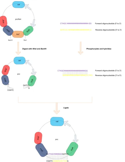
Figure 3-1: Insertion of oligonucleotide sequences corresponding to GPCR TM domains into the pccKan expression vector ... 62
Figure 3-2: Generation of point mutations in the Mam2 receptor ... 66
Figure 3-3: GFP-tagging of mutated pREP3x-Mam2 constructs ... 68
Figure 3-4: Creation of pKS-Rluc ... 70
Figure 3-5: Generation of Receptor-Rluc fusion constructs ... 72
Figure 3-6: Creation of the Rluc-Sxa2 and Rluc8-Sxa2 insertion cassettes ... 74
Figure 4-1: Comparison of endogenous and heterologous Mam2 activity ... 77
Figure 4-2: Comparison of the activity of Mam2 and Mam2-GFP ... 79
Figure 4-3: Comparison of the activity of Mam2-GFP and STE2-GFP expressed in Sz. Pombe ... 82
Figure 4-4: Representative images showing the localization of GFP, Mam2-GFP and STE2-GFP in Sz. pombe ... 85
Figure 4-5: Measured fluorescence intensities at the cell membrane of GFP, STE2-GFP and Mam2-STE2-GFP ... 86
Figure 4-6: Sequences of STE2 and Mam2 transmembrane domain 1 ... 89
Figure 4-7: Effect of conservative mutations in STE2 on the pheromone-response ... 91
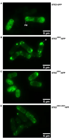
Figure 4-8: Representative images showing the localization of the conservatively mutated STE2-GFP receptors in Sz. pombe ... 93
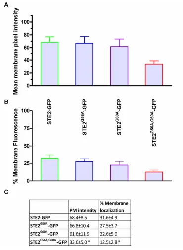
Figure 4-9: Measured fluorescence intensities at the cell membrane of STE2-GFP and the STE2-GFP constructs containing conservative mutations ... 94
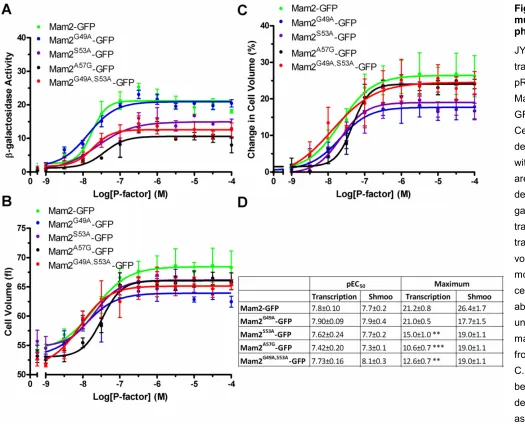
Figure 4-10: Effect of conservative mutations in Mam2 on the pheromone-response... 97
Figure 4-11: Representative images showing the localization of the conservatively mutated Mam2-GFP receptors in Sz. pombe ... 99
Figure 4-12: Measured fluorescence intensities at the cell membrane of Mam2-GFP and the Mam2-GFP constructs containing conservative mutations ... 100
Figure 4-13: Effect of disfavored mutations in STE2 on the pheromone-response... 102
Figure 4-14: Representative images showing the localization of the STE2-GFP receptors containing the disfavored mutations in Sz. pombe ... 104
Figure 4-15: Measured fluorescence intensities at the cell membrane of STE2-GFP and the STE2-GFP constructs containing the disfavored mutations ... 105
Figure 4-16: Effect of disfavored mutations in Mam2 on the pheromone-response ... 107
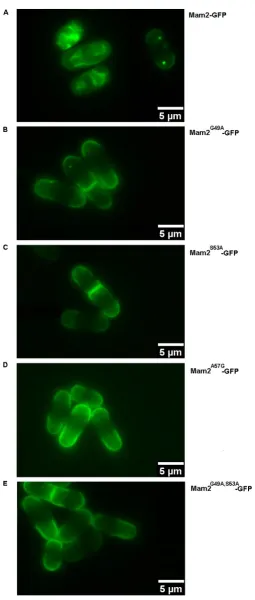
Figure 4-17: Representative images showing the localization of the Mam2-GFP receptors containing the disfavored mutations in Sz. pombe ... 109
Figure 4-18: Measured fluorescence intensities at the cell membrane of Mam2-GFP and the Mam2-GFP constructs containing the disfavored mutations ... 110
Figure 4-19: Cell viability in response to pheromone ... 112
Figure 4-20: Effect of Rluc and GFP tags on the activity of STE2 ... 115
Figure 4-21: Effect of Rluc and GFP tags on the activity of Mam2 ... 117
Figure 4-22: Luminescence intensity of cells expressing STE2-Rluc and Mam2-Rluc ... 118
Figure 5-1: Expression, insertion and orientation checks for the TOXCAT controls ... 125
Page xiii Figure 5-3: Expression, insertion, orientation and oligomerization of the STE2 TM
domains 1-7 TOXCAT Chimeras ... 131
Figure 5-4: Length optimization of transmembrane domain 1 from STE2 ... 134
Figure 5-5: Molecular modeling of STE2 TM domain 1 dimers ... 136
Figure 5-6: STE2 TM domain 2 in the context of the full-length protein ... 141
Figure 5-7: Structural models of wild-type STE2 TM domain 2 dimers ... 144
Figure 5-8: Structural dimeric models of STE2 TM domain 2 N84A mutants ... 145
Figure 5-9: Structural dimeric models of STE2 TM domain 2 H94A mutants ... 146
Figure 5-10: Mutations affecting STE2 TM domain 2 dimerization ... 148
Figure 5-11: Expression, insertion, orientation and oligomerization of the Mam2 TM domains 1-7 TOXCAT Chimeras ... 150
Figure 5-12: Length optimization of transmembrane domain 1 from Mam2 ... 153
Figure 5-13: Structural models of Mam2 TM domain 1 dimers ... 156
Figure 5-14: Mutations affecting wild-type Mam2 TM domain 1 dimer formation ... 157
Figure 5-15: Mutations affecting Mam2 TM domain 1 dimerization ... 159
Figure 5-16: TOXCAT assay and controls for Mam2 TM domain 2 mutants ... 162
Figure 5-17: Structural models of Mam2 TM domain 2 ... 164
Figure 5-18: Structural dimeric models of Mam2 TM domain 2 incorporating a N83A mutation ... 165
Figure 5-19: Structural dimeric models of Mam2 TM domain 2 incorporating a R93A mutation ... 166
Figure 5-20: Structural modeling of Mam2 TM domain 2 incorporating a C91A mutation ... 167
Figure 6-1: Sequence alignment of the rhodopsin, β2-adrenergic and STE2 receptors (Eilers, Hornak et al. 2005) ... 173
Figure 6-2: Sequence alignment of the rhodopsin, β2-adrenergic and STE2 receptors ... 176
Figure 6-3: Homology models of STE2 TM1 and TM7 on bovine rhodopsin and the human β2-adrenergic receptor ... 179
Figure 6-4: Homology models of STE2 TM2, TM3 and TM4 on rhodopsin and β2 -adrenergic receptor ... 181
Figure 6-5: Presence of a putative ionic lock in the homology models of STE2 ... 183
Figure 6-6: Effect of a conserved proline residue on hydrogen-bonding in the main-chain of TM6 of GPCRs ... 187
Figure 6-7: The GxxxG motif is accessible for interactions in the new models ... 189
Figure 6-8: CHI models of interactions between TM1 and TM2 in bovine rhodopsin ... 191
Figure 7-1: Activity of the GFP and β-galactosidase reporters ... 197
Figure 7-2: Creation of the sxa2::Rluc and sxa2::Rluc8 strains ... 199
Figure 7-3: Integration of Rluc or Rluc8 into the Sz. pombe genome ... 200
Figure 7-4: Time-dependent decay of luminescence ... 201
Figure 7-5: The different micro-well plates tested ... 203
Figure 7-6: The effects of using different micro-well plates on the luminescent signal ... 204
Figure 7-7: A comparison of the Rluc and Rluc8 Reporters ... 206
Figure 7-8: The number of dead cells increases in response to pheromone but do not signal ... 208
Figure 7-9: Comparison of the effects of different media on the luminescent reporters Rluc and Rluc8 ... 209
Figure 7-10: Rluc and Rluc8 expression levels ... 211
Figure 7-11: Time-course of Rluc reporter activity ... 214
Figure 7-12: Time-course of Rluc reporter activity with substrate addition at the start of the assay ... 216
Figure 7-13: The effects of varying the concentration of EnduRen on luminescent signal ... 218
Figure 7-14: Time courses in suspension ... 220
Figure 7-15: The effects of cells settling ... 222
Figure 7-16: Time-course on agarose plugs ... 224
Page xiv
LIST OF TABLES
Table 3-1: List of STE2 wild-type single TM domain TOXCAT constructs ... 63
Table 3-2: List of Mam2 wild-type single TM domain TOXCAT constructs ... 64
Table 3-3 Sequence of oligonucleotides to create Receptor-Rluc fusions ... 71
Table 4-1: BRET donor and acceptor pairs ... 119
Table 5-1: Sequence alignment of STE2 and Mam2 TM2 ... 160
Table 6-1: Comparison of TM domain lengths ... 172
Table 6-2: Comparison of STE2 TM boundaries ... 175
Table 6-3: Alignment of core residues in TM6 and TM7 of rhodopsin, β2-adrenergic receptor, STE2 and Mam2 ... 186
Table 8-1: The effects of conservative mutations on STE2 and Mam2 ... 230
Page xv
ABBREVIATIONS
AA Amino acid
Amp Ampicillin
AGS Activators of G protein Signalling ARF ADP-Ribosylation Factor
AUC Analytical Ultracentrifugation
BAR Bin–Amphiphysin–Rvs
BiFC Bimolecular Fluorescence Complementation
BRET Bioluminescence Resonance Energy Transfer
cAMP Cyclic AMP
CAM Constitutively Active Mutant
CAT Chloramphenicol Acetyl-Transferase
CBP CREB Binding Protein
CDC Cell Division Control
CHI CNS (Crystallography and NMR Searching) of Helix Interactions
CME Clathrin Mediated Endocytosis
CoA Coenzyme A
CREB cAMP Response Element Binding
CRLR Calcitonin Receptor-Like Receptor
DAG Diacylglycerol
EC50 Median Effective Concentration ERAD ER Associated Degradation
DAG Diacylglycerol
DMM Defined Minimal Media
DNA Deoxyribonucleic Acid
dodecylphosphocholine Dodecylphosphocholine
ECM Extracellular Matrix EGF Epidermal Growth Factor
E. coli Escherichia coli
EDTA Ethylenediaminetetraacetic Acid
ER Endoplasmic Reticulum
FACS Fluorescence Activated Cell Sorting
Page xvi
FRET Fluorescence Resonance Energy Transfer
GABA Metabotrophic γ-Aminobutyric Acid Receptor
GAP GTPase Accelerating Protein
GDI Guanosine Nucleotide Dissociation Inhibitors
GDP Guanosine Diphosphate
GEF Guanine nucleotide Exchange Factor
GFP Green Fluorescent Protein
G protein Guanosine nucleotide-binding protein
GTP Guanosine Triphosphate
GpA Glycophorin A
GRK G protein-coupled Receptor Kinase GTP Guanosine Triphosphate
GPCR G Protein-Coupled Receptor
HMG High Mobility Group
IP3 Inositol Triphosphate
kb kilo bases
kDa kilo Dalton
LB Luria-Bertani Broth
MAP Mitogen Activated Protein
MAPK Mitogen Activated Protein Kinase
MAPKK Mitogen Activated Protein Kinase Kinase
MAPKKK Mitogen Activated Protein Kinase Kinase Kinase
MBP Maltose Binding Protein MCS Multi-Cloning Site
MW Molecular Weight
NMR Nuclear Magnetic Resonance
OD Optical Density
ONPG o-Nitrophenyl-β-D-galactoside PAGE Polyacrylamide Gel Electrophoresis
PAK p21 Activating Kinase
PBS Phosphate Buffered Saline
PCA Protein fragment Complementation Assay
PCR Polymerase Chain Reaction
Page xvii
pEC50 Negative natural logarithm of EC50
PF Periplasmic Fraction
PKA Protein Kinase A / cAMP Dependent Protein Kinase
PKC Protein Kinase C
PLC Phospholipase C
PM Plasma Membrane
PVDF Polyvinylidene Fluoride
RAMP Receptor Activity-Modifying Protein
RET Förster Resonance Energy Transfer
RGS Regulator of G protein Signalling
Rluc Renilla luciferase
RMSD Root Mean Square Deviation
Sc. cerevisiae Saccharomyces cerevisiae
S.D Standard Deviation
SDS Sodium Dodecyl Sulphate
SDS-PAGE Sodium Dodecyl Sulphate Polyacrylamide Gel Electrophoresis
S.E.M Standard Error of the Mean SF Spheroplast Fraction
SPF Spheroplast Proteolysis Fraction
Sz. pombe Schizosaccharomyces pombe
TM Transmembrane
UTR Untranslated Region
WHIM Warts, Hypogammaglobulinemia, Infections and Myelokathexis
WT Wild-Type
w/v Weight per volume
Page xviii
AMINO ACID ABBREVIATIONS
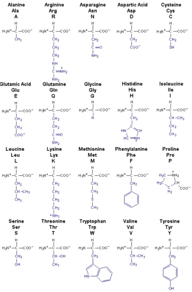
[image:22.595.76.460.160.742.2]Figure i-1 show the 20 amino acids encoded by the genetic code with their one letter and three letter abbreviations and structure. The R-group is highlighted in blue.
Page 1
1 GENERAL INTRODUCTION
1.1 Main Objectives
This thesis concerns the study of the oligomerization of G protein coupled receptors (GPCRs) and how specific amino acid interactions facilitate receptor interactions. In order to realize this objective, individual TM domains from two different GPCRs were studied, compared and contrasted using a range of diverse approaches. The development and optimization of a novel bioluminescent reporter assay is also discussed.
1.2 Membrane Proteins
Membrane proteins are involved in a range of biologically indispensable processes including; the transport of small molecules and ions, cell signalling, structural stability and cell-to-cell adhesion. Their correct functioning is critical to cells and organisms as implied by several diseases caused by malfunctioning membrane proteins, such as autoimmunity, diabetes and cancer (Sanders and Myers 2004; Bocharov, Volynsky et al. 2010). Membrane proteins targeted to the plasma membrane bridge the boundary between the exterior and interior of the cell and they are therefore amenable pharmacological targets. A remarkable ~60% of drugs are known to bind to the surface of cells (Overington, Al-Lazikani et al. 2006) with the majority binding to targets that include; GPCRs, ligand-gated ion channels, voltage-gated ion channels or protein kinases. The further understanding of membrane protein structure and function is therefore immensely important for pharmacological and economical reasons.
1.2.1 Integral Membrane Proteins
Integral membrane proteins span the lipid bilayer at least once and remain permanently attached to the membrane. The highly apolar environment of the membrane means that membrane proteins lose their 3D structure upon solubilisation, and consequently techniques used for the structural study of globular proteins are not readily applied to membrane proteins. Despite challenges in the structural characterization of membrane proteins, two main classes of integral membrane proteins have been described to date. These include the
Page 2
1.2.2 The Membrane Environment
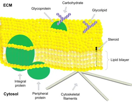
Biological membranes provide a boundary between the exterior and interior of cells as well as compartmentalizing intracellular organelles. The membrane is a highly heterogenous environment; the plasma membrane contains ~50% phospholipids and ~50% protein and other structures such as steroids and carbohydrates also associate with the membrane. The fluid mosaic model describes the current view of membrane organization (Singer and Nicolson 1972) and is illustrated in Figure 1-1. In this model phospholipids provide the fundamental structural organization of the membrane. Phospholipids consist of a hydrophilic phosphate-containing head-group linked to two fatty acid hydrocarbon tails. Due to their amphipathic nature, phospholipids can spontaneously form lipid bilayers in solution. The lipid:water interface does not provide a sharp division between a hydrophobic and a hydrophilic environment. Rather, an interfacial region exists spanning ~15 Å, which is thick enough to accommodate an amphipathic helix (White and Wimley 1999). The thickness of the hydrophobic core is ~30 Å (White 2003) but can vary depending on the lipid composition of the bilayer (Lewis and Engelman 1983). An appropriately thick bilayer surrounding a membrane protein is important due to the cost of exposing hydrophobic residues to water. It has therefore been proposed that lipids may stretch or compress to match the span of membrane proteins, and conversely membrane proteins may also structurally adjust to shield hydrophobic residues, for instance via tilting of transmembrane (TM) domains (Lee 2004).
Page 3 (Lindner and Naim 2009), although their roles, location and existence is debated. In another view, certain proteins favour interactions with certain lipids, although difficulties in determining the transient protein-lipid interactions make this phenomenon challenging to study (Lee 2004).
Figure 1-1: The fluid mosaic model of biological membranes
Page 4
1.2.3 Membrane Protein Biogenesis and Exit to the Plasma
Membrane
1.2.3.1 The Endoplasmic Reticulum
Not all biological membranes are able to translocate and integrate newly synthesised membrane proteins. Membranes that can include these of the endoplasmic reticulum (ER), mitochondria, chloroplasts and peroxisomes, whereas the plasma membrane and endocytic organelles are unable to do so (von Heijne 1990). Cells have therefore developed a secretory pathway that serves as a gateway for proteins destined for these organelles.
Polytopic membrane proteins (i.e. proteins that span the membrane multiple times) are synthesised by ribosomes attached to the cytosolic side of the ER and enter the ER co-translationally via the Sec61 translocation complex to which they associate via hydrophobic targeting sequences. Orientation signals within the protein (Higy, Junne et al. 2004) as well as molecular and lipid chaperones (van Voorst and De Kruijff 2000; Krebs, Noorwez et al. 2004) aid the correct insertion, folding and orientation of the membrane protein, and proteins may undergo certain post-translational modifications such as disulfide-bond formation in the ER (Achour, Labbe-Jullie et al. 2008). Incorrectly folded membrane proteins however are exported to the ER-associated degradation pathway (ERAD) in the cytosol for degradation (Tsai, Ye et al. 2002).
There is evidence that plasma-membrane signalling complexes may form in the ER. For instance the GABAB1 receptor forms a complex with the Gβγ subunits of G proteins and the
Page 5
1.2.3.2 Targeting to the Golgi Apparatus and Plasma Membrane
Export from the ER occurs at ER “exit sites” in a coat protein II (COPII)-dependent manner (Achour, Labbe-Jullie et al. 2008). Proteins destined for the Golgi accumulate at these sites that form buds when coated with the COPII associated proteins Sar1, Sec23-Sec24 and Sec13-Sec31. It has been proposed that integral membrane proteins interact with domains of Sar1 and Sec23-24 which labels them for transport (Barlowe 2003). Interaction sites vary but may involve a di-acidic motif composed of the amino acids DxE, a double phenylalanine motif, or other more complicated motifs, and is a rate-limiting step in COPII bud formation (Dong, Filipeanu et al. 2007). Multiple binding sites on Sec23 have been identified, which supports the theory that different interaction motifs on separate membrane proteins can bind to the same transport molecule (Miller, Beilharz et al. 2003).
Following budding, the COPII transport vesicles are transported and fused to the Golgi apparatus and subsequently migrate through the network of flattened cisternae; from the cis-Golgi network to the trans-cis-Golgi network via the medial-cis-Golgi and endo-cis-Golgi. Each of these regions contain different enzymes involved in selective post-translational modification of cargo, such as glycosylation (Alberts, Johnson et al. 2002). N-linked glycosylation may be important for the transport of some proteins from the Golgi to the plasma membrane, although for others it is less clear what the target signals are (Dong, Filipeanu et al. 2007).
It’s been suggested that in some cases, the same motifs involved in targeting proteins from the ER to the Golgi may be involved in targeting proteins from the Golgi to the plasma membrane, as has been found for the di-acidic motif (Nishimura, Plutner et al. 2002).
1.2.3.3 Regulation of the Secretory Pathway
Page 6 receptors are irreversibly activated and targeted to lysosomes following activation (Hein, Ishii et al. 1994). Thrombin is retained in the Golgi by another membrane protein, p24A, and following activation of G protein-bound receptor at the cell surface, the G protein dissociates and targets p24A to trigger the release of Golgi-bound thrombin (Luo, Wang et al. 2007). For other receptors in contrast, such as the β2-adrenergic receptor, plasma membrane expression following the secretory pathway is constitutive (Hein, Ishii et al. 1994).
1.2.4 Membrane Protein Internalization
1.2.4.1 Endocytosis
Endocytosis acts as a regulator of signalling through controlling the lipid-protein composition of the membrane. Three distinct cellular compartments are associated with the endocytic pathways; the early endosomes, late endosomes and lysosomes (or vacuoles in fungi). Internalized molecules first encounter the early endosomes. The mildly acidic pH of the lumen of these structures can cause ligands to dissociate from receptors, and receptors may therefore be recycled from the early endosomes via tubular structures back to the plasma membrane. Early endosomes mature into late endosomes, in which the lumen becomes increasingly acidic and there is a loss of the tubular structures needed for recycling, ultimately determining the fate of cargo for degradation by the lysosome (Lodish, Berk et al. 2000). Actin filaments have been strongly implicated in the movement of vesicles between these compartments in yeast and in some, but not all, mammalian cell types (Kaksonen, Sun et al. 2003; Gachet and Hyams 2005). Endosomal cargo destined for degradation is targeted to the lysosome where contents are broken down into component parts such as amino acids from proteins, or cholesterol and fatty acids from cholesterol esters (Lodish, Berk et al. 2000).
1.2.4.2 Pathways Mediating Endocytosis
Page 7 CME and it has been extensively reviewed in the literature (Mousavi, Malerod et al. 2004; Traub 2005; Roth 2006).
The most commonly reported non-clathrin coated buds are known as caveolae. These flask-shaped invaginations are rich in cholesterol, sphingolipids and the protein caveolin and are often observed in clusters, which can occupy as much as a third of plasma membrane area (Mayor and Pagano 2007). Caveolae are believed to perform multiple functions in the cell including lipid storage, responding to mechanical stress, and to play a role in cell signalling events such as Ca2+ signalling (Parton and Simons 2007). Caveolae can exist as immobile
plasma membrane domains (Thomsen, Roepstorff et al. 2002) but can also couple to the endocytic pathway through internalization of vesicles coated with caveolin; a process that can be exploited by pathogens to gain entry into cells (Pelkmans, Kartenbeck et al. 2001). Other endocytic pathways also exist (Sandvig, Pust et al. 2011) which may involve coat proteins other than caveolin and clathrin, flotilin is another example, or different GTPases; for instance RhoA, cell division control protein 42 (CDC42) or ADP-ribosylation factor 6 (ARF6). The mechanisms appear to vary greatly between cell-types making it difficult to propose specific general pathways (Mayor and Pagano 2007).
1.2.4.3 Regulation of Internalization
Page 8
1.2.5 Protein Folding in the Membrane
1.2.5.1 Integration of Proteins into the Membrane
The process by which membrane proteins are integrated into lipid bilayers is currently not fully understood. Mothes and colleagues (1997) proposed a model in which membrane proteins laterally integrate into the ER membrane via the ribosome-translocon channel. In this model, the appearance of a hydrophobic stretch of polypeptide within the translocon causes a closing of the channel, and the segment moves laterally into the bilayer. This process may be repeated when translating a polytopic protein (Mothes, Heinrich et al. 1997). The hydrophobic stretches found in membrane proteins and adjacent residues appear sufficient to drive the integration of a protein into the membrane, partly because the introduction of a TM domain into cytosolic proteins can convert them into membrane proteins and also because model peptides will only partition into the membrane if they are sufficiently hydrophobic (MacKenzie 2006). The structural comparison of the translocon in a peptide-bound form and inactive form provided further evidence for a lateral gate present in the translocon (Martinez-Gil, Sauri et al. 2011).
Membrane proteins insert into the membrane because it is energetically favourable to bury hydrophobic residues, even though dehydrating the hydrophilic backbone comes at a cost. For example, the free energy for the insertion of the α-helical protein glycophorin A (GpA) is estimated to be -36 kcal/mol-1 due to burial of hydrophobic residues. Conversely the cost of
dehydrating the backbone of GpA is +26 kcal/mol-1 (White 2003). Therefore, there is
favourable net free energy of 10 kcal/mol-1 when inserting the helix into the membrane
compared to leaving it in the cytosol (White 2003). The formation of secondary structures within the membrane is also energetically favourable, and most integral membrane proteins adopt α-helical structures across the lipid bilayer. The cost of transferring a non-H-bonded peptide bond from a hydrophilic to hydrophobic environment is +6.4 kcal/mol-1 whereas
transferring an H-bonded peptide bond to a hydrophobic environment is only +2.1 kcal/mol-1.
There is thus a free energy cost of +4 kcal/mol-1 of disrupting a single H-bond in the
Page 9 the membrane, and side-chain to main-chain hydrogen bonding may also increase the length of the helix (Lee 2004).
1.2.5.2 The Two-Stage Model of Membrane Protein Folding
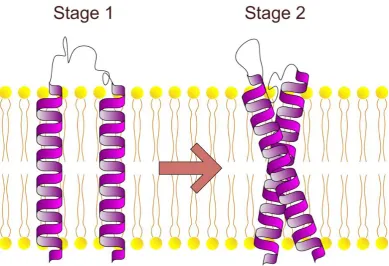
The two-stage model of membrane protein folding provides an important conceptual framework in which to understand protein folding into the membrane (Popot and Engelman 1990). According to the two-stage model (Figure 1-2) membrane proteins fold in two thermodynamically distinct stages. In the first stage, hydrophobic sequences insert into the lipid bilayer and form independently stable domains. This is driven by the hydrophobic effect. In the second stage, helical domains laterally interact to form the fully folded tertiary structure of the protein. This may be driven by energetically favoured bond-formation and packing between helices.
Page 10 Figure 1-2: The two-stage model of membrane protein folding
The two-stage model of membrane protein folding was proposed by Popot and Engelman in 1990. In the first stage, independently stable trans-bilayer helices form in response to the hydrophobic effect and main-chain hydrogen bonds form in the non-aqueous environment. In the second stage helices interact to form the tertiary fold of the protein that may be driven by energetically favorable polar contacts between helices, or packing.
Page 11
1.2.6 Transmembrane Domain Interactions
The two-stage model predicts that individual TM domains initially form independently stable structures, before laterally interacting to form a polytopic protein or form quaternary structures. The model does not however postulate how lateral interactions are formed. The hydrophobic effect is an important force driving the folding of globular proteins (Tanford 1978), but due to the low dielectric environment of the membrane other forces must drive the lateral association of membrane proteins. The study of the interactions within polytopic and/or oligomeric membrane proteins is not trivial and remains one of the biggest challenges in protein chemistry to this day. In the PDB only 2% of structures represent membrane proteins (http://pdbtm.enzim.hu/ 2011), however membrane proteins are believed to constitute 20-25% of all proteins (Boyd, Schierle et al. 1998; von Heijne 1999). Considering their pharmacological and economical importance, the lack of structural information of membrane proteins clearly reflects the challenges presented to structural biologists.
1.2.6.1 The Structural Importance of Transmembrane Domains
The TM spanning regions of membrane proteins were traditionally believed to merely anchor the protein to the membrane. Early studies on fragmented bacteriorhodopsin however showed that this is not the case. In bacteriorhodopsin a functional protein can be reconstituted only if all 7TM domains are expressed, and in contrast four of the six loop regions found outside of the hydrophobic core are dispensable, although they do influence folding stability and kinetics (Luneberg, Widmann et al. 1998; Marti 1998). This demonstrated that the transmembrane segments do not merely anchor membrane proteins to the lipid bilayer, but serve a purpose in maintaining protein structure.
1.2.6.2 Motifs Known to Mediate Membrane Protein-Protein Interactions
1.2.6.2.1
Motifs of Two Small Residues
The study of single-spanning membrane proteins and designed peptides has contributed to much of our current understanding in how transmembrane domains interact, and several motifs have been identified that appear important in driving associations. For instance, the single TM-spanning protein GpA which is expressed in erythrocytes, dimerizes via a motif of two glycines spaced four residues apart (GxxxG) (Lemmon, Flanagan et al. 1992; Adams, Engelman et al. 1996). Due to their relative spacing in the primary sequence, the two
Page 12
by a “ridges-into-grooves” packing of adjacent β-branched residues into the grooves created by the glycine residues (MacKenzie, Prestegard et al. 1997). The ridges-into-grooves, or knobs-into-holes, packing of residues is illustrated in Figure 1-3. Statistical analysis revealed that the GxxxG motif is overrepresented in genomes (Russ and Engelman 2000) and since
its discovery it’s been found to mediate the dimerization of several other membrane proteins
Page 13 Figure 1-3: Ridges-into-grooves packing of residues in dimeric glycophorin A
Page 14
1.2.6.2.2
Heptad Repeats
Heptad repeat motifs also drive the association of membrane proteins. In contrast to the GxxxG motif mainly found in right-handed helix interactions, this motif is generally found in left-handed helix interactions. The heptad motif is driven by repeats of seven residues and helices ultimately achieve a knobs-into-holes packing (Langosch and Heringa 1998; MacKenzie 2006).
1.2.6.2.3
Polar Amino Acids
The presence of polar amino acids in the hydrophobic core of a membrane is energetically unfavourable but can be compensated for by the surrounding hydrophobic residues. The high energetic cost of breaking H-bonds in the hydrocarbon core of a bilayer means that polar contacts provide a strong stabilising force for helix association. For instance, the introduction of a single asparagine residue into a hydrophobic model peptide can transform the peptide from monomeric species to dimeric species both in an in vitro and in an in vivo system (Zhou, Cocco et al. 2000). The strongly polar amino acids, including histidine, glutamic acid, arginine and aspartic acid, show the highest propensity to form inter-helical H-bonds in the membrane (Sodt and Head-Gordon 2010). Other polar amino acids also induce oligomerization, including asparagine and glutamine (Choma, Gratkowski et al. 2000; Zhou, Cocco et al. 2000; Zhou, Merianos et al. 2001). Threonine and serine residues tend to be overrepresented in transmembrane domains compared to other polar residues (Curran and Engelman 2003), however these residues, together with tyrosine, only appear to have a moderate effect on transmembrane domain oligomerization (Gratkowski, Lear et al. 2001; Zhou, Merianos et al. 2001). As with the smallxxxsmall motif, sequence context is important for associations. The membrane protein BNIP3 for instance, dimerizes strongly via a histidine residue but BNIP3 also contains a GxxxG motif on the same face of the helix as the histidine residue. Mutations of the GxxxG motif disrupts dimerization by not allowing the two monomers to come into close enough proximity for histidine to hydrogen bond (Sulistijo, Jaszewski et al. 2003; Lawrie, Sulistijo et al. 2010).
1.2.6.2.4
Aromatic Residues
Aromatic residues such as phenylalanine, threonine or tyrosine can also contribute to energetically favourable interactions between TM helices. Pairs of aromatic residues can π
Page 15 membrane proteins and interactions can be four-fold stronger than those arising from knobs-into-holes packing (Dougherty 1996; Johnson, Hecht et al. 2007). For each of these interactions two possible conformations are possible. For the interaction of two aromatic residues, the interaction can either be stacking or edge-to-face, the former typically involving extensive van der Waals contact whereas the latter involve a CH-π interaction. For the
cation-π interaction the positively charged residue can either be perpendicular or parallel to the aromatic ring (Johnson, Hecht et al. 2007). Cation-π interactions are interesting because they allow a polar, yet hydrophobic, interaction in the membrane i.e. they can bind ions but are composed of hydro-carbons (Dougherty 1996). This type of interaction is not exclusive to membrane protein-membrane protein interactions, but has also been observed between receptor and ligand (Tantry, Ding et al. 2010).
1.2.6.3 Effects of Proline Residues on Membrane Protein Structure
Prolines present in the transmembrane domains can also influence helix packing and their importance in membrane proteins is reflected by the phenotypic consequences of mutating prolines in human membrane proteins (Partridge, Therien et al. 2004). The amide nitrogen of proline cannot form backbone hydrogen bonds and proline residues therefore introduce distinct kinks in transmembrane domains. Proline is present at 60% of transmembrane helix deformation sites and it has been proposed that in cases where proline is not present, the proline could have been lost with time as the kink became stabilized by other molecular forces (Yohannan, Faham et al. 2003). Proline residues in transmembrane domains are most often found in the middle of the helices (Barlow and Thornton 1988).
On a molecular level, prolines are believed to reduce the stability and rigidity of transmembrane helices due to the loss of the NH...O=C bond. In one specific class of integral
membrane proteins, the 7TM spanning GPCR receptors, this allows a rotational switch upon activation (EIling, Frimurer et al. 2005). In this model, upon receptor activation the extracellular side of the receptor undergoes an inward movement which closes the ligand-binding site whereas the cytoplasmic side opens up to allow downstream signalling. This movement is achieved via a see-saw motion of TM6 and TM7 which pivots about the centre of the helices, and it has been suggested the pivoting is achieved via the proline residues (Schwartz, Frimurer et al. 2006). Mutations of transmembrane proline residues have been shown to have an effect on both ligand-binding and activation of GPCRs (Ladds, Davis et al. 2005; Reis, Santos et al. 2007; Mazna, Grycova et al. 2008)
Page 16 and hydrogen bonding) and the presence of certain motifs and residues in transmembrane domains can be strong predictors of transmembrane helix-helix interactions.
1.2.7 Methods for Studying Transmembrane Helix Interactions
1.2.7.1 Membrane Protein Structural Determination
Many biophysical techniques exist to study interactions between transmembrane peptides in vitro. These techniques include, among others, sodium dodecyl sulphate polyacrylamide gel-electrophoresis (SDS-PAGE), analytical ultracentrifugation (AUC), chemical cross-linking, solution-state nuclear magnetic resonance (NMR), or solid-state NMR and are often performed in detergent micelles or synthetic lipid bilayers (MacKenzie, Prestegard et al. 1997; Fisher, Engelman et al. 1999; Liu, Crocker et al. 2003; Doura, Kobus et al. 2004; Jenei, Borthwick et al. 2009). The biophysical characterization of polytopic proteins provides an even greater challenge. X-ray crystallography is the only currently available method for obtaining full-length polytopic membrane protein structures (Henderson, Baldwin et al. 1991) although it may be possible to piece together structural information on receptor fragments from NMR to generate an image of the full-length protein (Toyoshima, Nakasako et al. 2000).
Apart from being highly artificial systems, these biophysical techniques come with other challenges. Detergent micelles can destabilize helices for instance, through interactions between amino acids and the polar head-groups of the detergent, thereby allowing increased side-chain flexibility in the amino acid chain or disrupting inter-helical bonds thereby distorting the structure (Langosch, Brosig et al. 1996; MacKenzie 2006). Furthermore, only a handful of structures of large polytopic proteins have been solved to date by X-ray crystallography. This is largely due to difficulties in obtaining large quantities of protein and purifying them in their native form. Much research is therefore currently focused on improving techniques (Bill, Henderson et al. 2011).
1.2.7.2 Reporter Systems to Study Protein-Protein Interactions in the
Membrane
Page 17 complementation (BiFC) (Overton, Chinault et al. 2003; Milligan and Bouvier 2005; Gehret, Bajaj et al. 2006). Recently RET and PCA techniques have been combined to monitor the formation of large protein complexes (Vidi and Watts 2009). The BRET technique has also been used to monitor changes in protein conformation upon ligand binding (Percherancier, Yamina et al. 2005).
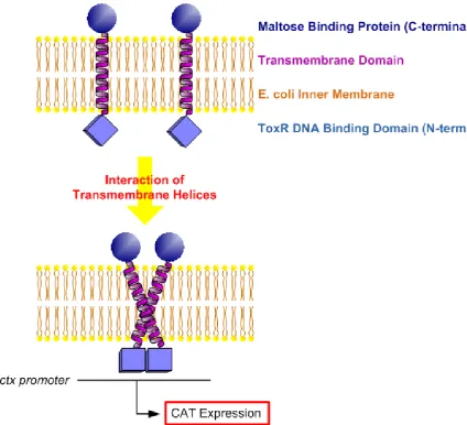
Oligomerization of single transmembrane spanning domains can be readily studied using the TOXCAT assay (Russ and Engelman 1999). In this technique a TM domain of interest is expressed as an MBP-TM-ToxR chimera in the E. coli inner membrane. The maltose binding protein (MBP) drives the construct to the membrane and ensures its correct orientation. Interaction of two TM domains bring their ToxR domains in close proximity, leading to expression of chloramphenicol acetyl-transferase (CAT), (Figure 1-4) and levels of CAT expression can directly be quantified by its activity on a substrate. Since TOXCAT can only
measure interactions between the same domains (“homo-oligomerization”) an alternative
technique was developed to allow the studying of “hetero-oligomers” i.e. oligomers formed
by the interaction of different transmembrane domains (Schneider and Engelman 2003). Other assays involving protein fusions and reporter complementation also exist, such as the split-ubiquitin assay (Johnsson and Varshavsky 1994).
Page 18 Figure 1-4: Principles of the TOXCAT assay.
Page 19
1.3 Cell to Cell Communication
The ability of cells to sense their environment and respond with appropriate cellular responses is crucial to survival. Social and multi-cellular organisms additionally require inter-cellular communication in order to formulate coordinated responses. Vast arrays of inter-cellular pathways have evolved to sense and respond to stimuli as diverse as nutrients, stress or developmental information. Some signalling pathways may involve the passive diffusion of signalling molecules into cells; estrogen for instance can cross the lipid bilayer. Most signalling pathways however rely on membrane bound receptors to sense external stimuli and to induce the appropriate response. In some cases, such as ligand- and voltage-gated ion channels or receptor tyrosine kinases, the receptor itself is involved in actively inducing the desired response. In other cases, such as GPCR signalling, the receptors merely relay the signal intracellularly.
Many diseases that plague modern society are associated with the disruption of normal cell-to-cell communication. These include, among many others, autoimmune disease, where an
individual’s immune system fails to recognise its own constituent parts, type-2 diabetes, where cells do not respond to insulin properly or cancer, where cells do not to respond to normal homeostatic control. The study of cell-to-cell communication is therefore important for understanding how to detect, and combat, such diseases.
1.3.1 G Protein-Coupled Receptors
GPCRs form a large ubiquitous family of signalling receptors; humans alone encode ~800 GPCRs (Fredriksson, Lagerstrom et al. 2003) and they are found in all eukaryotic organisms. Although GPCRs do not share a high degree of sequence similarity they do share a highly conserved topology. GPCRs are involved in sensing a wide array of stimuli, or ligands, including light, odours, proteins, peptides, lipids, sugars, nucleotides and ions, and the extracellular signal is typically relayed within the cell via the G protein to promote a response.