Odontogenic Cysts: Improved Imaging with a Dental CT
Software Program
James J. Abrahams1 and Patrick J. Oliverio1
PURPOSE: To evaluate a dental CT software program to determine whether it can provide a better means of assessing odontogenic cysts, lesions of the jaw derived from dental epithelium, than conventional techniques (orthopantomographic, intraoral, and mandibular films), which are
of limited usefulness because of the curved configuration of the mandible; and to provide a brief review of these lesions. METHODS: Nine odontogenic cysts were studied with conventional
radiographs and with the software program, which displays multiple cross-referenced axial, panoramic, and cross-sectional (unique to this program) views of the mandible. The two modalities
were compared for delineation of anatomy (inferior alveolar canal, mandibular foramen, mental foramen), detection of neurovascular bundle displacement, detection of cortical bone involvement,
and detection of root involvement. RESULTS: The software program rated higher regarding all four points. It was found to be superior for delineating anatomy and detecting mandibular canal
displacement and cortical and root involvement. CONCLUSIONS: This software program should be the study of choice when evaluating odontogenic cysts and other lesions of the mandible.
Index terms: Computed tomography, technique; Mandible, cysts; Computed tomography, software
AJNR 14:367-374, Mar/Apr 1993
Odontogenic cysts are lesions of the jaw that are derived from dental epithelium. The
classifi-cation of these lesions has undergone change in
the recent decades. Presently, the most widely
accepted and straightforward classification ( 1)
divides these lesions into the following six types: dentigerous cysts, radicular cysts, lateral peri-odontal cysts, gingival cysts, odontogenic
kera-tocysts, and calcifying odontogenic keratocysts. Although these lesions are histologically benign, some may be locally agressive or clinically per-sistent. Achieving a correct diagnosis and a clear
radiographic delineation of the cyst's extent can
significantly alter the treatment plan and surgical
approach. Determining the position of the neu-rovascular bundle in relation to the lesion is also
important. Recent attempts have therefore been
made to improve the radiographic assessment of these lesions.
Received October 31, 1991; revision requested February 6, 1992; revision received June 8 and accepted July 21.
1
Department of Diagnostic Radiology, Yale University School of Med-icine, 333 Cedar Street, New Haven, CT 06510. Address reprint requests
to James J. Abrahams.
AJNR 14:367-374, Mar/Apr 1993 0195-6108/93/1402-0367
© American Society of Neuroradiology
367
Typically, odontogenic cysts are studied in the dentist's office using orthopantomographic,
intra-oral, and mandibular films. These screening ex-ams are excellent but fail to provide the detailed
information necessary for appropriate manage-ment. They do not demonstrate internal anatomy
or the position of the lesion in relation to the
neurovascular bundle and cortical bone margins. MagQetic resonance, which provides excellent
soft-tissue contrast, is suboptimal for assessing osseous changes including cortical margins. It is
not surprising then that computed tomography
(CT) has been helpful in studying odontogenic cysts (2-4). Axial CT improves tissue contrast
and provides better delineation of the cortical
margins and internal structure of the mandible. It does not, however, clearly delineate the vertical
height of the buccal or lingual surfaces or the
neurovascular bundle since these structures run
parallel to the plane of the axial scan. Attempts at direct coronal CT have not met with success
because of the degree of hyperextension required
of the patient and the image degradation created
by metallic dental material.
To improve the radiographic assessment of
soft-368 ABRAHAMS AJNR: 14, March/ April 1993
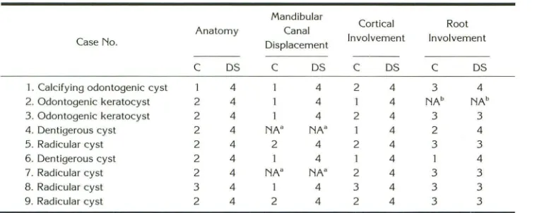
TABLE 1: Results: conventional films vs DentaScan
Mandibular
Anatomy Canal Cortical Root Case No. Displacement Involvement Involvement
c
DSc
DSc
DSc
DS1 . Calcifying odontogenic cyst 1 4 4 2 4 3 4 2. Odontogenic keratocyst 2 4 4 1 4 NAb NAb
3. Odontogenic keratocyst 2 4 4 2 4 3 3
4. Dentigerous cyst 2 4 NA" NA" 4 2 4
5. Radicular cyst 2 4 2 4 2 4 3 3
6. Dentigerous cyst 2 4 4 1 4 4
7. Radicular cyst 2 4 NA" NA" 2 4 3 3 8. Radicular cyst 3 4 4 3 4 3 3 9. Radicular cyst 2 4 2 4 2 4 3 3
Note.-C = conventional films; DS = DentaScan; 1 = poor; 2 = fair; 3 = good; 4 = excellent. • Lesion medial to mandibular canal.
b Edentulous in region of lesion.
ware program that
was
originally developed to
evaluate patients considering osseointegrated
dental implants (5-8) (metallic screw-like devices
that are surgically implanted in the mandible to
permit permanent fixation of dentures). The
pro-gram uses the axial scans to reformat
systemat-ically multiple cross-referenced panoramic and
cross-sectional images. The panoramic views are
reformatted parallel to a curved line
superim-posed on the axial image of the mandible; the
cross-sectional
images
are reformatted along
numbered lines drawn perpendicular to this (see
Figs. 2 and 3)
.
In this fashion the mandibular
canal and cortical margins are clearly delineated.
To assess the value of this program, we imaged
nine odontogenic cysts and compared the images
with the
conventional
films
.
Our findings and a
discussion of odontogenic cysts follow.
Methods
Nine proven odontogenic cysts of the mandible were
evaluated with conventional radiographs
(orthopantomo-graphic, intraoral, and/or mandibular films) and the dental CT software program (DentaScan, GE Medical, Milwaukee,
WI). The images from the two modalities were viewed separately and then simultaneously by a neuroradiologist
with expertise in this area. Films were evaluated for delin -eation of anatomy (inferior alveolar canal, mandibular for-amen, mental foramen), detection of neurovascular bundle
(inferior alveolar canal) displacement, detection of cortical involvement, and detection of root involvement. A grading
scale of 1 through 4 was used in which 1 = poor, 2
=
fair,3
=
good, and 4=
excellent.Image data for the software program was acquired on a GE 9800 CT scanner using a dynamic mode and bone
algorithm. Axial sections were acquired parallel to the
alveolar ridge of the mandible. One and one half millimeter
sections were obtained every millimeter, resulting in a 0.5-mm overlap.
Results
The nine lesions studied included one calcifying
odontogenic cyst, two odontogenic keratocysts
,
two dentigerous cysts
,
and four radicular cysts.
When the two modalities were compared,
DentaScan was found to be superior for
deline-ating anatomy and detecting mandibular canal
displacement, cortical involvement, and root
in-volvement. Results are summarized in Table 1
and described below.
Delineation of Anatomy (Mandibular Canal,
Mandibular Foramen, and Mental Foramen)
The mental foramen and inferior alveolar canal
were seen on the plain films but they were
visu-alized more clearly on the DentaScan images.
This is illustrated by comparing the plane film of
the calcifying odontogenic cyst (Fig. lA) to the
DentaScan (Figs
.
lC and
lD). Note how the
cross-sectional DentaScan images demonstrate the
ca-nal and establish its position
in
relation to the
lesion. Mandibular foramina were seen only on
the DentaScan images (Fig
.
2D).
Detection of Neurovascular Bundle (Mandibular
Canal) Displacement
Neurovascular bundle displacement was
ex-tremely difficult
to visualize
on plain films. The
plain
films
also
were
unable
to
determine the
position of the canal in
relation
to the lesion {Figs.
[image:2.612.126.506.91.242.2]AJNR: 14, March/ April 1993 ODONTOGENIC CYSTS 369
c
0
Fig. 1. Calcifying odontogenic cyst. Nine-year-old black girl with 3-month history of mandibular swelling.
A, Orthopantomogram. A double density reveals cortical expansion (small white arrows), but one cannot determine whether the buccal or lingual cortex is involved. The margins of the lesion are poorly defined and the neurovascular bundle is not identified. Superimpostion of the ectopic tooth (black arrows) makes it difficult to determine if resorption of the left first bicuspid root (large white arrow) has occurred. Note the normal follicle of the developing molar (black arrowhead).
B, Axial CT. What appears to be a single ectopic tooth on the orthopantomogram is actually two teeth (arrows). This is more clearly delineated on the cross-sectional images (D).
C, Panoramic DentaScan. Increased contrast permits clear delineation of the cortex (large arrowheads) and margins of the lesion (small arrowheads). Ectopic tooth (long arrow); left first bicuspid (wide arrow). Inferior alveolar canal (open straight arrows); follicle of developing molar (open curved arrow).
D, Cross-sectional DentaScan through lesion. Images clearly demonstrate expansion of the buccal cortex (short arrows), two teeth within the lesion (curved arrows), and displacement of the inferior alveolar canal (arrowheads).
images
,
however, clearly demonstrated
the
dis-placed neurovascular bundle
(Figs
.
lD
,
2C, and
3D)
.
DentaScan also established
the position of
the canal in relation to the lesion
.
In Figure lD,
note the d
i
splaced canal buccal to the ectopic
[image:3.612.53.561.78.560.2]370 ABRAHAMS
A
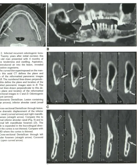
Fig. 2. Infected recurrent odontogenic kera-tocyst. Twenty years after initial excision this
52-year-old man presented with 4 months of
right jaw tenderness and swelling. Aspiration, which introduced air into the lesion, revealed gram positive organisms.
A, The curved line superimposed on the man-dible in this axial CT defines the plane and location of the reformatted panoramic images seen in B. The numbered lines drawn perpendic-ular to this define the plane and location of the
reformatted panoramic images seen in B. The
numbered lines drawn perpendicular to this de-fine the plane and location of the reformatted cross-sectional images in C and D. Odontogenic keratocyst (arrows).
B, Panoramic DentaScan. Lesion containing
air (large arrows); inferior alveolar canal (small arrows).
C, Cross-sectional DentaScan through lesion. Note the dramatic displacement of the inferior alveolar canal (curved arrows) and right mandib-ular foramen (straight arrow). Compare this to the normal inferior alveolar canal (Fig. 5) and to
the normal left mandibular foramen (D). The
mandible is expanded in the buccolingual direc-tion, but the cortex is not thinned. Compare with Figure 3D where the cortex is thinned.
D, Cross-sectional DentaScan through left
mandibular foramen (straight arrow). Coronoid
process (open curved arrow).
Detection of Cortical Involvement
B
c
D
Cortical involvement was more frequently and
clearly delineated on the DentaScan images. In a
calcifying odontogenic cyst, the
orthopantomo-gram (Fig. 1 A) demonstrates a double density in
AJNR: 14, March/ April 1993
[image:4.615.83.564.68.647.2]AJNR: 14, March/ April 1993
A
B
c
D
DentaScan (Figs. 1 B, 1 C, and 1 D), it was quite clear that only the buccal cortex was thinned. The degree of thinning and the size of the lesion was also more clearly demonstrated on the
DentaScan.
Similarly, in an odontogenic keratocyst, the cross-sectional images were instrumental in
de-ODONTOGENIC CYSTS 371
Fig. 3. Odontogenic keratocyst. Twenty-two-year-old man with an incidental left mandibular lesion discovered on films for a tripod fracture.
A, Orthopantomogram. The lesion (arrows) is clearly d e-lineated, but the neurovascular bundle is difficult to define.
B, Axial view. Note that the numbered lines superimposed on the image correspond to the numbers on the cr oss-sectional images in 0. Lesion (arrows).
C, Panoramic DentaScan. Lesion (arrows).
D, Cross-sectional DentaScan. The inferior alveolar canal is displaced to the bottom of the mandible (curved arrows)
and the buccal and lingual cortical margins are thinned
(straight arrows).
tecting buccolingual expansion (Fig. 2C). This is better seen by comparing the images of the unaffected left side (Fig. 2D) with the images of the right side, which contains the lesion (Fig. 2C).
In this case, it is of interest that despite the
372 ABRAHAMS
the odentogenic keratocyst in Figure 3D where
the buccal and lingual cortex is thinned. This type
of detail is not appreciated on the plain films. In Figure 4, a dentigerous cyst can be identified on the orthopantomogram but cortical
involve-ment cannot established. On the cross-sectional
DentaScan images (Fig. 4C), the thinning of the
buccal cortex is readily visualized.
Detection of Root Involvement
Both conventional films and DentaScan were
rated 'good' for evaluating root involvement.
When teeth were superimposed on plain films,
however, the DentaScan was superior. For ex
-ample, on the orthopantomogram of a dentiger-ous cyst, the unerupted cuspid within the cyst obscures the roots of the incisors (Fig. 4A) and
in the case of a calcifying odontogenic cyst the
ectopic tooth partially obscures the root of the first bicuspid (Fig. 1A). The multiplanar format of DentaScan alleviated these problems. In the
dentigerous cyst case, the cross-sectional images
(Fig. 4C) clearly separated the unerupted cuspid
from the incisors, and demonstrated that root
resorption was not present. The ectopic tooth in
the calcifying odontogenic cyst was actually
shown by the DentaScan to be two teeth (Fig.
1 D). The images also demonstrate that there was
no erosion of the first bicuspid root.
In addition to the above points, DentaScan was also judged to be superior because of its
higher-contrast resolution. For example, comparing the
orthopantomographic image of the calcifying
odontogenic cyst (Fig. 1A) with the DentaScan
panoramic image (Fig. 1 C), the increased contrast
of the DentaScan allowed better differentiation of
cortical from cancellus bone. This was
particu-larly evident on the cross-sectional DentaScan
images (Fig. 1D). In addition, the increased
con-trast allowed better resolution of the lesion and
better definition of its margins, as illustrated by
the calcifying odontogenic cyst (Fig. 1) and the
dentigerous cyst (Fig. 4). In both of these cases,
the margins and internal characteristics of the
lesions were noted on the DentaScan but not on
the orthopantomogram.
Discussion
We have demonstrated in this limited study
that dental CT software programs are useful for
evaluating odontogenic cysts. The cross-sectional
image, which is unique to these programs, was
AJNR: 14, March/ April 1993
B
c
Fig. 4. Dentigerous cyst of left cuspid. Fourteen-year-old boy with lesion discovered on routine dental films.
A, Orthopantomogram. The dentigerous cyst surrounding the
unerupted left cuspid is actually seen twice because of distortion near the midline (arrowheads). Note that the root of the primary cuspid on the left (thin arrow) is much shorter than the root of the permanent cuspid on the right (curved arrow).
B, Panoramic DentaScan. The increased contrast permits better
delineation between the surrounding tissue and the unerupted left cuspid and dentigerous cyst (large solid arrows). Primary left cuspid (solid curved arrow); permanent right cuspid (thin arrow);
mental foramina (open curved arrows).
C, Cross-sectional DentaScan. The relation of the lesion to the roots of the right (curved arrow) and left (straight arrow) central
[image:6.612.318.560.91.555.2]AJNR: 14, March/ April 1993
felt to be most valuable. It enables one to differ-entiate buccal from lingual cortical involvement and to determine the position of the neurovas-cular bundle in relation to the lesion. It should be pointed out that since these images are acquired using low technique, they are optimal only for evaluating the osseous jaw and its contents. Sur-rounding soft tissue is better studied with magnetic resonance.
In addition to supplying important diagnostic information, DentaScan was instrumental in
sur-gical management. The cortical margins of odon-togenic cysts may be remarkably thin or absent at various points along the cyst. Identifying these areas and planning the surgical approach through them will diminish the chance of postoperative fracture. Identification of the neurovascular bun-dle will help prevent intraoperative hemorrhage and/or postoperative parathesia.
Since some odontogenic cysts are more ag-gressive than others, it is important to develop an understanding of their classification and radio-graphic appearance. In the past, the classification (1, 9, 10) has undergone change, but the following groups are generally accepted today (1): dentig-erous cyst, radicular cyst, lateral periodontal cyst, gingival cyst, odontogenic keratocyst, and calci-fying odontogenic keratocyst. The gingival cyst is not radiographically apparent and will not be discussed.
Dentigerous cysts (follicular cysts) (1, 9, 10, 11) (Fig. 4) are well-circumscribed radiolucent lesions that develop around the crown of an impacted or unerupted tooth. Common sites are where teeth often impact-the upper and lower third molars and maxillar cuspids. The lesion has a thin sclerotic rim and is typically unilocular, but occasionally multilocular. The enlarging cyst,
which is benign, may show aggressive features including expansion of bone, displacement of teeth, and root resorption. A tooth within the cyst
helps differentiate it from other cystic lesions of the jaw. It should not be confused with the smaller (less than 3 mm) (11) follicular space, a normal radiolucency that surrounds developing teeth (Figs. 1A and 1C). Treatment consists of removal of the associated tooth and enucleation of the
cyst.
Radicular cysts (referring to the root) (1, 9),
also know as apical periodontal or periapical cysts, are the most common odontogenic cysts.
They appear as small periapical lucencies that
arise from preexisting infection. The infection typically enters the pulp chamber and root canal
ODONTOGENIC CYSTS 373
via a carious tooth. The cyst's proximity to the
root apex helps differentiate it from other cystic
lesions of the jaw. Treatment consists of
extrac-tion of the nonvital tooth and curetting of the
cyst. If the cyst is incompletely removed, a
"re-sidual cyst" may remain ( 1 0).
Lateral periodontal cysts are nonkeratinized,
noninflammatory developmental cysts that are
intimately related to the lateral root surface of an
erupted tooth. The borders are often well
margin-ated and frequently contain a fine sclerotic rim.
There is a predilection for the mandibular
bicus-pid, cuspid, or incisor (12), and there is a
male-to-female ratio of 2:1. Since the cysts rarely
exceed 10 mm, they usually do not displace teeth
and are considered relatively nonaggressive.
Sur-gery is curative, and recurrence is rare.
The odontogenic keratocyst (1, 9-11, 13-15) is a lesion that has stirred much controversy
re-garding its actual classification and etiology.
Ini-tially, the term was used to describe odontogenic
cysts containing keratin. This led to confusion
because other odontogenic cysts (dentigerous,
radicular, and residual) occasionally contain
ker-atin. Odontogenic keratocysts have now been
shown to contain a specific type of keratin
(pa-rakeratin) and to be histologically and clinically
distinct (1). Radiographically, they appear as uni-locular or multiuni-locular lucent lesions with sharply
demarcated borders. The cyst has propensity for
rapid growth and can be locally aggressive with
expansion of bone and displacement of teeth.
Most occur in the third molar region and often
involve the ramus as illustrated in Figures 2 and
3. Odontogenic keratocysts may be difficult to
differentiate from other odontogenic cysts, partic
-ularly when small. Distinguishing features are
their rapid growth, local aggressiveness, and fre -quent occurrence in the ramus. There is a very
high recurrence rate, even years after surgical
excision (9, 14, 15) (Fig. 2).
Finally, the calcifying odontogenic cyst (1, 9,
10, 16) (Fig. 1) is a unilocular or multilocuolar
radiolucent lesion containing variable amounts of
calcified material. The amount of calcium
in-creases with the age of the lesion. The margins
are well defined and about 70% occur in the
maxilla ( 1 ). Treatment is surgical because of their
tendency for continued growth.
In summary, odontogenic cysts form a
com-plex group of lesions that can be locally
aggres-sive as demonstrated by expansion of the
man-dible, erosion of the cortex, displacement of the
374 ABRAHAMS
of the teeth. Demonstrating these changes
radio-graphically is important for establishing a
diag-nosis and determining a
surgical
approach.
Tradi-tionally,
these lesions have been evaluated in the
dentist's office with standard
orthopantomo-graphic, intraoral, and mandibular films. We have
demonstrated, however,
that
dental CT software
programs are superior for evaluating these cysts.
They provide multiplanar images and are able to
detect subtle changes such as cortical
involve-ment and neurovascular bundle displaceinvolve-ment.
Therefore, we believe that this should be the
study of choice when evaluating these and other
osseous
lesions
of the mandible.
Acknowledgments
The authors thank Linda Abrahams for her assistance
with editing and Nancy Judd and Phyllis Festa for their
assistance in preparing this manuscript.
References
I. Regezi JA, Scrubbs JJ. Oral pathology: clinical-pathologic correla-tions. Philadelphia: Saunders, 1989:301-336
2. MacKenzie GD, Oatis GW, Mullen MP, Grisius RJ. Computed tomog-raphy in the diagnosis of an odontogenic keritocyst. Oral Surg Oral Med Oral Pathol 1985;59:302-305
3. DeiBalso AM, Wernig JT. The role of computed tomography in the evaluation of cemento-osseous lesions. Oral Surg Oral Med Oral Pathol 1986;62:354-357
AJNR: 14, March/ April 1993
4. Cohen MA, Hertzom Y, Mendelsohn DB. Computed tomography: the diagnosis and treatment of mandibular ameloblastoma. J Oral Max-illofac Surg 1985;43: 796-80 I
5. Abrahams JJ, Levine B. Expanded applications of DentaScan. lnt J Periodontics Restorative Dent 1990;10:465-471
6. Rothman SLG, Schwarz MS, Chafetz N, et al. CT in the preoperative assessment of the mandible and maxilla for endosseous implant
surgery. Radiology 1988;168:171-175
7. Schwarz MS, Rothman SLG, Rhodes ML, Chafetz N. Computed tomography. I. Preoperative assessment of the mandible for endos-seous implant surgery. lnt J Oral Maxillofac Implant 1987;2:137-141
8. Schwarz MS, Rothman SLG, Rhodes ML, Chafetz N. Computed tomography. II. Preoperative assessment of the maxilla for endos -seous implant surgery. lnt J Oral Maxillofac Implant 1987;2:143-148 9. Shaffer WG, Hine MK, Levy BM, eds. A textbook of oral pathology.
4th ed. Philadelphia: Saunders, 1983:258-317
10. Gibilisco JA, ed. Stafne's oral radiographic diagnosis. 5th ed. Phila-delphia: Saunders, 1985:159-171
II. Langlalis RP. Radiology of the jaws. In: Dalbalso AM, ed. Maxillofacial imaging. Philadelphia: Saunders, 1990:313-373
12. Fantasia JE. Lateral periodental cyst. Oral Surg Oral Med Oral Pathol
1979;48:237 -243
13. Toller P. Origin and growth of cysts of the jaws. Ann R Coli Surg
Engll987;40:306-336
14. Brannon MB. The odontogenic keratocyst: a clinicopathologic study of 312 cases. I. Clinical features. Oral Surg Oral Med Oral Pathol
1976;42:54-72
15. Payne TF. An analysis of the clinical and histopathologic parameters of the odonogenic keratocyst. Oral Surg Oral Med Oral Pathol 1972;33:538-546