(1
E
,2
E
)-1,2-Bis(2,3,4-trimethoxybenzyl-idene)hydrazine
Malai Haniti S. A. Hamid,aMohammad Akbar Ali,a Aminul Huq Mirza,aGan Ai Lenaand Ray J. Butcherb*
aFaculty of Science, Universiti Brunei Darussalam, Jln Tungku Link, BE 1410, Negara Brunei Darussalam, andbDepartment of Chemistry, Howard University, 525 College Street NW, Washington, DC 20059, USA
Correspondence e-mail: [email protected]
Received 25 August 2010; accepted 31 August 2010
Key indicators: single-crystal X-ray study;T= 100 K; mean(C–C) = 0.002 A˚; Rfactor = 0.048;wRfactor = 0.123; data-to-parameter ratio = 16.9.
The title compound, C20H24N2O6, was obtained as an
unexpected product by the reaction of hydrazinium dithio-carbazate with 2,3,4-trimethoxybenzaldehyde in refluxing ethanol. The molecule lies on a center of inversion. The crystal packing is stabilized by weak intermolecular C—H O interactions.
Related literature
The surprising formation of the title hydrazone was probably due to the decomposition of hydrazinium dithiocarbazate in solution resulting in the formation of hydrazine, which then reacted with 2,3,4-trimethoxybenzaldehdye. Hydrazinium dithiocarbazates are known to decompose on heating (Rudorf, 2007). For the biological activity of Schiff bases, see: Akbar Ali
et al. (2008); Chanet al.(2008). For a previous report of the title compound (the X-ray structure was not provided), see: Praefckeet al.(1991). For comparison bond lengths in an aroyl hydrazone, see: Jiet al.(2010).
Experimental
Crystal data
C20H24N2O6 Mr= 388.41
Monoclinic,P21=n
a= 10.0380 (9) A˚ b= 7.0713 (7) A˚ c= 13.9586 (14) A˚
= 102.800 (2) V= 966.18 (16) A˚3
Z= 2
MoKradiation
= 0.10 mm1 T= 100 K
0.600.360.04 mm
Data collection
Bruker SMART CCD area-detector diffractometer
Absorption correction: multi-scan (SADABS; Sheldrick, 1996) Tmin= 0.943,Tmax= 0.996
6576 measured reflections 2203 independent reflections 1846 reflections withI> 2(I) Rint= 0.033
Refinement
R[F2> 2(F2)] = 0.048
wR(F2) = 0.123 S= 1.05 2196 reflections
130 parameters
H-atom parameters constrained
max= 0.35 e A˚
3
min=0.20 e A˚
3
Table 1
Hydrogen-bond geometry (A˚ ,).
D—H A D—H H A D A D—H A
C7—H7B O3ii 0.98 2.54 3.3620 (18) 142 C8—H8B O1iii
0.98 2.62 3.3735 (19) 134 C8—H8B O2iii
0.98 2.62 3.5711 (18) 164
Symmetry codes: (ii)xþ1 2;yþ
1 2;zþ
1 2; (iii)xþ
3 2;y
1 2;zþ
3 2.
Data collection:SMART(Bruker, 1998); cell refinement: SAINT-Plus(Bruker, 1998); data reduction:SAINT-Plus; program(s) used to solve structure: SHELXS97(Sheldrick, 2008); program(s) used to refine structure:SHELXL97(Sheldrick, 2008); molecular graphics: SHELXTL(Sheldrick, 2008); software used to prepare material for publication:SHELXTL.
MHSAH, MAA, AHM and GAL thank Universiti Brunei Darussalam for support. The X-ray Diffraction Laboratory, Department of Chemistry, National University of Singapore, is acknowledged for the collection of the X-ray diffraction data.
Supplementary data and figures for this paper are available from the IUCr electronic archives (Reference: BT5339).
References
Akbar Ali, M., Abu Bakar, J. H., Mirza, A. H., Smith, S. J., Gahan, L. R. & Bernhardt, P. V. (2008).Polyhedron,27, 71–79.
Bruker (1998). SAINT-Plus and SMART. Bruker AXS Inc., Madison, Wisconsin, USA.
Chan, M.-H. E., Crouse, K. A., Tahir, M. I. M., Rosli, R., Umar-Tsafe, N. & Cowley, A. R. (2008).Polyhedron,27, 1141–1149.
Ji, N.-N., Shi, A.-Q., Zhao, R.-G., Zheng, Z.-B. & Li, Z.-F. (2010).Bull. Korean Chem. Soc.31, 881–886.
Praefcke, K., Kohne, B., Guendogan, B., Singer, D. & Demus, D. (1991).Mol. Cryst. Liq. Cryst.198, 393–405.
Rudorf, W.-D. (2007).J. Sulfur Chem.28, 295–339.
Sheldrick, G. M. (1996).SADABS. University of Go¨ttingen, Germany. Sheldrick, G. M. (2008).Acta Cryst.A64, 112–122.
Acta Crystallographica Section E
Structure Reports Online
supporting information
Acta Cryst. (2010). E66, o2557 [doi:10.1107/S160053681003518X]
(1
E
,2
E
)-1,2-Bis(2,3,4-trimethoxybenzylidene)hydrazine
Malai Haniti S. A. Hamid, Mohammad Akbar Ali, Aminul Huq Mirza, Gan Ai Len and Ray J.
Butcher
S1. Comment
The compound, C20H24N2O6 (I) was obtained by the reaction of hydrazinium dithiocarbazate and
2,3,4-trimethoxy-benzaldehyde in boiling ethanol. The surprising formation of the hydrazone was probably due to the decomposition of
hydrazinium dithiocarbazate in solution resulting in the formation of hydrazine, which then reacted with
2,3,4-trimeth-oxybenzaldehdye to form the corresponding hydrazone (I). Hydrazinium dithiocarbazates are known to decompose on
heating (Rudorf, 2007).
Schiff bases have attracted considerable attention because they can act as chelating agents for metal ions and many of
them also exhibit useful biological activities (Akbar Ali et al., 2008; Chan et al., 2008). Although the compound has
previously been reported its X-ray structure has not been provided (Praefcke et al., 1991). Hydrazones derived from the
reactions of hydrazines with aldehydes or ketones are common but bis-hydrazones are not.
The molecular structure of (I) is shown in Figure 1 and its selected bond lengths and angles are given in Table 1. Like
most thiosemicarbazones and Schiff bases, the imine moiety in [I] shows an E configuration about the C10—N1
[1.283 (2) Å] and N1A—C10A bonds. The C10—N1 and N1A—C10A bond distances also compare well with C=N
double bonds in other related compounds. A comparison of the N(1)—N(1 A) distance [1.413 (3) Å] with that in an aroyl
hydrazone [1.377 (3) Å] (Ji et al. 2010) shows that the bond is shorter than a single N—N bond (1.44 Å) indicating that a
significant π-charge delocalization occurs along the C—N—N—C moiety. As the bond angles C6—C10—N1 (121.68°)
and C6A—C10A—N1A (121.68°) are close to that of a sp2-hybridized carbon atom (ca 120°), the molecule does not
have a distorted geometry. Due the fact that the molecule lies on a center of inversion the dihedral angle between the two
phenyl rings is 0.0°.
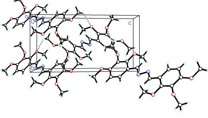
Figure 2 shows the packing of (I) in the unit cell. The packing diagram shows that there are intermolecular hydrogen
bonds between one of the CH3 hydrogen atoms of one molecule with an ether oxygen of another molecule.
S2. Experimental
2,3,4-trimethoxybenzaldehyde (0.24 g, 1.24 mmol) dissolved in absolute ethanol (5 ml) was mixed with a solution of
hydrazinium dithiocarbazate (0.93 g, 0.66 mmol) in the same solvent (45 ml). After refluxing for two hours, the resulting
clear yellow solution was left to stand at room temperature for five days to afford crystalline yellow plates. The crystals
were filtered, washed with cold absolute ethanol and dried in vacuo. Yield: 0.152 g (63%); m.p. 192–194 °C; IR (KBr,
cm-1): 2968, 2937, 2832, 1614, 1590, 1494, 1457, 1431, 1410, 1286, 1229, 1199. 1166, 1090, 1023, 1008, 943, 898, 809,
699, 667, 594, 540, 433; 1H NMR (400 MHz, CDCl
3, 30 °C): δ 8.92 (2H, s, CH=N), 7.84 (2H, d, ArH), 6.76 (2H, d,
ArH), 3.96 (6H, s, OCH3), 3.92 (6H, s, OCH3), 3.90 (6H, s, OCH3); Anal. Calcd. for C20H24N2O6 (388.42): C 61.85, H
IR spectrum was recorded as a KBr disc with 13 mm KBr discs SPECAC accessory on a Perkin-Elmer 1600 F T IR
spectrometer. 1H NMR spectrum was run in CDCl
3 on a Varian 400-NMR spectrometer at Universiti Brunei Darussalam.
Elemental analysis for C, H and N was done by the Elemental Analysis Laboratory, Department of Chemistry, National
University of Singapore. The X-ray data were collected at the X-ray Diffraction Laboratory, Department of Chemistry,
National University of Singapore using a Bruker-AXS Smart Apex CCD single-crystal diffractometer.
S3. Refinement
H atoms were placed in geometrically idealized positions and constrained to ride on their parent atoms with a C—H
[image:3.610.125.480.204.381.2]distances of 0.95 Å and 0.98 Å, Uiso(H) = 1.2Ueq(C).
Figure 1
The title compound, C20H24N2O6 with atom labeling. Displacement ellipsoids are at the 50 % probability level.
Figure 2
[image:3.610.128.480.426.626.2](1E,2E)-1,2-Bis(2,3,4-trimethoxybenzylidene)hydrazine
Crystal data
C20H24N2O6
Mr = 388.41 Monoclinic, P21/n
a = 10.0380 (9) Å
b = 7.0713 (7) Å
c = 13.9586 (14) Å
β = 102.800 (2)°
V = 966.18 (16) Å3
Z = 2
F(000) = 412
Dx = 1.335 Mg m−3
Mo Kα radiation, λ = 0.71073 Å Cell parameters from 1770 reflections
θ = 2.8–27.1°
µ = 0.10 mm−1
T = 100 K Plate, yellow
0.60 × 0.36 × 0.04 mm
Data collection
Bruker SMART CCD area-detector diffractometer
Radiation source: fine-focus sealed tube Graphite monochromator
ω scans
Absorption correction: multi-scan (SADABS; Sheldrick, 1996)
Tmin = 0.943, Tmax = 0.996
6576 measured reflections 2203 independent reflections 1846 reflections with I > 2σ(I)
Rint = 0.033
θmax = 27.5°, θmin = 2.3°
h = −13→12
k = 0→9
l = 0→18
Refinement
Refinement on F2
Least-squares matrix: full
R[F2 > 2σ(F2)] = 0.048
wR(F2) = 0.123
S = 1.05 2196 reflections 130 parameters 0 restraints
Primary atom site location: structure-invariant direct methods
Secondary atom site location: difference Fourier map
Hydrogen site location: inferred from neighbouring sites
H-atom parameters constrained
w = 1/[σ2(F
o2) + (0.0608P)2 + 0.353P]
where P = (Fo2 + 2Fc2)/3
(Δ/σ)max < 0.001
Δρmax = 0.35 e Å−3
Δρmin = −0.20 e Å−3
Special details
Geometry. All e.s.d.'s (except the e.s.d. in the dihedral angle between two l.s. planes) are estimated using the full covariance matrix. The cell e.s.d.'s are taken into account individually in the estimation of e.s.d.'s in distances, angles and torsion angles; correlations between e.s.d.'s in cell parameters are only used when they are defined by crystal symmetry. An approximate (isotropic) treatment of cell e.s.d.'s is used for estimating e.s.d.'s involving l.s. planes.
Refinement. Refinement of F2 against ALL reflections. The weighted R-factor wR and goodness of fit S are based on F2,
conventional R-factors R are based on F, with F set to zero for negative F2. The threshold expression of F2 > σ(F2) is used
only for calculating R-factors(gt) etc. and is not relevant to the choice of reflections for refinement. R-factors based on F2
are statistically about twice as large as those based on F, and R- factors based on ALL data will be even larger.
Fractional atomic coordinates and isotropic or equivalent isotropic displacement parameters (Å2)
x y z Uiso*/Ueq
O1 0.62846 (10) 0.45681 (14) 0.85047 (7) 0.0180 (2)
O2 0.61624 (9) 0.20990 (14) 0.70188 (7) 0.0165 (2)
O3 0.38623 (10) 0.18335 (14) 0.55439 (7) 0.0175 (2)
C10 0.15962 (14) 0.4221 (2) 0.54359 (10) 0.0154 (3)
C1 0.29226 (14) 0.5791 (2) 0.69604 (11) 0.0172 (3)
H1 0.2190 0.6649 0.6944 0.021*
C2 0.40646 (15) 0.5899 (2) 0.77231 (10) 0.0180 (3)
H2 0.4113 0.6834 0.8218 0.022*
C3 0.51481 (14) 0.4635 (2) 0.77677 (10) 0.0153 (3)
C4 0.50841 (13) 0.33003 (19) 0.70148 (10) 0.0146 (3)
C5 0.39310 (14) 0.32122 (19) 0.62483 (10) 0.0147 (3)
C6 0.28211 (14) 0.44466 (19) 0.62113 (10) 0.0149 (3)
C7 0.64403 (15) 0.6012 (2) 0.92428 (11) 0.0202 (3)
H7A 0.6488 0.7252 0.8938 0.030*
H7B 0.7282 0.5789 0.9740 0.030*
H7C 0.5657 0.5983 0.9555 0.030*
C8 0.60816 (16) 0.0407 (2) 0.75753 (11) 0.0218 (3)
H8A 0.6081 0.0745 0.8256 0.033*
H8B 0.6871 −0.0401 0.7563 0.033*
H8C 0.5238 −0.0274 0.7286 0.033*
C9 0.44808 (17) 0.2382 (2) 0.47521 (11) 0.0250 (4)
H9A 0.3970 0.3440 0.4393 0.038*
H9B 0.4466 0.1309 0.4305 0.038*
H9C 0.5428 0.2769 0.5017 0.038*
N1 0.05345 (12) 0.52567 (17) 0.53984 (8) 0.0165 (3)
Atomic displacement parameters (Å2)
U11 U22 U33 U12 U13 U23
O1 0.0149 (5) 0.0161 (5) 0.0206 (5) 0.0020 (4) −0.0011 (4) −0.0043 (4)
O2 0.0135 (5) 0.0140 (5) 0.0223 (5) 0.0028 (4) 0.0046 (4) 0.0010 (4)
O3 0.0191 (5) 0.0148 (5) 0.0188 (5) −0.0001 (4) 0.0043 (4) −0.0027 (4)
C10 0.0161 (6) 0.0143 (7) 0.0160 (6) 0.0000 (5) 0.0043 (5) 0.0016 (5)
C1 0.0152 (7) 0.0160 (7) 0.0206 (7) 0.0040 (5) 0.0045 (5) 0.0010 (5)
C2 0.0199 (7) 0.0159 (7) 0.0183 (7) 0.0008 (6) 0.0043 (6) −0.0041 (5)
C3 0.0119 (6) 0.0155 (7) 0.0175 (7) −0.0021 (5) 0.0014 (5) 0.0012 (5)
C4 0.0130 (6) 0.0130 (7) 0.0189 (7) 0.0015 (5) 0.0055 (5) 0.0020 (5)
C5 0.0167 (6) 0.0128 (7) 0.0155 (6) −0.0011 (5) 0.0055 (5) 0.0002 (5)
C6 0.0136 (6) 0.0146 (7) 0.0164 (6) −0.0006 (5) 0.0028 (5) 0.0025 (5)
C7 0.0203 (7) 0.0190 (7) 0.0196 (7) −0.0001 (6) 0.0009 (6) −0.0035 (6)
C8 0.0277 (8) 0.0176 (8) 0.0212 (7) 0.0069 (6) 0.0082 (6) 0.0040 (6)
C9 0.0320 (8) 0.0244 (8) 0.0206 (7) 0.0016 (7) 0.0098 (6) −0.0024 (6)
N1 0.0146 (6) 0.0185 (6) 0.0154 (6) −0.0004 (5) 0.0012 (5) 0.0020 (5)
Geometric parameters (Å, º)
O1—C3 1.3572 (16) C3—C4 1.4036 (19)
O1—C7 1.4344 (17) C4—C5 1.3926 (19)
O2—C4 1.3750 (16) C5—C6 1.4072 (19)
O2—C8 1.4383 (17) C7—H7A 0.9800
O3—C5 1.3755 (17) C7—H7B 0.9800
C10—N1 1.2846 (19) C8—H8A 0.9800
C10—C6 1.4556 (19) C8—H8B 0.9800
C10—H10 0.9500 C8—H8C 0.9800
C1—C2 1.383 (2) C9—H9A 0.9800
C1—C6 1.400 (2) C9—H9B 0.9800
C1—H1 0.9500 C9—H9C 0.9800
C2—C3 1.398 (2) N1—N1i 1.411 (2)
C2—H2 0.9500
C3—O1—C7 117.32 (11) C1—C6—C10 122.64 (13)
C4—O2—C8 112.18 (11) C5—C6—C10 119.40 (13)
C5—O3—C9 113.41 (11) O1—C7—H7A 109.5
N1—C10—C6 121.62 (13) O1—C7—H7B 109.5
N1—C10—H10 119.2 H7A—C7—H7B 109.5
C6—C10—H10 119.2 O1—C7—H7C 109.5
C2—C1—C6 121.54 (13) H7A—C7—H7C 109.5
C2—C1—H1 119.2 H7B—C7—H7C 109.5
C6—C1—H1 119.2 O2—C8—H8A 109.5
C1—C2—C3 120.20 (13) O2—C8—H8B 109.5
C1—C2—H2 119.9 H8A—C8—H8B 109.5
C3—C2—H2 119.9 O2—C8—H8C 109.5
O1—C3—C2 124.83 (13) H8A—C8—H8C 109.5
O1—C3—C4 115.81 (12) H8B—C8—H8C 109.5
C2—C3—C4 119.36 (12) O3—C9—H9A 109.5
O2—C4—C5 119.67 (12) O3—C9—H9B 109.5
O2—C4—C3 120.47 (12) H9A—C9—H9B 109.5
C5—C4—C3 119.85 (12) O3—C9—H9C 109.5
O3—C5—C4 118.84 (12) H9A—C9—H9C 109.5
O3—C5—C6 119.97 (12) H9B—C9—H9C 109.5
C4—C5—C6 121.13 (13) C10—N1—N1i 111.38 (15)
C1—C6—C5 117.88 (13)
C6—C1—C2—C3 −0.8 (2) O2—C4—C5—O3 4.25 (19)
C7—O1—C3—C2 −6.1 (2) C3—C4—C5—O3 −176.89 (12)
C7—O1—C3—C4 174.78 (12) O2—C4—C5—C6 −178.71 (12)
C1—C2—C3—O1 −177.00 (13) C3—C4—C5—C6 0.2 (2)
C1—C2—C3—C4 2.1 (2) C2—C1—C6—C5 −0.9 (2)
C8—O2—C4—C5 −93.85 (15) C2—C1—C6—C10 175.88 (13)
C8—O2—C4—C3 87.29 (15) O3—C5—C6—C1 178.16 (12)
O1—C3—C4—O2 −3.77 (19) C4—C5—C6—C1 1.1 (2)
C2—C3—C4—O2 177.08 (13) O3—C5—C6—C10 1.3 (2)
O1—C3—C4—C5 177.38 (12) C4—C5—C6—C10 −175.69 (12)
C2—C3—C4—C5 −1.8 (2) N1—C10—C6—C1 0.1 (2)
C9—O3—C5—C4 −87.17 (15) N1—C10—C6—C5 176.74 (13)
C9—O3—C5—C6 95.76 (15) C6—C10—N1—N1i −178.28 (13)
Hydrogen-bond geometry (Å, º)
D—H···A D—H H···A D···A D—H···A
C7—H7B···O3ii 0.98 2.54 3.3620 (18) 142
C8—H8B···O1iii 0.98 2.62 3.3735 (19) 134
C8—H8B···O2iii 0.98 2.62 3.5711 (18) 164
![Crystal structure of benzyl N′ [(1E,4E) 1,5 bis(4 methoxyphenyl)penta 1,4 dien 3 ylidene]hydrazine 1 carbodithioate](data:image/gif;base64,R0lGODlhAQABAIAAAP///wAAACH5BAEAAAAALAAAAAABAAEAAAICRAEAOw==)