COMPARISON OF AC IMPEDANCE SPECTROSCOPY TECHNIQUE WITH CONVENTIONAL METHODS
FOR DETECTION OF OCCLUSAL CARIES IN PRIMARY TEETH
*1
Dr. Farhin Katge,
1
Professor and Head, Department of Paedodontics and Preventive Dentistry,
2Lecturer, Department of Pedodontics and Preventive Dentistry, DY Patil University
3
Reader, Department of Paedodontics and Preventive Dentistry,
ARTICLE INFO ABSTRACT
Purpose: (CarieScan PRO in primary teeth. Method:
occlusal caries by tactile examination, bitewing radiography a Results:
caries. The highest sensitivity was seen with CarieScan PRO
radiography (79.41%) and tactile examination (76.47%).95.24% specificity wa radiography, 92.86% specificity was seen with CarieScan PRO
with 71.43% which was least specific.
radiography (16.68), followed by CarieScan PRO
examination (0.33) showedhighest negative likelihood ratio followed by bitewing radiography (0.22) and least with CarieScan PRO
was signifi Conclusions:
better than tactile examination and bitewing radiography in early caries detection on non cavitated pits and fissures of primary
Copyright©2017, Dr. Farhin Katge et al. Thisis an open access article distributed under the Creative Commons Att use, distribution, and reproduction in any medium, provided the original work is properly
INTRODUCTION
Occlusal surfaces of both primary and permanent dentitions in children account for majority of new carious lesions 1988). These surfaces are susceptible to dental caries due to the complex anatomy of pits and fissures. Secondly, due to the minute dimensions of the pits and fissures, a toothbrush cannot access the area to disturb the microorganisms and clean the area (Ekstrand 2001). Accurate detection of depth of occlusal caries is challenging unless cavitation is visible. Non cavitated dentinal lesions are more prevalent as a result of little to no demineralization on occlusal enamel surfaces making detection much more difficult (Sawle 1988; Weerheijm 1989).
*Corresponding author: Dr. Farhin Katge,
Professor and Head, Department of Paedodontics and Preventive Dentistry, TPCT’s Terna Dental College and Hospital, Navi Mumbai
ISSN: 0975-833X
Vol.
Article History:
Received 09th August, 2017
Received in revised form 20th September, 2017
Accepted 21st October, 2017 Published online 30th November, 2017
Citation: Dr. Farhin Katge, Dr Ashveeta J. Shetty and
conventional methods for detection of occlusal caries in primary teeth
Key words:
Dental caries, Primary teeth, CarieScan, Bitewing radiography, Tactile examination.
RESEARCH ARTICLE
COMPARISON OF AC IMPEDANCE SPECTROSCOPY TECHNIQUE WITH CONVENTIONAL METHODS
FOR DETECTION OF OCCLUSAL CARIES IN PRIMARY TEETH
Dr. Farhin Katge,
2Dr Ashveeta J. Shetty and
3Dr. Manohar Poojari
Professor and Head, Department of Paedodontics and Preventive Dentistry, Terna Dental College,
Navi Mumbai, India
Lecturer, Department of Pedodontics and Preventive Dentistry, DY Patil University
Navi Mumbai, India
Department of Paedodontics and Preventive Dentistry, Terna Dental College,
Navi Mumbai, India
ABSTRACT
Purpose: To compare effectiveness of alternating current impedance spectroscopy technique (CarieScan PROTM) with tactile examination and bitewing radiography in detection of occlusal caries in primary teeth.
Method: 110 primary molars in 40 patients aged 6 to 12 years were examined for presence of occlusal caries by tactile examination, bitewing radiography and CarieScan PRO
Results: Out of 110 teeth, 68 showed presence of dentinal caries and 42 showed presence of enamel caries. The highest sensitivity was seen with CarieScan PROTM
radiography (79.41%) and tactile examination (76.47%).95.24% specificity wa radiography, 92.86% specificity was seen with CarieScan PROTM
with 71.43% which was least specific. Highest positive likelihood ratio was seen with bitewing radiography (16.68), followed by CarieScan PROTM (12.97) and tactile examination (2.68). Tactile examination (0.33) showedhighest negative likelihood ratio followed by bitewing radiography (0.22) and least with CarieScan PROTM (0.079). The diagnostic accuracy of CarieScan PRO
was significant (AUC = 0.928, P < 0.001).
Conclusions: In terms of sensitivity, likelihood ratios and accuracy, CarieScan PRO
better than tactile examination and bitewing radiography in early caries detection on non cavitated pits and fissures of primary teeth.
is an open access article distributed under the Creative Commons Attribution License, which reproduction in any medium, provided the original work is properly cited.
Occlusal surfaces of both primary and permanent dentitions in children account for majority of new carious lesions (Ripa, susceptible to dental caries due to Secondly, due to the the pits and fissures, a toothbrush bristle disturb the microorganisms and clean urate detection of depth of occlusal caries is challenging unless cavitation is visible. Non cavitated dentinal lesions are more prevalent as a result of little to no demineralization on occlusal enamel surfaces making 1988; Weerheijm 1989).
Professor and Head, Department of Paedodontics and Preventive Dentistry, TPCT’s Terna Dental College and Hospital, Navi Mumbai
Even when occlusal caries are detected, the extent of caries progression and whether it should be treated is debated (Ashley P 2000). It is of increasing clinical importance that early lesions are detected to prevent further progression of dental caries into the tooth (Hill
carious lesions is focused on the identification of early mineral changes. Various techniques of caries detection are available like visual examination, tactile examination, conventional radiography, digital radiography, transill
Imaging Fiber-Optic Transillumination
illumination, near-IR light imaging, quantitative light fluorescence, DIAGNOdent, endoscope, ultra sound imaging, caries detection dyes, electronic caries meter and alternating current impedance spectroscopy technique (ACIST). CarieScan PROTM device is based on the technology of ACIST. It involves passing of an insensitive level of electrical current through the tooth to identify the presence and location
International Journal of Current Research
Vol. 9, Issue, 11, pp.60577-60582, November, 2017
Dr. Farhin Katge, Dr Ashveeta J. Shetty and Dr. Manohar Poojari, 2017. “Comparison of ac impedance spectroscopy technique with conventional methods for detection of occlusal caries in primary teeth”, International Journal of Current Research, 9, (11),
COMPARISON OF AC IMPEDANCE SPECTROSCOPY TECHNIQUE WITH CONVENTIONAL METHODS
FOR DETECTION OF OCCLUSAL CARIES IN PRIMARY TEETH
Dr. Manohar Poojari
Terna Dental College,
Lecturer, Department of Pedodontics and Preventive Dentistry, DY Patil University- School of Dentistry,
Terna Dental College,
To compare effectiveness of alternating current impedance spectroscopy technique ) with tactile examination and bitewing radiography in detection of occlusal caries
110 primary molars in 40 patients aged 6 to 12 years were examined for presence of CarieScan PROTMdevice.
Out of 110 teeth, 68 showed presence of dentinal caries and 42 showed presence of enamel
TM
(92.65%) followed by bitewing radiography (79.41%) and tactile examination (76.47%).95.24% specificity was seen with bitewing
TM
followed by tactile examination Highest positive likelihood ratio was seen with bitewing (12.97) and tactile examination (2.68). Tactile examination (0.33) showedhighest negative likelihood ratio followed by bitewing radiography (0.22) (0.079). The diagnostic accuracy of CarieScan PROTM measurement
In terms of sensitivity, likelihood ratios and accuracy, CarieScan PROTM performed better than tactile examination and bitewing radiography in early caries detection on non cavitated
ribution License, which permits unrestricted
caries are detected, the extent of caries progression and whether it should be treated is debated It is of increasing clinical importance that early lesions are detected to prevent further progression of dental caries into the tooth (Hill IN 1967).The detection of carious lesions is focused on the identification of early mineral changes. Various techniques of caries detection are available like visual examination, tactile examination, conventional radiography, digital radiography, transillumination, Digital Optic Transillumination (DIFOTI), UV IR light imaging, quantitative light fluorescence, DIAGNOdent, endoscope, ultra sound imaging, caries detection dyes, electronic caries meter and alternating mpedance spectroscopy technique (ACIST).
device is based on the technology of ACIST. It involves passing of an insensitive level of electrical current through the tooth to identify the presence and location
OF CURRENT RESEARCH
of demineralisation. The present study aims to compare effectiveness of alternating current impedance spectroscopy technique (CarieScan PROTM) with tactile examination and bitewing radiography in detection of occlusal caries in primary teeth.
MATERIALS AND METHODS
Study approval was obtained from the Institutional Review Board of Ethics of Terna Dental College, Navi Mumbai. Visually sound maxillary and mandibular first and second primary molars with stained pits and fissures in six to twelve year old cooperative children were selected. Each tooth was cleaned using a prophylactic brush after application of prophylactic paste. Every tooth was evaluated by two trained and independent examiners to avoid inter examiner bias. For tactile examination, the tooth was first dried with compressed air for 5 seconds and examined under standard operating light. Occlusal surface of each selected tooth was examined using EXS-9 probe (Hu-Friedy, Chicago, US) having a tip diameter of 22 micron. Readings were recorded according to Moller’s criteria (1966) (Goel A 2009).
Criteria specified for pit and fissure surfaces; Moller (1966)
Score Interpretation 0 Sound(normal)
1 Discolouration (no definite sticking of probe) 2 Sticking of probe with or without discolouration 3 Definite cavity with dentin involvement 4 Probable pulp complication
Bitewing radiographs were taken to detect the presence or absence of occlusal caries using criteria given by Ekstrand et al (1997). Size 0 radiograph dental film (E-Speed, Kodak, New York, USA) held in bitewing film holders (Hawes-Neos, Bioggio, Switzerland) were used. A freshly prepared solution was used each time for film development. The same investigators examined the films on a viewing box (View-IOPA, Surat, India) without magnification. The radiological examination was conducted blindly and independently by the two examiners. This was done so that the examiners could not associate the visual or tactile examination of the tooth and the radiographic film.
Radiographic criteria given by Ekstrand et al (1997)
Score Interpretation 0 No radiolucency visible
1 Radiolucency visible in the enamel
2 Radiolucency visible in the dentine but restricted to the outer third of the dentin.
3 Radiolucency extending to the middle third of the dentin. 4 Radiolucency in the pulpal third of the dentin.
CarieScan PROTM was used as per the manufacturer’s instructions to record caries score on the occlusal surface. To detect caries it requires the placement of a lip hook to complete the circuit. The lip hook was placed and the tooth was isolated by cotton rolls followed by air drying for 5 seconds. The ends of the sensor tip were lightly pressed into the fissures with the same pressure as when writing with a pen, ensuring that the tip was not moving during measurement. The test value was registered when the device showed a stable score for three consecutive readings at the preselected site. Red, yellow and green LED pyramids were illuminated on the device to correspond with the numerical score.
Interpretation of Carie Scan PROTM readings
Score Interpretation CarieScan PRO reading 0 Sound tooth 0
1 Enamel caries 1 - 99 2 Dentinal caries 100
Reference Standard
The validation method for diagnosis (gold standard)was determined by fissure eradication or enameloplasty. The decision about invasive treatment was made when at least one diagnostic method showed score 2 (dentinal caries) by both examiners. Recordings of early occlusal caries by tactile, radiographic and CarieScan PROTMwas validated by gold standard invasive method to calculate sensitivity, specificity and accuracy of the caries diagnostic techniques (Bahrololoomi, 2015).
The scores were given as follows (Bahrololoomi Z 2015):
Score Interpretation 0 No caries
1 Caries confined to enamel 2 Caries extending into dentin
Data collected was entered into Microsoft Office Excel 2010. Twenty entries were chosen at random and were rechecked to verify accuracy of data entry. Statistical analysis was done using the SPSS version 17 software (SPSS Inc., Chicago IL). Significance level was set at 0.05.
All the three methods assessed were compared with the following outcome variables:
Inter examiner agreement between two independent examiners for all three caries detection systemsusing Cohen’s unweighted kappa test.
Sensitivity for all three caries detection systems.
Specificity for all three caries detection systems.
Positive and negative likelihood ratios for all three systems.
Receiver Operating Characteristic (ROC) curves and Area under ROC Curve (AUC) for all three systems.
RESULTS
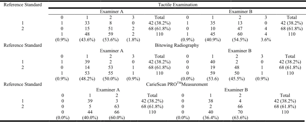
In the present study, 110 primary molars in 40 patients aged 6 to 12 years were examined by tactile examination, bitewing radiography and CarieScan PROTMdevice. Out of the 110 teeth, 7 were maxillary 1st primary molars, 33 were maxillary 2nd primary molars, 14 were mandibular 1st primary molars and 56 were mandibular 2nd primary molars. Out of the 110 teeth, 68 showed the presence of dentinal caries by any one method by both examiners and 42 showed the presence of enamel caries. The teeth with dentinal caries were further subjected to validation by reference standard method. The scores allotted by two examiners and results of reference standard validation were tabulated (Table 1).
Inter examiner agreement between two independent examiners for all three caries detection systems
Inter examiner agreement of 0.499 was seen with tactile examination which indicates moderate agreement according to classification given by Landis and Koch in 1977. Bitewing
radiography showed an agreement of 0.690 which is categorized as substantial agreement. CarieScan Pro had an agreement of 0.846 which is almost perfect agreement according to the above mentioned classification.
Sensitivity, Specificity, Positive Likelihood Ratio, Negative Likelihood Ratiofor all three caries detection systems
2)
With tactile examination, sensitivity was 76.47 %, specificity 71.43 %, positive likelihood ratio 2.68 and negative likelihood ratio 0.33. With bitewing radiography, sensitivity was 79.41 %, specificity 95.24 %, positive likelihood ratio 16.68 and negative likelihood ratio 0.22. With CarieScan PRO measurement, sensitivity was 92.65 %, specificity 92.86 %, positive likelihood ratio 12.97 and negative likelihood ratio 0.079.
Receiver Operator Characteristic (ROC) curves and Area under ROC Curve (AUC) for all three systems
Figure1)
[image:3.595.49.554.138.340.2]The diagnostic accuracy of tactile examination was significant (AUC = 0.751, Z = 5.886; P < 0.0001).
Table 1. Scores according to tactile exami
Reference Standard
Examiner A
0 1
1 1 33
2 0 15
1 (0.9%) 48 (43.6%) Reference Standard Examiner A
0 1
1 1 39
2 0 14
1 (0.9%) 53 (48.2%) Reference Standard Examiner A
0 1
1 0 39
2 0 5
0 (0.0%)
[image:3.595.39.559.385.473.2]44 (40.0%)
Table 2. Sensitivity, specificity, positive likelihood ratio (+LR), negative likelihood ratio ( radiography and
Parameter Sensitivity
(95% Confidence Interval)
Tactile Examination 76.47
(64.6 - 85.9) Bitewing Radiography 79.41
(67.9 - 88.3) CarieScan PROTMMeasurement 92.65
(83.7 - 97.6)
Table 3. Area under the curve (AUC) of tactile examination, bitewing radiography and
Parameter
Tactile Examination Bitewing Radiography CarieScan PROTMMeasurement
radiography showed an agreement of 0.690 which is categorized as substantial agreement. CarieScan Pro had an agreement of 0.846 which is almost perfect agreement according to the above mentioned classification.
Sensitivity, Specificity, Positive Likelihood Ratio, Negative Likelihood Ratiofor all three caries detection systems (Table
With tactile examination, sensitivity was 76.47 %, specificity 71.43 %, positive likelihood ratio 2.68 and negative likelihood ratio 0.33. With bitewing radiography, sensitivity was 79.41 %, specificity 95.24 %, positive likelihood ratio 16.68 and likelihood ratio 0.22. With CarieScan PROTM measurement, sensitivity was 92.65 %, specificity 92.86 %, positive likelihood ratio 12.97 and negative likelihood ratio
Receiver Operator Characteristic (ROC) curves and Area all three systems(Table 3,
The diagnostic accuracy of tactile examination was significant
The diagnostic accuracy of bitewing radiography was significant (AUC = 0.876, Z = 12.825; P < 0.0001) diagnostic accuracy of CarieScan PRO
significant (AUC = 0.928, Z = 16.659; P < 0.0001)
Figure 1. Receiver Operator Characteristic (ROC) curves and Area under ROC Curve (AUC) for all three systems tactile examination, bitewing radiography, CarieScan PROTM
validation using reference standard method
Tactile Examination
Examiner A Examiner B
2 3 Total 0 1
8 0 42 (38.2%) 1 35
51 2 68 (61.8%) 0 10
59 (53.6%)
2 (1.8%)
110 1
(0.9%)
45 (40.9%) Bitewing Radiography
Examiner A Examiner B
2 3 Total 0 1
2 0 42 (38.2%) 0 40
53 1 68 (61.8%) 0 19
55 (50.0%)
1 (0.9%)
110 0
(0.0%)
59
(53.6) (45.5%) CarieScan PROTMMeasurement
Examiner A Examiner B
2 Total 0 1
3 42 (38.2%) 0 38
63 68 (61.8%) 0 2
66 (60.0%)
110 0
(0.0%)
40 (36.4%)
Sensitivity, specificity, positive likelihood ratio (+LR), negative likelihood ratio (-LR) of tactile examination, bitewing radiography and CarieScan PROTMmeasurement
Sensitivity (95% Confidence Interval)
Specificity (95% Confidence Interval)
+ LR (95% Confidence Interval)
85.9)
71.43 (55.4 - 84.3)
2.68 (1.6 - 4.4)
88.3)
95.24
(83.8 - 99.4)
16.68
(4.3 - 64.8)
97.6)
92.86 (80.5 - 98.5)
12.97 (4.4 - 38.7)
Table 3. Area under the curve (AUC) of tactile examination, bitewing radiography and CarieScan PRO
AUC Standard Error
95% Confidence Interval
Z statistic
0.751 0.0426 0.659 - 0.828 5.886 0.876 0.0293 0.800 - 0.931 12.825 0.928 0.0257 0.862 - 0.968 16.659
The diagnostic accuracy of bitewing radiography was significant (AUC = 0.876, Z = 12.825; P < 0.0001). The CarieScan PROTMmeasurement was significant (AUC = 0.928, Z = 16.659; P < 0.0001).
Receiver Operator Characteristic (ROC) curves and rve (AUC) for all three systems
TM
measurement and
Examiner B
2 3 Total
13 0 42 (38.2%) 47 4 68 (61.8%) 60 (54.5%) 4 3.6% 110 Examiner B
2 3 Total
2 0 42 (38.2%)
48 1 68 (61.8%)
50 (45.5%) 1 (0.9%) 110 Examiner B
2 Total
4 42 (38.2%)
66 68 (61.8%)
70 (63.6%)
110
LR) of tactile examination, bitewing
(95% Confidence Interval)
-LR
(95% Confidence Interval) 0.33
(0.2 - 0.5) 0.22
(0.1 - 0.3) 0.079 (0.03 - 0.2)
CarieScan PROTMmeasurement
Pairwise comparison of ROC curves (Graph 1)
When AUC of tactile examination and radiography were compared, the difference between the areas was 0.125 and the difference was statistically significant (P <0.05). Difference of AUC between tactile examination and CarieScan PROTMmeasurement was 0.177 which was statistically significant (P <0.05). Difference of AUC between bitewing radiography and CarieScan PROTMmeasurement was 0.015 and this was statistically insignificant (P=0.1603).
DISCUSSION
The conventional and validated techniques for detecting early caries lesions include visual-tactile examination and bitewing radiography. Amongst the newer technologies, electrical measurement devices are one of them. Electrical caries measurement device uses multiple frequencies (ACIST), as different substrates respond differently to the resistance test at different frequencies (Chalas RE 2013; Eldamat AH 2007).CarieScan PROTM device uses this technology. It is non invasive; causes no pain, sensation or ionising radiation; with each measurement taken in a very short time per site (Amaechi BT 2009). Another feature is the appearance of the device which may be aesthetically pleasing to the patient. The present study was done to compare this new technology device with commonly used traditional methods of tactile examination and bitewing radiography. Previous studies have tested the accuracy of this device, in vitro, on primary and permanent teeth (Teo TK 2014; Singh R 2016). In vitro impedance measurements are affected by various external factors like size of electrode, electrode contact, surface area of the contact electrode, temperature changes, changing concentration of fluid in storage solution, their irregularities, distribution of minerals, post eruptive mineralization, maturation time of the tooth in the oral environment, thickness of enamel and dentin (Chalas RE 2013). In vivo studies on permanent teeth have shown good results with this device (Teo TK 2014; Jablonski-Momeni 2015; Melo M 2015). Hence, this study was taken up to test the validity of this device, in vivo, on non cavitated occlusal surfaces of primary teeth and to compare it with conventional methods. Two independent trained examiners performed the caries detection using tactile examination, bitewing radiography and CarieScan PROTM. Inter examiner agreement between the two independent examiners showed highest agreement of 0.846 by CarieScan PROTM measurement. This indicates almost perfect agreement between the two examiners.
This was followed by bitewing radiography which had a substantial agreement of 0.690 and the least was seen with tactile examination which showed moderate agreement with a score of 0.499. This is in accordance with previous studies conducted by Katge F et al (2016) and Singh R et al (2016) where perfect agreement scores were seen in caries detection using CarieScan PROTM in primary molars in vivo and in vitro when compared with conventional visual and radiographic methods. Agreement and reproducibility results obtained from bitewing radiography and tactile examination may be affected by the examiner’s clinical expertise and experience (Mileman PA 1992; Diniz MB 2010). The sensitivity of a procedure is its ability to correctly detect people who have the disease, expressed as the percentage of diseased people who are correctly diagnosed(Pretty JA 2004).Highest sensitivity was seen with CarieScan PROTM (92.65%) followed by bitewing
radiography (79.41%) and least was with tactile examination (76.47%). The specificity of a diagnostic procedure is the percentage of disease free individuals who are diagnosed correctly(Pretty JA 2004). In the present study, 95.24% specificity was seen with bitewing radiography, 92.86% specificity was seen with CarieScan PROTM followed by tactile examination with 71.43% which was least specific.In vitro studies on permanent teeth using CarieScan PROTM, Jablonski-Momeni A et al (2014) obtained a sensitivity of 68% and specificity of 90.8% whereas Melo M et al(2015), found 92% sensitivity, 75% specificity (Jablonski-Momeni 2015; Melo M 2015).
However, in vivo studies on primary teeth, showed a high sensitivity of 97% and specificity of 82% (Katge F 2016) whereas 88.89% sensitivity, 37% low specificity (Singh R 2016). The sensitivity results of this present study are in accordance with previous studies but the specificity obtained is highest as compared to the previous studies on primary teeth. Jablonski-Momeni A et al have suggested that physiological differences between primary teeth and permanent teeth affect the impedance measurement. Primary teeth have more conducting electrolytes and offer lesser resistance when alternating current is passed through it. The likelihood ratio (LR) is defined as the ratio between the probability of a defined test result given in the presence of a disease and the probability of the same test result given in the absence of a disease (Choi BC 1998).Highest positive likelihood ratio was seen with bitewing radiography (16.68), followed by CarieScan PROTM (12.97) and tactile examination (2.68). The highest negative likelihood ratio was with tactile examination (0.33), followed by bitewing radiography (0.22) and least with CarieScan PROTM (0.079). The likelihood ratio is useful in clinical decision making because it is also the ratio of the post-test odds of disease (odds of disease among persons with a given test result) to the pretest odds of disease (odds of disease among all persons) (Choi BC 1998). Maximum AUC is observed with CarieScan PROTM (0.928) with the ROC curve closest to the upper left corner (orange line); followed by bitewing radiography (0.876, green line) and least with tactile examination (0.751, blue line). These results indicate that the overall diagnostic accuracy of CarieScan PROTM is better than the conventional methods of bitewing radiography and tactile examination. Difference in AUC between tactile examination and CarieScan PROTM was statistically significant. However, difference in AUC between bitewing radiography and CarieScan PROTM measurement was not statistically significant. It is, thus, observed from this study that in terms of sensitivity, likelihood ratios, ROC and AUC CarieScan PROTM performed better than tactile examination and bitewing radiography in early caries detection on non cavitated pits and fissures of primary teeth. The specificity of bitewing radiography is higher than CarieScan PROTM. However, when the overall combination of sensitivity and specificity (AUC) is seen, CarieScan PROTM performs better than the conventional techniques. The other major advantage of this device is that it is not operator dependent unlike tactile examination or bitewing radiography interpretation. This does not offset the role of tactile examination or bitewing radiography as diagnostic aids. CarieScan PROTM has limited application on proximal surfaces and secondary caries detection (Amaechi BT 2009). In such situations, a combination of visual-tactile examination and radiography are the preferred diagnostic aids. The other limitation of the device is that it requires absolute isolation and even a slight amount of moisture does not give
reading with the device. It may be suggested that following visual examination, this device may be used to accurately detect the lesion depth on non cavitated occlusal surfaces of primary teeth. The need for bitewing radiography as diagnostic procedure may be eliminated in such cases. This has the advantage of reducing radiation exposure to the patients as well as clinicians.
Conclusion
From the present study, the following conclusions were drawn:
CarieScan PROTM – AC impedance spectroscopy technique has higher inter examiner agreement as compared to tactile examination and bitewing radiography in detecting non cavitated occlusal caries in the primary molars.
CarieScan PROTM has higher overall combination of sensitivity, specificity and Area under Curve as compared to tactile examination and bitewing radiography in detecting non cavitated occlusal caries in the primary molars.
Positive likelihood ratio was higher with bitewing radiography, followed by CarieScan PROTM and tactile examination.
Negative likelihood ratio was higher with tactile examination, followed by bitewing radiography and least with CarieScan PROTM.
Why this paper is important to paediatric dentists?
This paper suggests that AC Impedance spectroscopy technique is effective in detection of occlusal caries in primary molars.
Early detection of non cavitated lesions will help in rendering prompt treatment and prevent further progression of lesion.
Paediatric dentists can use this device for early caries detection, especially in younger or uncooperative patients in whom taking radiographs may be difficult.
Compliance with Ethical Standards:
Funding: Nil
Conflict of Interest: Nil
Ethical approval: All procedures performed in studies
involving human participants were in accordance with the ethical standards of the institutional and/or national research committee and with the 1964 Helsinki declaration and its later amendments or comparable ethical standards.
Informed consent: Informed consent was obtained from all
individual participants included in the study.
REFERENCES
Amaechi, B.T. 2009. Emerging technologies for diagnosis of dental caries: The road so far. J Appl Phys 105(10):102047. Ashley, P. 2000. Diagnosis of occlusal caries in primary teeth.
Int J Paediatr Dent., 10(2):166-71.
Bahrololoomi, Z., Ezoddini, F., Halvani, N. 2015. Comparison of Radiography, Laser Fluorescence and Visual Examination for Diagnosing Incipient Occlusal Caries of
Permanent First Molars. Journal of dentistry., 12(5):324-32.
Chałas, R.E., Piatek, D., Wojcik-Checinska, I.L., Zubrzycka-Wrobel, J.O., Bachanek, T. 2013. AC-impedance spectroscopy and caries detection. Curr Iss Pharm Med Sci
26(3):344-6.
Choi, B.C. 1998. Slopes of a receiver operating characteristic curve and likelihood ratios for a diagnostic test. American Journal of Epidemiology., 148(11):1127-32.
Diniz, M.B., Rodrigues, J.A., Neuhaus, K.W., Cordeiro, R.C., Lussi, A. 2010. Influence of examiner’s clinical experience on the reproducibility and accuracy of radiographic examination in detecting occlusal caries. Clinical oral investigations., 14(5):515-23.
Ekstrand, K.R., Ricketts, D.N., Kidd, E.A. 1997. Reproducibility and accuracy of three methods for assessment of demineralization depth on the occlusal surface: an in vitro examination. Caries Res., 31(3):224-31. Ekstrand, K.R., Ricketts, D.N., Kidd, E.A. 2001. Occlusal caries: pathology, diagnosis and logical management. Dent Update., 28(8):380-7.
Eldarrat, A.H. 2007. Age-related changes in ac- impedance spectroscopy studies of normal human dentine. J Mater Sci Mater Med., 18(6):1203-10.
Goel, A., Chawla, H.S., Gauba, K., Goyal, A. 2009. Comparison of validity of DIAGNOdent with conventional methods for detection of occlusal caries in primary molars using the histological gold standard: An in vivo study. J Indian Soc Pedod Prevent Dent., 27(4):227-34.
Hill, I.N., Blayney, J.R., Zimmerman, S.O., Johnson, D.E. 1967. Deciduous teeth and future caries experience. J Am Dent Assoc., 74:430-8.
Jablonski-Momeni, A., Klein, S.M. 2015. In-Vivo Performance of the CarieScan Pro Device for Detection of Occlusal Dentine Lesions. The Open Access Journal of Science and Technology., 3:1-6.
Katge, F., Wakpanjar, M., Rusawat, B., Shetty, A. 2016. Comparison of three diagnostic techniques for detecting occlusal dental caries in primary molars: An in vivo study.
Indian J Dent Res., 27(2):174-7.
Landis, J.R., Koch, G. 1977. The measurement of observer agreement for categorical data. Biometrics., 33(1):159–74. Melo, M., Pascual, A., Camps, I., Del Campo, Á. 2015. In vivo
study of different methods for diagnosing pit and fissure caries. J Clin Exp Dent., 7(3):387-91.
Mileman, P.A., Mulder, E., Weele, L. 1992. Factors influencing the likelihood of successful decisions to treat dentin caries from bitewing radiographs. Community Dent Oral Epidemiol., 20(4):175-80.
Pretty, J.A., Maupome, G. 2004. A closer look at diagnosis in clinical dental practice: Part 1. Reliability, validity, specificity and sensitivity of diagnostic procedures. J Can Dent Assoc., 70(4):251-6.
Ripa, L.W., Leske, G.S., Varma, A.O. 1988. Longitudinal study of the caries susceptibility of occlusal and proximal surfaces of first permanent molars. J Public Health Dent
48:8–13.
Sawle, R.F., Andlaw, R.J. 1988. Has occlusal caries become more difficult to diagnose? A study comparing clinically undetected lesions in molar teeth of 14-16-year old children in 1974 and 1982. Br Dent J., 164(7):209-11.
occlusal caries in primary molars. J Indian Soc Pedod Prev Dent., 34(2):152-8.
Teo, T.K., Ashley, P.F., Louca, C. 2014. An in vivo and in vitro investigation of the use of ICDAS, DIAGNOdent pen and CarieScan PRO for the detection and assessment of occlusal caries in primary molar teeth. Clinical oral investigations., 18(3):737-44.
Weerheijm, K.L., Van Amerongen, W.E., Eggink, C.O. 1989. The clinical diagnosis of occlusal caries: a problem. ASDC
Journal of Dentistry for Children., 56(3):196-200.