RELATION BETWEEN COMBINED WIDTH OF MAXILLARY ANTERIOR TEETH AND
INTERPUPILLARY WIDTH, INTERCOMMISSURAL WIDTH AMONG DENT
Post Graduate Student, Department
ARTICLE INFO ABSTRACT
Background & Objectives:
esthetic importance of dentofacial beauty. Over the years, guidelines for teeth selection and arrangement have been suggested. However, no universally accepted single parameter currently exists.
from 21
maxillary anterior teeth were measured using digital vernier caliper (Mitutoyo Ltd 505 results were statistically analyzed using Pearson correlation test and student t test. correlation was found between i
teeth and a non
maxillary anterior teeth. There is co
teeth
among Dakshina Kannada population. There is no correlation between
teeth
among Dakshina Kannada population.
Copyright © 2019, Sarath Sasidharan. This is an open distribution, and reproduction in any medium, provided
INTRODUCTION
Face is the most expressive part of the human body which decides an individual's social acknowledgment. Loss of teeth influences facial appearance as well as makes psychological trauma to the individual; subsequently it is crucial that an esthetically satisfying and practically agreeable substitution of the missing teeth ought to be provided. Selection of ideal anterior teeth for edentulous patient becomes a difficult task in the absence of pre-extraction records. The size, form and color
of teethshould be in harmony with surrounding oral and facial
structures. One of the important landmarks every dental
professional strive to achieve after delivering treatment is “improvement in patient esthetic value”. Very often, while constructing denture prosthesis in absence of pre
records dentist face great difficulty in choosing the ideal analog of missing teeth.
*Corresponding author: Dr. Sarath Sasidharan,
Post Graduate Student, Department of Prosthodontics, K.V.G College and Hospital, Sullia, Karnataka.
ISSN: 0975-833X
Article History:
Received 24th September, 2019
Received in revised form 18th October, 2019
Accepted 15th November, 2019 Published online 30th December, 2019
Citation:Dr. Sarath Sasidharan. 2019. “Relation Between Among Dentate Dakshina Kannada Population.”, International Key Words:
Interpupillary width, Intercommissural width,
Combined width of Maxillary Anterior Teeth.
RESEARCH ARTICLE
COMBINED WIDTH OF MAXILLARY ANTERIOR TEETH AND
INTERPUPILLARY WIDTH, INTERCOMMISSURAL WIDTH AMONG DENT
KANNADA POPULATION
*
Dr. Sarath Sasidharan
Department of Prosthodontics, K.V.G Dental College and Hospital, Sullia, Karnataka
ABSTRACT
Background & Objectives: The maxillary anterior teeth are the key elements contributing to the esthetic importance of dentofacial beauty. Over the years, guidelines for teeth selection and arrangement have been suggested. However, no universally accepted single parameter currently exists. Methodology: Interpupillary width and intercommissural width of
from 21-30 years were obtained. Interpupillary width, intercommissural width and maxillary anterior teeth were measured using digital vernier caliper (Mitutoyo Ltd 505 results were statistically analyzed using Pearson correlation test and student t test.
correlation was found between intercommissural width and combined width of maxillary anterior and a non-significant correlation was found between interpupillary width and combined width of maxillary anterior teeth. Interpretation & Conclusion: It can be concluded that,
There is correlation between Intercommissural width and combined width of maxillary anterior teeth which can be used as a guide for selection of combined width of maxillary anterior teeth among Dakshina Kannada population.
There is no correlation between Interpupillary width and combined width of maxillary anterior teeth which cannot be used as a guide for selection of combined width of
among Dakshina Kannada population.
access article distributed under the Creative Commons Attribution the original work is properly cited.
Face is the most expressive part of the human body which decides an individual's social acknowledgment. Loss of teeth influences facial appearance as well as makes psychological trauma to the individual; subsequently it is crucial that an hetically satisfying and practically agreeable substitution of Selection of ideal anterior teeth for edentulous patient becomes a difficult task in
The size, form and color should be in harmony with surrounding oral and facial
One of the important landmarks every dental
delivering treatment is improvement in patient esthetic value”. Very often, while
constructing denture prosthesis in absence of pre-extraction records dentist face great difficulty in choosing the ideal
,
Post Graduate Student, Department of Prosthodontics, K.V.G Dental
Several anatomic measurements, including bizygomatic width (BZW), inter-pupillary distance (IPD), interalar width (IAW), innercanthal distance (ICD), and inter
been suggested to aid in the estimation of a combined width the maxillary anterior teeth. Moreover most of the standard guideline present in literature uses soft tissue landmarks, which may be misleading due to soft tissue changes with ageing; weight & build of person. The maxillary anterior teeth are the key elements contributing to the esthetic
we call dento- facial beauty. However the selection and arrangement of maxillary anterior teeth for edentulous patients in a natural and esthetically pleasing form has remained an elusive and challenging endeavor.
criteria and guidelines for esthetic tooth selection and arrangement have been suggested by the artisans of the dental profession. However, no universally accepted parameter currently exists for selection of anterior teeth in local population, and no such studies have been carried out previously. The maxillary anterior teeth are considered the most important in denture esthetics.
factors contributing to a harmonious anterior dentition are the size, shape, and arrangement of the maxillary anterior teeth. By using anatomic references, it is possible to place artificial teeth International Journal of Current Research
Vol. 11, Issue, 12, pp.8702-8707, December, 2019
DOI: https://doi.org/10.24941/ijcr.37086.12.2019
Relation Between Combined Width Of Maxillary Anterior Teeth And Interpupillary Width,
International Journal of Current Research, 11, (12), 8702-8707.
COMBINED WIDTH OF MAXILLARY ANTERIOR TEETH AND
INTERPUPILLARY WIDTH, INTERCOMMISSURAL WIDTH AMONG DENTATE DAKSHINA
nd Hospital, Sullia, Karnataka
The maxillary anterior teeth are the key elements contributing to the esthetic importance of dentofacial beauty. Over the years, guidelines for teeth selection and arrangement have been suggested. However, no universally accepted single parameter currently Interpupillary width and intercommissural width of 400 subjects of age ranging obtained. Interpupillary width, intercommissural width and combined width of maxillary anterior teeth were measured using digital vernier caliper (Mitutoyo Ltd 505-633-50). The results were statistically analyzed using Pearson correlation test and student t test. Results: A positive ntercommissural width and combined width of maxillary anterior significant correlation was found between interpupillary width and combined width of
It can be concluded that,
combined width of maxillary anterior which can be used as a guide for selection of combined width of maxillary anterior teeth
combined width of maxillary anterior which cannot be used as a guide for selection of combined width of maxillary anterior teeth
License, which permits unrestricted use,
Several anatomic measurements, including bizygomatic width pupillary distance (IPD), interalar width (IAW), innercanthal distance (ICD), and inter-commissural width have been suggested to aid in the estimation of a combined width of . Moreover most of the standard guideline present in literature uses soft tissue landmarks, which may be misleading due to soft tissue changes with ageing; The maxillary anterior teeth are the buting to the esthetic importance of what facial beauty. However the selection and arrangement of maxillary anterior teeth for edentulous patients in a natural and esthetically pleasing form has remained an elusive and challenging endeavor. Over the years, norms, criteria and guidelines for esthetic tooth selection and arrangement have been suggested by the artisans of the dental However, no universally accepted parameter currently exists for selection of anterior teeth in local opulation, and no such studies have been carried out The maxillary anterior teeth are considered the most important in denture esthetics. The most influential factors contributing to a harmonious anterior dentition are the ngement of the maxillary anterior teeth. By using anatomic references, it is possible to place artificial teeth INTERNATIONAL JOURNAL
OF CURRENT RESEARCH
within the bounds, or “average of nature,” permitting individual esthetics and physiologic variations.
Objectives of the study
To study the relation between combined width of maxillary anterior teeth and inter-pupillary width.
To study the relation between combined width of maxillary anterior teeth and inter-commissural width.
MATERIALS AND METHODS
The study was carried out in the Department of Prosthodontics, K.V.G. Dental College and Hospital, Sullia. A total of 400 subjects with age ranging between 21-30years were selected among the population belonging to Dakshina population on the basis of defined inclusion and exclusion criteria.
Inclusion criteria
Angle Class I molar relationship, pleasing profile, and sound morphologically normal permanent dentition up to the second molar.
Subject’s age ranging between 21-30 years.
Should be a native of Dakshina Kannada
No evidence of apparent loss of tooth structure due to attrition, erosion, abrasion or trauma
Exclusion criteria for the subject in this study:
Subjects with history of orthodontic treatment
Gingival inflammation and hypertrophy in the upper anterior region
Severe attrition; crowns or proximal restorations placed in the anterior teeth
History of congenital anomaly, orbital disease, trauma, or facial surgery
Methods of Collecting Data
Instruments and materials that were used during course of study
Irreversible hydrocolloid impression material (Tropicalgin ISO
1563:1990/ADA18/BS )
ADA type III dental stone
Wooden tongue spatula
Dentulous rim lock perforated stock tray (SS WHITE)
Digital vernier caliper (MITUTOYO Ltd 505-633-50)
Curved and straight stainless steel spatula
Flexible Rubber bowl
Base former
Water & Powder measuring device (supplied by manufacturer)
Dental floss (Colgate-waxed dental floss)
METHODOLOGY
Following the written approval of the ethical committee, K.V.G.
Dental College and Hospital, Sullia, the subjects were screened according to the defined inclusion and exclusion criteria. The subjects qualifying for the study were selected.
Impression & cast making procedures: After doing a brief
oral examination, proper size perforated rim lock stock tray(S S WHITE) was selected, utility/beading wax was used for customization of tray if required in any specific area. Alginate impression powder (Tropicalgin ISO 1563:1990/ADA 18/BS 4269) was mixed with water according to manufacturer instructions with 1:2.6 (proportion by weight) water / powder ratio in flexible rubber bowl with curved stainless steel spatula by spatulating it in figure of eight motion then swiping and stroping it against the rubber bowl walls. Impression tray was loaded and maxillary impression was made. The cast was poured immediately with hard setting dental stone (TYPE III).Dental stone was taken in proper powder liquid ratio (according to manufacturer) and spatulated in rubber bowl with wide blade straight stainless steel spatula .After one hour, casts was recovered & damaged casts were discarded & impression was remade. Base was made for the casts.
To measure the combined width of the maxillary anterior:
Mesiodistal dimension of maxillary six anterior teeth was measured with the help of dental floss passing through distal surface of the canine in the maxillary cast . Dental floss was placed at the greatest curvature of the maxillary teeth, and a mark on each side was placed at the distal surface of the canines. Dental floss was made straight, and the distance was measured between the marks using digital vernier calliper (Fig.1).
To measure the Inter-pupillary distance: After seating the
subject comfortably on the dental chair , subject was asked to look straight, then the midpoint of the pupils was marked on a wooden tongue spatula held horizontally at the forehead. The distance between the markings were measured using digital vernier caliper to get the inter-pupillary distance. (Fig.2)
To measure the Inter-commissural width: The
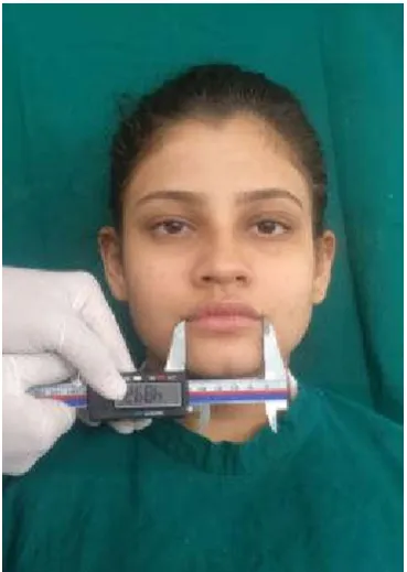
inter-commissural width was determined by measuring the maxillary lip vermilion from commissure to commissure. Distances were measured using a digital vernier caliper without the application of pressure. Subjects were asked to widely open and close their mouth several times and to move their lips to allow fatigue of the surrounding muscles to relax during the measurement. (Fig.3). The measurements were obtained from the patient by two independent observers and average value was tabulated and analyzed statistically. All measurements were obtained in millimeters. The study results were analyzed using Pearson’s correlation coefficient and Student ‘t’-test
RESULTS
Table 1. Measurement of interpupillary distance, intercommissural distance and combined width of
maxillary anterior teeth of study participants
IPD ICM COMWIDTH
N 400 400 400
Mean 61.70 50.123 52.27
Std. Deviation 3.352 3.4989 2.154
Minimum 50 42.5 47
[image:3.595.47.276.102.163.2]Maximum 69 69.5 63
Table 2. Mean ratio of relative interpupillary distance, intercommissural distance and combined width of maxillary
anterior teeth among males
IPD ICM COMWIDTH
N 200 200 200
Mean 62.07 49.886 52.25
Std. Deviation 3.336 3.1333 2.158
Minimum 54 42.6 47
[image:3.595.33.277.219.281.2]Maximum 69 60.1 63
Table 3. Mean ratio of relative interpupillary distance, intercommissural distance and combined width of maxillary
anterior teeth amomg females
IPD ICM COMWIDTH
N 200 200 200
Mean 61.33 50.360 52.29
Std. Deviation 3.335 3.8230 2.156
Minimum 50 42.5 49
Maximum 69 69.5 63
Table 4- Correlation between combined width of maxillary anterior teeth and interpupillary distance
Mean Std. Deviation
IPD 61.70 3.352
COMWIDTH 52.27 2.154
IPD COMWIDTH
Pearson Correlation( r ) 1 .067
Sig. (2-tailed) . .180
Table 5- Correlation between combined width of maxillary anterior teeth and intercommissural distance
Mean Std. Deviation
COMWIDTH 52.27 2.154
ICM 50.123 3.4989
ICM COMWIDTH
Pearson Correlation( r ) 1 .454
Sig. (2-tailed) . .000
Table 6- Mean values of interpupillary width intercommissural width and combined width of maxillary anterior teeth among
males and females
GENDER Mean Std.
Deviation
Std. Error Mean
IPD Male 62.07 3.336 .236
Female 61.33 3.335 .236
ICM Male 49.886 3.1333 .2216
Female 50.360 3.8230 .2703
COMWIDTH Male 52.25 2.158 .153
[image:3.595.337.531.263.479.2]Female 52.29 2.156 .152
[image:3.595.38.260.340.401.2]Figure 1- Measurement Of Combined width of Maxillary Anterior Teeth Using Dental Floss
[image:3.595.342.527.510.770.2]Figure 2- Measurement of Interpupillary Width
[image:3.595.41.283.724.804.2]The mean intercommissural width as recorded by the observer is 50.123 ±3.4989mm. Minimum value recorded for combined width of maxillary anteriors was 47mm and maximum record value of 63mm. The mean value for the combined width of maxillary anteriors was 52.27±2.154mm.Table 2 shows Mean ratio of relative interpupillary width, intercommissural width
and combined width of maxillary anterior teeth among males (n = 200). The minimum values obtained for the interpupillary
width, intercommissural width and combined width of maxillary anteriors are 54mm, 42.6mm and 47mm respectively. The maximum value obtained for the interpupillary width, intercommissural width and combined width of maxillary anteriors are 69mm, 60.1mm and 63mmrespectively. The mean ratio obtained for the interpupillary width, intercommissural width and combined width of maxillary anteriors are 62.07mm, 49.886mm and 52.25mm respectively.
Table 3 shows the mean ratio of relative interpupillary width , intercommissural width and combined width of maxillary anterior teeth amomg females (n = 200). The minimum values obtained for the interpupillary width, intercommissural width and combined width of maxillary anteriors are 50mm, 42.5mm and 49mm respectively.The maximum values obtained for the interpupillary width, intercommissural width and combined width of maxillary anteriors are 69mm, 69.5mm and 63mm respectively. The mean ratio obtained for the interpupillary width, intercommissural width and combined width of maxillary anteriors are 61.33mm, 50.360mm and 52.29mm respectively. Table 4 shows the correlation between combined width of maxillary anterior teeth and interpupillary width. Pearson correlation indicates that there is no positive correlation between interpupillary width and combined width of maxillary anterior teeth since the value of significance is greater than 0.05 ( p > 0.05 ) .Table 5 shows the correlation between combined width of maxillary anterior teeth and intercommissural width . Pearson’s correlation indicates that there is a high correlation between combined width of maxillary anterior teeth and intercommissural width ( p < 0.001 ; R= 0.454 ; R2 = 0.206 ) and R2 = 0.206 , this means 20.6 % of ones intercommissural width is directly accounted for ones combined width of maxillary anterior teeth. Table 6 shows the mean values of interpupillary width, intercommissural width and combined width of maxillary anterior teeth among males and females. The mean valules for interpupillary width (IPD) among males and females are 62.07±3.336mm and 61.33±3.335mm.
The mean valules for intercommissural width (ICM ) among males and females are 49.88 ±3.122mm and 50.30 ±3.823mm .The mean valules for combined width of maxillary anterior teeth among males and females are 52.25±2.158mm and 52.29±2mm. Table 7 shows the mean difference between interpupillary width, intercommissural width and combined width of maxillary anterior teeth among Males and Females using independent sample t- test. Levene’s tests of equality of variance in different samples were used to determine the assumption of equal variances or not. From the test it can be concluded that there is a significant difference in interpupillary width among males and females and there is no significant difference in intercommissural width and combined width of maxillary anterior teeth among males and females.
DISCUSSION
[image:4.595.37.565.85.251.2]The artificial teeth selection and arrangement is always a challenge to dentist. It becomes more difficult in the resorbed residual ridges. Boucher has stated, “The only correct position of a tooth is the one in which it was placed by nature. Pre extraction records serves as an aid in arranging teeth in nature’s position .Various intra oral landmarks like Incisive papilla, Inter-canine distance has also been taken into consideration for selection and arrangement of teeth. Many of the sof tissue landmarks are subjective to change as age progress .The interpupillary distance of human remains fairly static throughout the life. Mishra MK et alconducted a study to find out the relationship between the inner inter-canthal distance, inter-pupillary distance, inter-commissural width, inter-alar width, and the width of maxillary anterior teeth in Aryans and Mongoloids. The inter-alar width was measured as the distance between the widest points on the outer surface of the alae of the nose on either side . For the measurement of inter-pupillary distance, the midpoint of the pupils was marked on a wooden tongue spatula. The inter-commissural width was determined by measuring the maxillary lip vermilion from commissure to commissure. The inter-canthal distance was measured as a distance between the medial angle of the palpebral fissure of the eyes. Distances were measured using a Boley’s gauge without the application of pressure. . A highly significant correlation was found between inter-pupillary distance and combined width of maxillary anterior in the total population, Aryans, and Mongoloids. For Aryans, inter-alar width, inter-pupillary distance, and inter-commissural width can be used to determine anterior teeth widths.
Table 7- Mean difference between interpupillary distance ,intercommissural distance and combined width of maxillary anterior teeth among Males and Females using independent sample t- test.
Levene's Test for
Equality of Variances t-test for Equality of Means
F Sig. t df Sig. (2-tailed) Mean
Difference
Std. Error Difference
95% Confidence Interval of the Difference
Lower Upper
IPD Equal variances
assumed .040 .842 2.223 398
.027
(<0.05) .74 .334 .086 1.397
Equal variances
not assumed 2.223 398.000 .027 .74 .334 .086 1.397
ICM Equal variances
assumed 4.432 .036 -1.356 398
.176
(>0.05) -.474 .3495 -1.1611 .2131
Equal variances
not assumed -1.356 383.224 .176 -.474 .3495 -1.1612 .2132
COMWIDTH Equal variances
assumed .019 .890 -.188 398
.851
(>0.05) -.04 .216 -.465 .384
Equal variances
For Mongoloids, inter-pupillary can be used to determine anterior teeth widths. A study was performed by Kini AY., Angadi GS in 2013 to correlate dental measurements i.e. combined mesio-distal width of six maxillary anterior teeth with facial measurements i.e. inner canthal distance, interpupillary distance and intercommissural width . Two standardized digital photographs of the face were generated; one, when the facial muscles were relaxed and the other, when the subject was smiling; thereby, revealing the maxillary anterior teeth up to the canine tip. Inner canthal distance, interpupillary distance, intercommissural distance, distance between the tips of the maxillary canines and distance between the distal surfaces of the canines were measured. On the cast, the distance between tips of maxillary canines and distance between distal surfaces of maxillary canines were noted. Extra oral anthropometric measurements of the interpupillary distances and the intercommissural distances with the help of standardized photographs can help us determine the combined widths of the anterior teeth accurately, thus aiding their selection in the absence of pre-extraction records. A study was
conducted by Glynis Anita Miranda, Mariette D’Souza in 2016
to evaluate the reliability of interalar width and intercommissural width as guide in selection of artificial maxillary anterior teeth. Total of 200 samples were selected. The four parameters such as interalar width, intercanine distance, intercommissural width, and distance between distal aspects of canines were measured. The data obtained were statistically analyzed. A correlation was observed between the interalar distance and the intercanine distance and also between the intercommissural distance and the distance between the distal aspects of canines. With the review of literature it is clear that, in the absence of pre-extraction records, there is however, no accurate objective method in order to select the width of maxillary anterior teeth for edentulous patients. Although many methods have been utilized, none of these methods are absolutely reliable for teeth selection. The anatomical -landmark as a guide for selecting maxillary anterior teeth will vary from population to population. Development of a more scientific or objective method of teeth selection would greatly assist dentist in delivering their service for patient care and satisfaction confidently and will also enable lab technician to accurately pick the teeth without seeing the patient on dental chair, computer would also be used to scan and measure cast to give image of patient teeth in the proper size relationship.
Conflict of Interest: No conflict of interest
Funding statement: Self Funded
REFERENCES
Sharma SS, Nagpal A, Verma PR. 2012. Correlation between facial measurements and the mesiodistal width of the maxillary anterior teeth. Indian Journal of Dental
Sciences., 4(3).
Patel JR, Sethuraman Rajesh, G Naveen Y, ShaMitul H. 2011. A comparative evaluation of the relationship of inner canthal distance and inter-alar width to the inter-canine width amongst the Gujarati population. Journal of
Advanced Oral Research.,2(3):31-38.
Pound E. 1962. Applying harmony in selecting and arranging teeth. Dent Clin North Am., 6:241-258.
Krajicek D. Guides for natural facial appearance as related to complete denture construction. J Prosthet Dent 1969;21(6):654-62.
Guldag, Sebnem, Fatih. 2010. Relationship between pterygomaxillary notch & maxillary anterior teeth. J Prosthodontics; 19: 231-234
LaVere AM., Marcroft KR., Smith RC., Sarka RJ. 1994. Denture tooth selection: size matching of natural anterior tooth width with artificial denture teeth. J Prosthet Dent., 72(4):381-384.
Al Wazzan KA. 2001. The relationship between intercanthal dimension and the widths of maxillary anterior teeth. J
Prosthet Dent., 86: 608-12.
Varjão FM., Nogueira SS. 2005. Intercommissural width in 4 racial groups as a guide for the selection of maxillary anterior teeth in complete dentures. Int J Prosthodont., 18: 513-5.
Varjão FM., Nogueira SS. 2006. Nasal width as a guide for the selection of maxillarycomplete denture anterior teeth in four racial groups. J Prosthodont., 15:353-8.
Varjão FM., Nogueira SS. 2006. Nasal width as a guide for the selection of maxillarycomplete denture anterior teeth in four racial groups. J Prosthodont., 15:353-8.
French FA. 1951. Selection & arrangement of the anterior teeth in prosthetic denture. J Prosthet Dent., 1: 587-93 Hasanresisoglu, Berksun, Kerem, Arsalan. 2005. An analysis
of maxillary anterior teeth: facial &dental proportion. J
Prosthet Dent., 94 530-38.
Smith DE. 1971. The reliability of pre-extraction records for complete dentures. J Prosthet Dent., 25(6):592-608. Cesario, V. A., and Latta. G. H. 1984. "Relationship between
the mesiodistal width of the maxillary central incisor and interpupillary distance." Journal of Prosthetic
Dentistry 52.5: 641-643.
Murphy WK., Laskin DM. 1990. Intercanthal and interpupillary distance in the black population. Oral
Surgery, Oral Medicine, Oral Pathology and Oral
Radiology. Jun 1;69(6):676-80.
Latta GH., Weaver JR., and Conkin JE. 1991. The relationship between the width of the mouth, interalar width, bizygomatic width, and interpupillary distance in edentulous patients. J prosthet dent., 65:250-4.
Gheriani. 1992. A new guide for positioning of maxillary posterior denture teeth. J Oral Rehabil., 19:535-38. Lavere et al. 1992. Denture tooth selection: An analysis of the
natural maxillary central incisor compared to the length and width of the face. J Prosthet dent., 67:661-3.
Al-el-sheikh H M., Al-athel M S. 1998. The relationship of interalar width, interpupillary width and maxillary anterior teeth width in saudi population. Odontostomatol trop. dec;21(84):7-10
Pivnick EK., Rivas ML., Tolley EA., Smith SD., Presbury GJ. 1999. Interpupillary distance in a normal black population. Clinical genetics. Mar;55(3):182-91.
Ibrahimagić L., Jerolimov V., Čelebić A., Carek V., Baučić I., Knezović Zlatarić D. 2001. Relationship between the face and the tooth form. Collegium antropologicum. Dec 17;25(2):619-26.
Filipović T. 2003. Changes in the interpupillary distance (IPD) with ages and its effect on the near convergence/distance (NC/D) ratio. Collegium antropologicum. Dec 15;27(2):723-7.
anterior teeth in complete dentures. Int J Prosthodont., 18: 513-5.
Gomes VL., Gonçalves LC., Do Prado CJ., Junior IL., De Lima Lucas B. 2006. Correlation between facial measurements and the mesiodistal width of the maxillary anterior teeth. Journal of Esthetic and Restorative Dentistry. Jul;18(4):196-205.
Etezad-Razavi M., Jalalifar S. 2008. Correlation between interpupillary and inner-outer intercanthal distances in individuals younger than 20. Journal of ophthalmic & vision research. Jan;3(1):16.
Gomes VL., Gonçalves LC., Costa MM., Lucas Bde L. 2009. Interalar distance to estimate the combined width of the six maxillary anterior teeth in oral rehabilitation treatment. J EsthetRestor Dent., 21(1):26-35.
DV N. 2009. Interpupillary distance as a guide for the selection of upper anterior teeth. The Internet Journal of Dental Science. 2009;9(1).