Original Article
Novel real-time polymerase chain reaction assay for
quantitative detection of
Chlamydia trachomatis
in vaginal and cervical secretions
Jinji Jin1, Jianxin Tu2, Shanli Zhu3, Xiaochun Zhu2, Xiangyang Xue3, Lifang Zhang3
Departments of 1General Surgery, 2Rheumatology, The First Affiliated Hospital of Wenzhou Medical University, Wenzhou, China; 3Department of Medical Microbiology and Immunology, Wenzhou Medical University, Wenzhou, China
Received September 4, 2018; Accepted February 11, 2019; Epub May 15, 2019; Published May 30, 2019
Abstract: Chlamydia trachomatis (C. trachomatis) is an important sexually transmitted disease pathogen associated with infertility and several urogenital infectious diseases. A variety of detection methods have been developed for C. trachomatis, including the isolated cell culture method, PCR, and real-time polymerase chain reaction (RT-PCR). These methods have inherent limitations, however. The current study developed a novel RT-PCR assay specific for quantitative detection of C. trachomatis elementary bodies (EBs) in vaginal and cervical secretions without DNA extraction. Based on homology analysis between Chlamydia and other common genital tract infectious pathogens, oligonucleotide primers were designed to target the C. trachomatis ompA70-213 (ompA1) gene. These primers were confirmed in HeLa 229 cells infected with C. trachomatis and in a mouse model of genital C. trachomatis infection. Moreover, RT-PCR was used to detect C. trachomatis in vaginal and cervical secretions from 86 female patients. These results were compared with those of the traditional C. trachomatis isolated cell culture method, evaluating the sensitivity and specificity of this novel RT- PCR assay. The ompA1 gene was found to have high homology across several Chlamydia serotypes. The newly developed RT-PCR assay provided highly sensitive detection of EBs in geni-tal tracts of the mouse model and in vaginal and cervical secretions of the 86 female patients, compared with the traditional detected method.
Keywords: Chlamydia trachomatis, elementary bodies, quantitative detection, real-time PCR, OmpA1
Introduction
Chlamydia trachomatis (C. trachomatis) is an intracellular bacterium implicated in reproduc-tive tract infections. It is also the pathogen re- sponsible for the most common sexually trans-mitted disease, worldwide, in humans [1]. Alth- ough antibiotics are an effective treatment for
C. trachomatis infections, its incidence has still increased steadily in recent years [2]. Each ye- ar, approximately 131 million new cases, world-wide, are reported by the WHO [3]. Moreover, repeated infections with C. trachomatis result in serious diseases, such as chronic pelvic pa- in, pelvic inflammatory disease (PID), tubal fac-torinfertility (TFI ), ectopic pregnancy [1, 4], uro-genital infections [5], lymphogranuloma vene-reum (LGV) [6], and even autoimmune diseases [7]. Therefore, there is an urgent need for
devel-opment of a rapid and accurate method for detection of C. trachomatis.
C. trachomatis encompasses 15 major serovar-iants. Of these, serovars E-K are the most com-mon causes of genital infection, worldwide [1].
Chlamydia are obligate intracellular parasites that have a similar cell wall structure like that of gram-negative bacteria. They also have a uni- que biphasic developmental cycle that involves an extracellular form, known as the elementary body (EB), and an intracellular form, known as the reticulate body (RB) [8]. Various serovars of
C. trachomatis and its unique life cycle make diagnosis of C. trachomatis infection difficult.
the isolated cell culture method, despite its re- quirements for staining and microscopic count-ing of inclusion-formcount-ing units (IFUs). It also has inherent limitations. Recently, real-time poly-merase chain reaction (real-time PCR, RT-PCR), which offers high sensitivity and specificity, has been successfully applied for quantitative de- tection of many pathogens, such as human im- munodeficiency virus (HIV) [9], hepatitis C virus (HCV) [10], and C. trachomatis. However, these methods have their own limitations and do not satisfy the requirement of more convenient and sensitive research.
The current study developed a highly sensitive real-time quantitative polymerase chain reac-tion (RT-PCR) assay to detect Chlamydia in vagi-nal swab samples from women without DNA
gene in the C. trachomatis genome because there is only one copy that encodes the major outer membrane protein (MOMP), one of the main target antigens for development of vac-cines and diagnostic reagents [11]. The gene
ompA70-213 was selected as a special target for development of a novel RT-PCR assay to quan-titatively detect ompA1 copies of C. trachoma -tis. It was confirmed in an isolated cell culture, mouse model, and female genital tract secre- tions.
Methods
Standardized plasmid generation and primer design
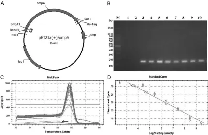
[image:2.612.90.520.70.348.2]The ompA1 region was selected from the con-served region of ompA and is located at 70-213
Figure 1. Standard plasmid curve and melt curve for real-time PCR assay. A. Schematic diagram of the pET21a (+)/ompA standard plasmid. The recombinant plasmid pET21a (+)/ompA is the standard plasmid for the real-time PCR. ompA is a gene encoding the major outer membrane protein (MOMP) from C. trachomatis and ompA1 can be amplified by a pair of specific primers (Ozlf-335, Ozlf-336) to yield a single fragment (144 bp) within ompA (17-161). B. Real-time PCR products from 8-fold standard plasmid dilutions were separated on 1% agarose at 100 V in 1 × TAE and then the gel was stained with 0.3 μg/mL ethidium bromide (EB). Lane M: 1 Kb DNA marker; Lane 1 to Lane 2: Negative controls (ddH2O, HeLa 229 DNA as the template), Lane 3 to lane 12: real-time PCR products of 8-fold plasmid dilutions (109 to 102). C. Melting curves of products amplified by the specific primers (Ozlf-335, Ozlf-336)
from 8 dilutions of the standard plasmid, showing the presence of a single peak at a melting temperature of 84°C. The negative control does not have the single peak at 84°C (indicated by the arrow). D. Dilutions of the plasmid standard from 109 to 102 plasmid copies. Each dilution was amplified in triplicate using PCR and plotted as a mean
amplified from C. trachomatis strain E, then inserted into the pET21a (+) vector using
BamHI and SacI. A diagram of the pET21a (+)/
ompA standard plasmid is shown in Figure 1. The construct was verified by PCR, restriction enzyme digestion, and sequencing. The con-centration of the pET21a (+)/ompA plasmid was 166 ng/mL, according to spectrophotom-eter assay. Based on the OD260 nm and Avo- gadro’s constant, the number of plasmid cop-ies was calculated to be 2.5 × 10¹⁰ copies/μl.
The formula is 6.02 × 10²³ (copies/mol) × nucl-
eic acid concentration (ng/μl)/ (MW g/mol) = copies/ml; nucleic acid concentration (ng/μl) = OD260 nm × dilution factor × 50. A standard cu- rve was prepared using 10-fold serial dilutions of the plasmid standard: 2.5 × 10⁹, 2.5 × 10⁸,
2.5 × 10⁷, 2.5 × 10⁶, 2.5 × 10⁵, 2.5 × 10⁴, 2.5
× 103, and 2.5 × 10².
Based on C. trachomatis ompA sequences, the primers were designed using Primer 5.0, ob- tained from Sangon, Shanghai. The sense prim-er was 5’-CCTGTGGGGAATCCTGCTGAA-3’ and the antisense primer was 5’-GTCGAAAACAAA- GTCACCATAGTA-3’. This pair of primers ampli-fied a 144-bp fragment (ompA1) of Chlamydia
ompA (70-213), which appears as a single copy in the C. trachomatis genome (Figure 1A).
Real-time PCR
The real-time PCR reaction system consisted of 10 μl 2 × SYBR Green Mix (Toyobo), 0.2 μl of 10 mM solutions of the primers 335 and Ozlf-336, 2 μl template DNA, and enough ddH2O to reach a total volume of 20 μl. Moreover, HeLa 229 DNA was used as the template in the nega-tive control group and included in each real-time PCR trial run. Analysis of each sample and the negative control was performed in tripli-cate. Reaction conditions included the follow-ing steps: 1 cycle at 94°C for 10 minutes, fol-lowed by 40 cycles at 94°C for 30 seconds, 54°C for 30 seconds, and 72°C for 1 minute. Thermal cycling, fluorescence data collection, and data analysis were all performed according to Bio-Rad CFX96 Touch™ Real-time PCR quan-titative detection instructions. A total of 8 serial dilutions of the plasmid standard, as the target DNA, were detected using the above methods. A plasmid standard curve was obtained via real-time PCR assay by analyzing the initial plasmid standard copies and threshold cycles (Ct value).
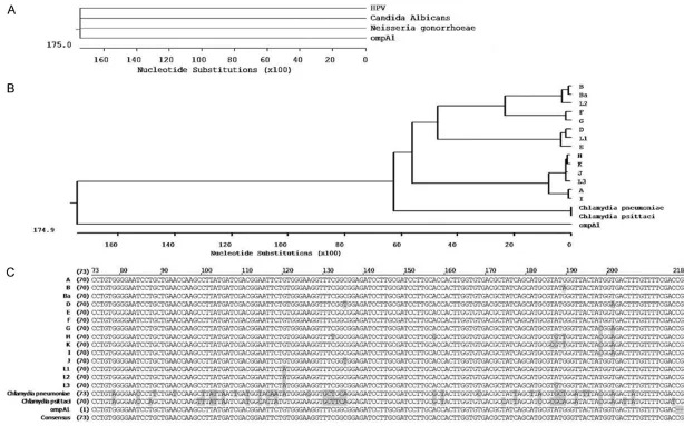
Cluster analysis of ompA1
A sequence of ompA1 was obtained from se- quencing. DNA sequences of multiple C. tra -chomatis serovars (A, B, Ba, D-K, L1, L2, L3),
Chlamydia pneumoniae and Chlamydia psitta -ci, Candida albicans, Neisseria gonorrhoeae,
and human papilloma virus (HPV) wereobtained from the National Center of Biotechnology In- formation (NCBI) source GenBank (http://www. ncbi.nlm.nih.gov/). Multiple alignments of omp- A1 were performed using the ClustalW method. Multiple sequence alignments were carried out using the MegAlign application of the DNAStar software program.
C. trachomatis detection in cell culture super
-natant by real-time PCR
C. trachomatis strain E was obtained from the American Type Culture Collection (ATCC: VR. 348B) and HeLa 229 cells were obtained from the Chinese Academy of Sciences Institute of Cell Biology, Shanghai Cell Bank.
HeLa 229 cells were grown in six-well plates with RPMI-1640 medium (HyClone) and 10% FBS (HyClone) at 37°C in 5% CO2 for 16 hours. The aim was to achieve a confluent monolayer. The cells were then infected with 106 IFU C. tra
-chomatis and centrifuged at 2000 rpm at room temperature for 1 hour. Finally, 1 mL/well of C.
trachomatis growth medium containing 10% FBS and 2 μg/mL actidione (Sigma) was added to six-well plates for culturing of C. trachomatis. After culturing for 24 hours, 48 hours, and 72 hours, the cell culture supernatant was collect-ed and quantitatively assesscollect-ed by real-time PCR assay, directly, without centrifugation or DNA extraction.
C. trachomatis detection for murine model genital tract secretions by real-time PCR
sites of the pET21a (+) vector (Figure 1A), resulting in recombinant plasmid pET21a (+)/
ompA as the standard plasmid for real-time PCR. The melting-curve of ompA1 was sharply defined with a narrow peak, indicating that a pure and homogeneous PCR product was pro-duced (Figure 1C). A single band at the appro-priate position of 144 bp on an electrophoresis gel indicated that the PCR product was as expected (Figure 1B). The combination of melt-ing curves and gel electrophoresis confirmed that the established PCR method had sufficient specificity to perform quantitative detection of the ompA1 gene of C. trachomatis. In the plas-mid DNA standard curve, the log of the initial copies of 10-fold plasmid dilutions was plotted on the x-axis and threshold cycles (Ct) were plotted on the y-axis. Results showed that the plasmid DNA standard curve was linear from 102 to 109 (Figure 1D). This plasmid DNA
stan-dard curve enables quantitative detection of C.
trachomatis in unknown samples.
Homology analysis of ompA1 DNA sequence
The ompA1 gene had high homology among various Chlamydia strains (14 strains of Chla-mydia trachomatis, Chlamydia pneumoniae, and Chlamydia psittaci) (Figure 2B), especially the 14 strains of C. trachomatis (Figure 2C). It had low homology with other common genital tract infectious pathogens, such as Candida albicans, Neisseria gonorrhoeae, and human papillomavirus (HPV) (Figure 2A).
Quantitative detection in cell culture superna
-tant and the mouse model
Cell culture supernatants and mouse model specimens containing Chlamydia EBs were used to verify the feasibility of Chlamydia EB detection using this novel real-time PCR meth-od. Figure 3 indicates that the level of Chlamydia
EBs in the extracellular environment was 5.3 × 10³ copies/µl, 5.1 × 10⁵ copies/µl and 5.8 ×
105 copies/µl at 24 hours, 48 hours, and 72
hoiurs, respectively, after Chlamydia trachoma -tis infection of HeLa 229 cells. Furthermore, Figure 4 shows the quantitation of Chlamydia
EBs in wash specimens from the mouse infec-tion model. The mice had positive genital tract environments continuously through the 27th
day after C. trachomatis infection. Data are rep-Mice were administered progesterone (2.5 mg/
dose) subcutaneously. After 10 days, they were challenged intravaginally by direct instillation of 20 μl of SPG containing 106 IFU C. trachomatis/
mouse. Genital tract secretion samples were collected every 3 days (0, 3, 6, 9, 12, 15, 18, 21, 24, and 27 days) and stored at -80°C until use.
Collected samples were filtered through 0.45 μm sterile filters, then used to quantitatively de- tect ompA1 copies by real-time PCR. All detec-tion methods were performed as previously described.
Female genital tract secretion collection and detection
Genital tract secretions of the eighty-six outpa-tients (the Second Affiliated Hospital of Wen- zhou Medical University, Gynecology Depart- ment) were swabbed. Each swab was placed into a 1.5 mL Eppendorf tube containing 1 mL SPG and stored at -80°C until use. Informed written consent was obtained from each patient and the study was approved by the Human Research Ethics Committee at Wenzhou Me- dical University.
Viable thawed samples were filtered through 0.45 μm sterile filters, then used to infect a monolayer of HeLa 229 cells. After 48 hours of culturing, the infected monolayer of cells was stained with Giemsa. Chlamydia inclusion form-ing units (IFUs) in 30 visual fields of a 400 × microscope were counted by multiplying the mean number of inclusions per visual field, the ratio of the coverslip area to the visual field area, and the dilution factor. Simultaneously, 2 μl of the filtered secretion sample was used to quantitatively detect ompA1 copies via real-time PCR assay. All detection methods were the same as previously described.
Statistics
Differences in C. trachomatis infection rates in female specimens were assessed by Chi-sq- uared test. The level of statistical significance is defined as P < 0.05 and all calculations were performed using SPSS 21.0 Software (IBM, USA).
Results
Plasmid standard curve by real-time PCR for quantitative C. trachomatis detection
syn-5046 Int J Clin Exp Med 2019;12(5):5042-5050
Quantitative detection for female genital tract
secretions
Rounded deep-blue inclusion bodies of C. tra -chomatis were observed in infected HeLa 229 monolayer cells by microscopic examination (Figure 5). Table 1 shows that all 22 samples identified as positive using IFU enumeration could also be positively identified using real-time PCR detection. There were also 8 samples that were positive on real-time PCR detection but negative on IFU enumeration. The infection rate was 34.9% (30/86) by real-time PCR detec-tion and 25.6% (22/86) by IFU enumeradetec-tion detection for the 86 female outpatients (Table 1). The C. trachomatis infection rate of female genital tract secretions, according to real-time PCR, was significantly different from that de- tected by IFU enumeration (χ2 = 55.95, P <
0.05). Discussion
The method of cell culturing for IFU enumera-tion of C. trachomatis has been widely used for detection in animal models and patients infect-ed with C. trachomatis. However, this method is subjective, cumbersome, and has low sample throughput. The current study developed an RT- PCR assay based on the ompA gene for quanti-tative and direct detection of Chlamydia EBs in genital secretions, aiming to improve the accu-racy and sensitivity of detection of C. trachoma -tis infection.
Recently, traditional PCR and real-time PCR as- say applications for quantitative detection of C.
trachomatis have been reported. These meth-ods are based on various target genes, such as the 16S ribosomal RNA gene, ompA, and a cryptic plasmid [12-15]. PCR assay based on the cryptic plasmid is regarded sensitive for detection of C. trachomatis. However, the cryp-tic plasmid has multiple copies in different st- rains [16] and the 16S ribosomal RNA gene has 2 or 3 copies in a single C. trachomatis genome [12]. Although the 16S ribosomal RNA gene is conserved across Chlamydia strains [17], the multicopy characteristics of this target gene may affect the correct numbers of C. trachoma -tis, thus reducing the accuracy ofquantitative detection [18]. Since ompA is present in a sin-gle copy in the C. trachomatis DNA sequence [19, 20], RT-PCR assay based on the ompA tar-get gene could solve the quantitative detection problem caused by multi-copy genes, to a cer-tain extent. However, there is variance through-out the ompA gene in various C. trachomatis
strains [21], perhaps limiting the broad applica-bility of this real-time PCR assay.
The current study used multiple alignment me- thodology to analyze the homology of 14 strains of C. trachomatis, Chlamydia pneumoniae, and
[image:6.612.90.289.73.201.2]Chlamydia psittaci. Results revealed that the construct ompA70-213 (ompA1) had high homol-ogy specifically among C. trachomatis infec-tions in the reproductive tract and low homolo-gy with other common reproductive infectious pathogens, such as C. albicans, N. gonorrhoe -ae, and human papillomavirus (Figure 2). Hen- ce, results indicate that ompA1 is appropriate
Figure 3. Real-time PCR assay with Chlamydia EBs in culture medium. Culture medium was collected from one well of infected monolayer HeLa 229 cells at 24 hours, 48 hours, and 72 hours. The number of Chla
-mydia EBs in the culture medium was quantitatively assessed by real-time PCR assay in triplicate.
[image:6.612.326.523.75.216.2]for specific and quantitative detection of C. tra -chomatis in genital tract secretions.
An ompA1 standard plasmid (Figure 1) was constructed and a novel real-time PCR method was developed based on ompA1, aimingto su- ccessfully quantitatively detect C. trachomatis
incell culture supernatants. After 48 hours of culturing, the number of EBs in the extracellu-lar environment reached a maximum (Figure 3). Present results demonstrate that the novel RT-PCR for C. trachomatis detection had a
cer-tain accuracy, consistent with a previous report [22]. Identical results were obtained during detection in an infected mouse model (Figure 4). C. trachomatis copies in mouse genital tract secretions could be quickly and accurately detected by this novel real-time PCR. As in oth- er experiments involving C. trachomatis clear-ance, similar results were obtained by IFU en- umeration.
In addition, compared with traditional detection methods, this newly developed real-time PCR method is more sensitive and specific, espe-cially in terms of quantitative detection of C.
trachomatis (Figure 5). According to ompA PCR analysis, one published study [20] showed C.
[image:7.612.91.526.71.251.2]trachomatis infections in 27% of sexually active women (SAW). In 86 female genital tract secre-tion samples, real-time PCR analysis revealed a positive rate of 34.9% (30/86) for C. trachoma -tis infection. This is significantly higher (P < 0.05) than the 25.6% (22/86) rate detected by the cell culture method. The current study pro-vides additional evidence that this novel real-time PCR may be more sensitive than conven-tional PCR [23] and that application of a real-time PCR assay that directly detects genital tract secretions, without DNA extraction, may reduce the loss of C. trachomatis DNA [12]. In this study, 8 samples were determined to be positive via the real-time PCR assay that were negative via the IFU enumeration method
Figure 5. HeLa 229 monolayer cellsinfected with C. trachomatis stained with Giemsa. HeLa 229 cells were infected for 48 hours with 200 μl genital tract secretion samples from clinical patients and stained with Giemsa.A. HeLa 229 cells infected with genital tract secretion samples, C. trachomatis inclusion-forming units can be observed as bodies that appear deeper blue than the cytoplasm under a 400 × microscope (indicated by arrows); B. HeLa 229 cells without genital tract secretion sample as a control after 48 hours of cell culture.
Table 1. Comparison of real-time PCR analy-sis and IFU enumeration (gold standard) of 86 C. trachomatis specimens tested in clini-cal human female genital tract secretions Real-time PCR Positive Negative culture (IFUs) Positive 22 0
Negative 8 56
[image:7.612.90.290.385.424.2](Table 1). A possible reason may involve the sensitivity of the real-time PCR method. Real-time PCR can amplify DNA from both infectious and noninfectious EBs, whereas the traditional culture method detects DNA only from infec-tious EBs. It is also possible that samples that yielded a positive real-time PCR result and a negative IFU result may reflect low levels of via-ble infection that are not detectavia-ble by IFU staining. Another possibility for the results by real-time PCR assay is that some of the infec-tious C. trachomatis EBs were lost because of transport, storage of samples, and/or filtering before testing by IFU staining was performed [24]. Thus, the currently developed real-time PCR method could provide feasible enumera-tion to assist in C. trachomatis infectious ass- essment.
Approximately 70%-95% of genital tract infec-tions are asymptomatic in women [25]. This assay, therefore, allows the detection of very low levels of Chlamydia in genital tracts. It can be applied in certain particularly critical situa-tions, such as when evaluating infection clear-ance for determination of vaccine efficacy in mouse infection models and when assessing infections in clinical patients.
In conclusion, this novel real-time PCR assay, based on the ompA1 gene, to quantitatively detect C. trachomatis is especially suitable for genital secretion specimens.
Acknowledgements
This work was supported by grants from the National Natural Science Foundation of China (30972669, 81172463, 31700160); The Sci- entific Research Incubation Project of The First Affiliated Hospital of Wenzhou Medical Unive- rsity (FHY2014052); The Science and Techno- logy Planning Project of Wenzhou (Y20170056).
Disclosure of conflict of interest
None.
Address correspondence to: Dr. Lifang Zhang, Depa- rtment of Medical Microbiology and Immunology, Wenzhou Medical University, Chashan College Town, Wenzhou, China. Tel: 577-86689910; Fax: +86-577-86689910; E-mail: [email protected]
References
[1] Witkin SS, Minis E, Athanasiou A, Leizer J and Linhares IM. Chlamydia trachomatis: the
per-sistent pathogen. Clin Vaccine Immunol 2017; 24.
[2] Choroszy-Krol IC, Frej-Madrzak M, Jama-Kmi-ecik A, Bober T and Jolanta Sarowska J. Cha- racteristics of the Chlamydia trachomatis spe-cies - immunopathology and infections. Adv Clin Exp Med 2012; 21: 799-808.
[3] Newman L, Rowley J, Vander Hoorn S, Wijeso-oriya NS, Unemo M, Low N, Stevens G, Gottlieb S, Kiarie J and Temmerman M. Global esti-mates of the prevalence and incidence of four curable sexually transmitted infections in 20- 12 based on systematic review and global re-porting. PLoS One 2015; 10: e0143304. [4] Land JA, Van Bergen JE, Morre SA and Postma
MJ. Epidemiology of Chlamydia trachomatis in-fection in women and the cost-effectiveness of screening. Hum Reprod Update 2010; 16: 189-204.
[5] Kelly H, Coltart CEM, Pant Pai N, Klausner JD, Unemo M, Toskin I and Peeling RW. Systematic reviews of point-of-care tests for the diagnosis of urogenital Chlamydia trachomatis infec-tions. Sex Transm Infect 2017; 93: S22-S30. [6] Halse TA, Musser KA and Limberger RJ. A mul
-tiplexed real-time PCR assay for rapid detec-tion of Chlamydia trachomatis and identifica -tion of serovar L-2, the major cause of lympho-granuloma venereum in New York. Mol Cell Probes 2006; 20: 290-297.
[7] Rizzo A, Domenico MD, Carratelli CR and Pao-lillo R. The role of Chlamydia and Chlamydophila infections in reactive arthritis. Intern Med 2012; 51: 113-117.
[8] Omsland A, Sixt BS, Horn M and Hackstadt T. Chlamydial metabolism revisited: interspecies metabolic variability and developmental stage-specific physiologic activities. FEMS Microbiol Rev 2014; 38: 779-801.
[9] Supadej K and Intorasoot S. Establishment of cheap and reliable real-time PCR for quantita-tion of HIV-1 viral load in plasma. J Med Assoc Thai 2012; 95: 1563-1568.
[10] Raza A, Ali Z, Irfan J, Murtaza S and Shakeel S. Analytical variables influencing the HCV RNA determination by TaqMan real-time PCR in rou-tine clinical laboratory practice. Mol Biol Rep 2012; 39: 7421-7427.
[11] Zhu S, Chen J, Zheng M, Gong W, Xue X, Li W and Zhang L. Identification of immunodomi -nant linear B-cell epitopes within the major outer membrane protein of Chlamydia tracho-matis. Acta Biochim Biophys Sin (Shanghai) 2010; 42: 771-778.
[13] Lehmusvuori A, Tapio AH, Maki-Teeri P, Ranta-kokko-Jalava K, Wang Q, Takalo H and Soukka T. Homogeneous duplex polymerase chain re -action assay using switchable lanthanide fluo -rescence probes. Anal Biochem 2013; 436: 16-21.
[14] Hopkins MJ, Ashton LJ, Alloba F, Alawattegama A and Hart IJ. Validation of a laboratory-devel -oped real-time PCR protocol for detection of Chlamydia trachomatis and Neisseria gonor-rhoeae in urine. Sex Transm Infect 2010; 86: 207-211.
[15] Last AR, Roberts C, Cassama E, Nabicassa M, Molina-Gonzalez S, Burr SE, Mabey DC, Bailey RL and Holland MJ. Plasmid copy number and disease severity in naturally occurring ocular Chlamydia trachomatis infection. J Clin Micr- obiol 2014; 52: 324-327.
[16] Ferreira R, Borges V, Nunes A, Borrego MJ and Gomes JP. Assessment of the load and tran-scriptional dynamics of Chlamydia trachomatis plasmid according to strains’ tissue tropism. Microbiol Res 2013; 168: 333-339.
[17] Lienard J, Croxatto A, Aeby S, Jaton K, Posfay-Barbe K, Gervaix A and Greub G. Development of a new Chlamydiales-specific real-time PCR and its application to respiratory clinical sam-ples. J Clin Microbiol 2011; 49: 2637-2642. [18] Vetrovsky T and Baldrian P. The variability of
the 16S rRNA gene in bacterial genomes and its consequences for bacterial community an- alyses. PLoS One 2013; 8: e57923.
[19] Stevens MP, Twin J, Fairley CK, Donovan B, Tan SE, Yu J, Garland SM and Tabrizi SN. Develo-pment and evaluation of an ompA quantitative real-time PCR assay for Chlamydia trachomatis serovar determination. J Clin Microbiol 2010; 48: 2060-2065.
[20] Hoque SM, Hossain MA, Paul SK, Mahmud MC, Ahmed S, Mahmud NU, Khan ER, Sakib MA, Ghosh S and Kobayashi N. Detection of Chlamydia trachomatis by immunological and genetic methods in female sex workers and the local female population of reproductive age in Mymensingh, Bangladesh. Jpn J Infect Dis 2013; 66: 256-259.
[21] Turingan RS, Kaplun L, Krautz-Peterson G, Norsworthy S, Zolotova A, Joseph SJ, Read TD, Dean D, Tan E and Selden RF. Rapid detection and strain typing of Chlamydia trachomatis us-ing a highly multiplexed microfluidic PCR as -say. PLoS One 2017; 12: e0178653.
[22] Manam S, Chaganty BK, Evani SJ, Zafiratos MT, Ramasubramanian AK, Arulanandam BP and Murthy AK. Intranasal vaccination with Chlamydia pneumoniae induces cross-species immunity against genital Chlamydia murida- rum challenge in mice. PLoS One 2013; 8: e64917.
[23] Zemtsova GE, Montgomery M and Levin ML. Relative sensitivity of conventional and real-time PCR assays for detection of SFG Rickettsia in blood and tissue samples from laboratory animals. PLoS One 2015; 10: e0116658. [24] Mahony JB and Chernesky MA. Effect of swab
type and storage temperature on the isolation of Chlamydia trachomatis from clinical speci-mens. J Clin Microbiol 1985; 22: 865-867. [25] Lanjouw E, Ouburg S, de Vries HJ, Stary A,