Development of Automated Classifier of Diabetic
Retinopathy using Datasets by Machine Learning
S. Sharon Rose1,Dr. B. Kezia Rani Madam2
1
M. Tech (CST with BDA), 2Assistant Professor, Department of Computer Science, Adikavi Nannaya University, Rajamahendravaram, Andhra Pradesh, India
Abstract: The conspicuous medical condition that occurs in the majority of population enlarges in diabetic. The condition is caused due to varying amount of insulin and glycogen secreted by endocrine gland of pancreas. At the higher stage of diabetes patients may start suffering from some sight disorders. It is due to bleeding or the accumulation of fluid in the retina which is a symptom of Diabetic Retinopathy (DR). Diabetic Retinopathy (DR) is one such abnormality affects the eye due to the significant amount of insulin present in the blood stream. When this condition is detected at the earliest stage of inception this abnormality can be controlled easily. if the blood vessels of the retina are damaged, it results in Diabetic Retinopathy (DR). The earliest stage of diabetic retinopathy will be visible on the surface of retina as Micro aneurysm, haemorrhage, exudates.
Keywords: Micro aneurysms, exudates, support vector machine, machine learning.
I. INTRODUCTION
Diabetes is a gathering of metabolic illnesses in which an individual has high glucose, either in light of the fact that the body does not deliver enough insulin, or in light of the fact that the pancreas Don't react to the insulin that is produced. Diabetic retinopathy is one of the normal complexities of diabetes. It is a serious and generally spread eye ailment. It harms the little veins in the retina bringing about loss of vision. The danger of the infection increments with age and in this manner, moderately aged and more seasoned diabetics are inclined to Diabetic Retinopathy. Non-proliferative diabetic retinopathy is a beginning period of diabetic retinopathy. In this stage, small veins inside the retina spill blood or liquid. The releasing liquid makes the retina swell or to frame stores called exudates. Proliferative diabetic retinopathy, PDR is an endeavour by the eye to develop or resupply the retina with fresh Recruits vessels (neovascularization), because of far reaching conclusion of the retinal blood supply. Unfortunately, the new, anomalous veins don't resupply the retina with ordinary blood stream, however drain effectively and are frequently joined by scar tissue that may wrinkle or isolate the retina. In this paper, a mechanized methodology for grouping of the ailment diabetic retinopathy utilizing fundus pictures is introduced. We utilized the retinal fundus pictures gathered from the online informational indexes. The retinal picture is taken in the RGB shape by fundus camera. A fundus camera or retinal camera is a specific low power magnifying instrument with a connected camera intended to photo the inside surface of the eye, including the retina, optic circle, macula, and back pole. The pictures were caught utilizing a Canon Top Con TRC-50 EX with Nikon retinal camera at a field-of-vision (FOV) of 50. The gained picture goals are1280 ×1024 in 24bit JPEG design. The assessment of the proposed computerized analysis arrangement of diabetic retinopathy has been performed by utilizing a lot of 250 fundus pictures which is a mix of ordinary, NPDR and PDR influenced pictures. The first picture is changed over to dark scale picture. From that point forward, versatile histogram evening out is connected to enhance the complexity of the picture. At that point, Discrete Wavelet Transform (DWT) is connected and the extent of the picture is decreased into half as 640 × 512. At that point Matched channel reaction (MFR) is connected to lessen the commotion in the picture. At last, SVM and Decision Tree based grouping is connected to portion the veins in the picture. In the wake of pre-processing of pictures is finished, highlights, for example, Radius, Diameter, Area, Arc length, Centre Angle and Half zone are determined for each image. Finally, the pictures are characterized into three gatherings specifically, ordinary picture, Non-Proliferative Diabetic Retinopathy (NPDR) and Proliferative Diabetic Retinopathy (PDR).
II. SVM IMPLEMENTATION FOR DIABETIC RETINOPATHY
Support Vector Machine (SVM) was first heard in 1992, presented by Baser, Guyon, and Vapnik in COLT-92. Bolster vector machines (SVMs) are a lot of related administered learning strategies utilized for order and relapse.
space, prepared with a taking in calculation from streamlining hypothesis that actualizes a taking in inclination got from measurable learning Theory. It is likewise being utilized for some applications, for example, hand composing examination, face investigation, etc, particularly for example characterization and replace based applications. SVM were created to take care of the order issue, yet as of late they have been reached out to tackle replace issues.
Figure:1 Support vector machine
III. IMAGE SEGMENTATION
Image Segmentation is implemented using SVM classifiers Firstly region are determined by processing pixel classification approach where channels in a particular image were classified. Intensity levels of pixels are being compared Generally the pixel In the image with highest intensity level are set to be having hue value in a yellow range and lowest intensities having white values are also selected. A clustering mechanism is used to gather these group of pixels which are having low intensity and high intensity into their respective clusters. This classification mechanism also forms various clusters falling in between the highest and lowest Range. The region falling in lowest range and in between range are taken into consideration and highest intensity ranges are omitted as This Regions are generally non - effected region of DR.
The candidate region for a cluster is defined in square form of 110/110 pixels with the Centroid lying in the middle of the cluster.
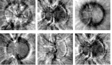
Figure 2: The eigen discs obtained for training set
The classified clusters of high and medium intensities are processed and converted into their respective Alpha Channels. This process is otherwise called propagation where image patch is being classified as Alpha channels intensity values and distance between each image patch is being calculated these Calculated Image patch is compared of that a original image
Based on the comparison values a boundary will be drawn on the cluster of lower intensities to highlight the effected region. Then this boundary regions are enhanced using lab colour morphology. In this process regions are allowed to involve towards boundary under the constraints.
F
int
F
ext
0
Equ (1)where
)
(
)
(
''''''
int
X
s
x
s
F
and ext
F
[image:2.612.201.381.423.528.2]
dxdy
f
v
f
v
v
x yy
x
)
|
|
|
|
}
{(
2
2
2
2
2
2
Equ(2)
where f is an edge map defined as
Equ(3)
[image:3.612.173.396.297.389.2]where G is a gaussian filter with standard deviation s. µ is set according to the amount of noise present in the image.
Figure 3: Results of image localization.
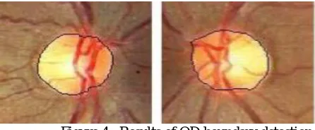
The black square represents the localized image among all the candidate regions (which are shown in red squares) s The results of image boundary extraction are shown in figure 4.
Figure 4: Results of OD boundary detection.
The segmented OD region is not considered in the later steps for detection of exudates.
IV. RESEARCH METHODOLOGY
Fleming et al.(2010) have demonstrated the job of microaneurysm and discharge in programmed reviewing of diabetic retinopathy. A standout amongst the most essential strides in the robotized location of DR is the discovery of microaneurysms. Microaneurysms are among the soonest recognizable indications of the nearness of diabetic retinopathy. Because of countless, the accessible ophthalmologists are not adequate in dealing with every one of the patients, particularly in country territories. In this manner, computerized early discovery of microaneurysms could facilitate the weight of ophthalmologists. Robotized microaneurysms identification can likewise help the ophthalmologists in exploring and treating the infection more
In [10] the present status of diabetic retinopathy has been examined. The circumstances and end results of diabetes and DR have been examined. The distinctive kinds of DR have been talked about. The distinctive element extraction strategies and identification techniques has been quickly clarified. An ophthalmoscope has been utilized by an ophthalmologist to distinguish, examine and envision the modest veins and the diverse DR stages. A framework has been proposed where computerized pictures are taken and broke down after this screening and DR location is finished utilizing a robotized framework and couple of calculations.
V. PRE-PROCESSING
1) Colour Normalization: The retinal tissue pigmentation crosswise over patients shift broadly. Thus the shade of exudates may not seem more splendid than the foundation in some picture areas. Consequent, The pictures are standardized to a reference picture browsed the test informational collection utilizing histogram detail. Shading standardization is accomplished by joining the Normalized R, G, and B parts.
2) Local Contrast Enhancement: Another perception is the debasement of value conversely from the Centre towards the limit of the retinal picture. Therefore, Local differentiate upgrade method is connected to the information pictures [4]. The pictures are first changed over to HSI shading space and the improvement strategy is connected on the force channel. For every pixel x (I, J)
in the picture, a M x M window W with x as the middle is considered. Let w µ and w σ be the mean and standard deviation of
the forces inside the window.
2
2
(
(
,
)
)
1
w
x w
w
x
i
j
M
Let Max and Min represent the Maximum and minimum intensity values for the whole image.
The new intensity value n
x
n( i, j) is obtained as:Equ(6)
Equ(7)
A sample results shown in figure 5.
(a) (b)
(c) (d)
w x
x
x
i
j
M
1
2(
,
)
Equ(4)
Exudates appear significantly contrasted in the green channel. Density threshold (DT) algorithm is applied on the green component of the pre-processed image. Only the candidate pixels found are considered for DT. The image is divided into blocks of 64x64 pixels each such that there is 50% overlap between adjacent pairs. Histogram of each of these blocks is computed. If the histogram is unimodal a high threshold value is used to set, otherwise Otsu’s thresholding algorithm is used to find the threshold for the block. Threshold values for all the blocks are calculated and stored. Each pixel belongs to four blocks. The threshold ‘t’ of the candidate pixel is interpolated from the threshold values of the four blocks to which it belongs.
Let1
c
1
(
x
1,
y
1);
c
2
(
x
2,
y
1);
c
3
(
x
2,
y
2);
c
4(
x
1,
y
2)
represent the Centre of four blocks whose thresholds are 1t, 2 t, 3t and4 t respectively. Then t is calculated as:
The candidate pixel is classified as an exudates pixel if its intensity value is greater than its threshold t.
VI. DETECTION OF OBJECTS WITH SHARP EDGES
Our aim in this step is to detect all objects with sharp edges, as exudates are found to have sharp edges.
Canny edge detector is applied on the green component of the pre -processed image. Then a global threshold value is set to obtain a binary image (B2) containing sharp edges. Fig 6(c) shows the result on a test image.
i. (b)
(c) (d) Figure:6
VII. IDENTIFICATION OF FUNDUS IMAGES USING ALPHA CHANNEL EXTRACTION
VIII. RESULTS
The work carried out in the proposed methodology detects the presence of proliferative diabetic Retinopathy. By applying digital image processing and fundus images taken from medical camera the investigation was carried out in the input dataset of images. The techniques was used were to extract Alpha channel, edge detection to enhancement of fundus Images, Colour normalization was used for enhancement of Colour fundus images.
By using the above techniques exudates were detected generally. There exudates are embedded into the nerve Centre of eyeball. There were differentiate using the above techniques and were highlighted accordingly to predict the area effected by DR.
IX. CONCLUSION
The above methodology is used to detect in the condition of DR using image processing techniques area effected by DR is pointed out, in the level of the severity was also Analyzed by taking all parameters of DR consideration.
Eyeball images of normal person are also compared with that of person effected with DR and the level of damage was Analyzed through the above methodology and efficient way was proposed to detect Diabetic Retinopathy.
REFERENCES
[1] D. Fleming, K. A. Goat man, and J. A. Olson, “The role of haemorrhage and exudate detection in automated grading of diabetic retinopathy,” British Journal of Ophthalmology, vol 94, no. 6, pp. 706- 711, 2010.
[2] Hip well JH, Strachan F, Olson JA, McHardy KC, Sharp PF, Forrester JV. Automated detection of microaneurysms in digital red-free photographs: a diabetic retinopathy screening tool. Diabetes Med 2012; 17: 588–594.
[3] A. Sopherim Mathew N. Dailey, Bunya Ullyanov, Sarah Barman, Tom Williamson, Yin Aye Moe, “Machine Learning approach to automatic Exudates detection in retinal images from diabetic patients”, Journal of Modern optics, Vol. 57, No. 2, pp. 124-135, Nov 2015.
[4] Mathai, Daniela, and R. Matai., “Detection of diabetic symptoms in retina images using analog algorithms,” International Journal of Biological and Life Sciences, pp. 224-227, 2016.
[5] W. Hsu, P.M.D.S Pallawala, Mong Li Lee, Kah-Guan Aueong, The Role of Domain Knowledge in the Detection of Retinal Hard Exudates, Proc. 2013 IEEE Computer Society Conf. on Computer Vision and Pattern Recognition, 2, 2001, II-246 - II-251. Classification and Localization of Diabetic-Related Eye Disease by Alireza Osareh Majid Mirmehdi, Barry Thomas, and Richard Markham.
[6] U.R. Acharya, C.M. Lim, E.Y. K. Ng, C.Chee and T.Tamura. “Computer-based detection of diabetes retinopathy stages using digital fundus images” Journal of Engineering in Medicine, Vol. 223, Issue no. 5, Feb. 2009.
[7] A. Sopharak, B. Uyyanonvara, S. Barman, TH. Williamson. “Automatic detection of diabetic retinopathy exudates from non-dilated retinal images using mathematical morphology methods” Computerized medical imaging and graphics 32 (8), pp.720-727, Aug. 2008
[8] B. Wu, W. Zhu, F. Shi, S. Zhu, and X. Chen, "Programmed recognition of microaneurysms in retinal fundus pictures," Computerized Medical Imaging and Graphics, vol. 55, pp. 106– 112, 2017.
[9] H.H.Crokell,“Specialization and sInternational Competitiveness,” in Managing the Multinational Subsidiary, H.E Temad and L.S.Saluda (eds.), Croom-Helm, London, 1986. (book chapter style)