Original Article
The protective effects of
ischemic postconditioning on renal
tubular epithelial Epithelial-mesenchymal transition
Xiaodong Weng, Guanjun Jiang, Xiuheng Liu, Hui Chen, Zhiyuan Chen
Department of Urology, Renmin Hospital of Wuhan University, Wuhan University, Wuhan, China
Received October 10, 2015; Accepted December 25, 2015; Epub February 15, 2016; Published February 29, 2016
Abstract: Renal tubular Epithelial-mesenchymal transition (EMT) plays an important role in the process of renal fibrosis. The aim of this study is to observe the effects of ischemia post-conditioning on renal tubular Epithelial-mesenchymal transition in vitro. A valid in vitro ischemia post-conditioning model was established by using NRK-52E cell line. The ischemic postconditioning (IPO) model was established by placing the cells in an ischemic condition for 3 hours and then followed by exposing the cells to three cycles of a reperfusion state for 10 minutes and an ischemic state for another 10 minutes. Flow cytometry and Hoechst were used to gain access to the apoptosis that took place. The protein expression levels of α-SMA, TGF-β, CTGF and Ecadherin were detected by western blot. The simulating I/R injury results in severe injury in NRK-52E cells as evidenced by increased apoptotic in-dex, which may be significantly attenuated by IPO treatment applied before the abrupt reperfusion (P<0.05 vs I/R group). Meanwhile, I/R increase the expression levels of α-SMA, TGF-β, CTGF, and decrease the expression levels of E-cadherin. However, IPO inhibts the expression changes of these proteins. The results offer evidence that I/R injury causes cell apoptosis, and IPO can effective to attenuate renal cell apoptosis. Meanwhile, IPO could inhibit the occurrence of EMT potentially mediated by decreasing the expression of α-SMA, TGF-β, CTGF and protetecting the expression of E-cadherin.
Keywords: Epithelial-mesenchymal-transition (EMT), fibrosis, ischemic postconditioning, ischemic reperfusion
Introduction
Renal ischemia/reperfusion (I/R), which is an important cause of renal dysfunction. Moreover, severe renal I/R not only cause renal failure, but also leads to chronic tubulointerstitial fibro-sis, which is a necessary part of the various causes of kidney disease progressed to end stage renal disease. During the process of renal fibrosis, the renal tubular Epithelial-me- senchymal transition (EMT) plays a major role. The process of EMT in embryonic development is for primitive epithilial cells to form the meso-derm and primitive neuroepithelial cells to form neural crest cells. The conversion of epithelial cells to mesenchymal cells is fundamental for embryonic development and profound pheno-typic changes may occur including the loss of cell-cell adhesion, the loss of cell polarity, and the acquisition of migratory and invasive
prop-erties [1, 2]. In the field of cancer cell biology, EMT is an important molecular mechanism of tumor invasion and metastasis [3]. However, it is worth noting that the cancer cell of EMT does not lead to the formation of metastases of fibro-blasts and only epithelial cells acquire mesen-chymal characteristics, which had significant difference from the ordinary myofibroblasts. Over the course of the past decade, emerging evidence suggests that EMT is a source of myo-fibroblast recruitment in the fibrosis process [4].
are a series of studies to support this hypothe-sis: Firstly, under the culture conditions of Transforming Growth Factor-beta 1 (TGF-β1) in vitro, the original tubular epithelial cells can experience phenotypic transformation. The ch- aracteristics of this transition include the loss of epithelial features such as E-cadherin, zonu-la occludens-1 (ZO-1), cytokeratin, and activat-ed the markers of interstitial or myofibroblast cells including the expression of α-smooth mu- scle actin (α-SMA), vimentin, fibroblast-specific protein-1 (FSP-1) and interstitial matrix compo-nents [5]. Secondly, Double-labeling immuno-histochemistry co-localization of epithelial and mesenchymal cell markers to detect kidney damage showed simultaneous expression of two intermediate cell markers, this indicating the presence of EMT. Finally, the most impor-tant and a landmark study published in 2002, which proposed using the Cre/Lox technology and the analysis of cell lineage to trace the renal tubular epithelial fate in the process of renal fibrosis, for the first time in vivo, proved the existence of EMT in renal tubular epitheli-um [6]. However, our primary study showed that IPO may inhibit the EMT of renal tubular epithe-lial which induced by ischemic renal fibrosis [7]. In the study α-SMA and TGF-β1 expression were inhibited by IPO in vivo. Therefore, the present study utilized an in vitro model we pre-established to evaluate the effects of ischemic postconditioning (IPO) on the renal tubular epi-thelial-mesenchymal transition, to verify wheth-er the IPO in vitro can inhibit EMT in the renal tubule.
Materials and methods
Cell culture
The renal tubular epithelial cell line, NRK-52E cells, was purchased from Cell Resource Center of Shanghai Institutes for Biological Sciences, Chinese Academy of Sciences. The cells were cultured on culture dishes and gassed with 95% air/5% CO2 and maintained at pH 7.4 and 37°C. The medium was changed once every two days, and the cells were used for experi-ments at day 10 after seeding. All experiexperi-ments were performed with cells that were cultured in serum-free medium for 24 hours prior to the experiments. Cells were seeded on 6 well plates or culture dishes as appropriate.
In vitroischemic postconditioning model
The complete medium is replaced with serum free DMEM one day before the experiment to synchronize the cells. The preparation of con-trol buffer and ischemic buffer refference method for the preparation of C Sauvant [8]. Then, the cells are randomly divided into three treatment groups as follows: 1) Control group: NRK-52E cells are first incubated in 1 mL con-trol buffer (NaHCO3 24.0 mM, Na2HPO4 0.8 mM, NaH2PO4 0.2 mM, NaCl 86.5 mM, KCl 5.4 mM, CaCl2 1.2 mM, MgCl2 0.8 mM, HEPES 20 mM; pH adjustment to 7.4 with 1N NaOH) under normoxic conditions (95% air-5% CO2) for 3 hours (h), and then are incubated in 2 mL fresh complete DMEM medium (DMEM-10% FBS-streptomycin 100 g/ml and penicillin 100 U/ml) under normal conditions (95% air-5% CO2) for 24 h; 2) Ischemic and reperfusion injury (I/R) group: NRK-52E cells are washed with 1 mL serum free DMEM medium pH 7.4, following with 1 mL sugar free DMEM medium pH 7.4, and then are incubated in 1 mL ischemia buffer (NaHCO3 4.5 mM, Na2HPO4 0.8 mM, NaH2PO4 0.2 mM, NaCl 106.0 mM, KCl 5.4 mM, CaCl2 1.2 mM, MgCl2 0.8 mM, MES 20 mM; pH
adjust-ment to 6.6 with 1N NaOH) under hypoxic con-ditions (0.5% O2-5% CO2-94.5% N2) for 3 h. After this phase, the serum free and sugar free DMEM medium is replaced with complete DMEM medium to simulate reperfusion, fol-lowed by 3 h, 6 h, 12 h and 24 h in normal con-ditions. 3) Ischemic postconditioning by “add-ing” medium group (IPO): NRK-52E cells are first incubated in 1 mL ischemia buffe under hypoxia conditions for 3 h. After this phase, the culture dishes are transferred to a normoxic conditions incubator in a humidified atmo-sphere with adding another 0.5 mL fresh com-plete DMEM medium for 10 min. Subsequently, the cells are returned to hypoxia conditions for 10 min. The postconditioning cycle is repeated three times and is described as IPO. Finally, the cells are incubated in 2 mL fresh complete DMEM medium under normoxic conditions for 3 h, 6 h, 12 h and 24 h.
Fluorescein isothiocyanate (FITC)-conjugated annexin-V-propidium iodide (PI) dual staining
The 5×10⁵ NRK-52E cells were cultured in a
h later, all cells were processed in according with the above grouping. After an additional 24 h culture, NRK-52E cells were collected and stained with FITC-conjugated annexin V and propidium iodide according to the steps given in the apoptosis detection kit, and testing com-pleted within an hour after dyeing. Then we per-formed flow cytometry for apoptosis analysis (Apoptosis Kit, BD Pharmingen, Germany).
Hoechst 33342 assay
Exponentially growing cells are plated in 6-well plates at a density of 2×10⁵ cells/well and
cul-tured for 72 h. Following simulated I/R and IPO procedures, cells are fixed for 8 min with the precooled (-20°C) formaldehyde and acetone solution (1:1, v/v) and washed with PBS 3×3 min, followed by staining with Hoechst33342 solution (50 mmol/L) at 37°C in darkness for 5 min. Apoptotic cells are observed and images are taken using fluorescence microscope (OlympusBX51, Tokyo, Japan), with excitation wavelength of 350 nm and emission wave-length of 460 nm.
Protein analysis by western blot
The protein expression levels of α-SMA, TGF-β, CTGF and E-cadherin were examined by west-ern blotting. Briefly, proteins were extracted from NRK-52E, subjected to SDS-PAGE on 10% polyacrylamide gels (20 μg/lane), and then electrotransferred to nitrocellulose membrane. The membranes were blocked with 5% nonfat milk in TBST buffer (10 mmol/L Tris-HCl, 0.15 mol/L NaCl, and 0.05% Tween 20, pH 7.2) and incubated with the following rabbit polyclonal primary antibodies. Subsequently, the mem-branes were incubated with secondary horse-radish peroxidase-conjugated anti-rabbit IgG antibody. The membrane was washed exten-sively with TBST, and the immunoreactive ba- nds were detected with ECL-detecting reagents.
Statistical analysis
[image:3.629.100.529.81.391.2]numbers of apoptotic cells, comparisons am- ong groups were compared using one-way ANOVA3 Student-Newman-Keuls test. P<0.05 was used to determine statistical significance.
Results
Evaluation of apoptosis rates by fluorescein
isothiocyanate (FITC)-conjugated annexin-V-propidium iodide (PI) dual staining
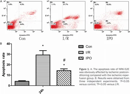
NRK-52E subjected to synchronization was treated in accordance with the above grouping. After 24 h of culture, NRK-52E were collected
and stained with FITC-conjugated annexin V and propidium iodide for detecting apoptosis. As shown in Figure 1, the rate of apoptosis of the control group was significantly lower than the I/R and IPO groups (P<0.05). Compared with both I/R and IPO groups, the rate of apop-tosis of I/R group is significantly higher than IPO (P<0.05).
Hoechst 33342 staining
[image:4.629.97.533.80.187.2]Effects of postconditioning on the I/R induced cell apoptosis are assayed by Hoechst 33342 staining. As shown in Figure 2, few apoptotic Figure 2. Effect of postconditioning on ischemia/reperfusion induced apoptosis of NRK-52E cells with Hoechst 33342 staining. A. Control, NRK-52E cells culture in control medium under normal condition. B. I/R, cells cultured in ischemic condition and then reperfusion for 24 h. C. IPO, cells cultured in ischemic condition, immediately for three cycle postconditioning and then reperfusion up to 24 h. (“→” Indicates the apoptotic cells. All fluorescence photomicrographs original magnification ×200).
[image:4.629.101.531.272.516.2]cells are present in the normal group. As ex- pected, there are many apoptotic (Hoechst 33342-positive) cells in the I/R group and in the IPO group. However, compared with the I/R group, fewer apoptotic cells are observed in the IPO group.
Protein expression
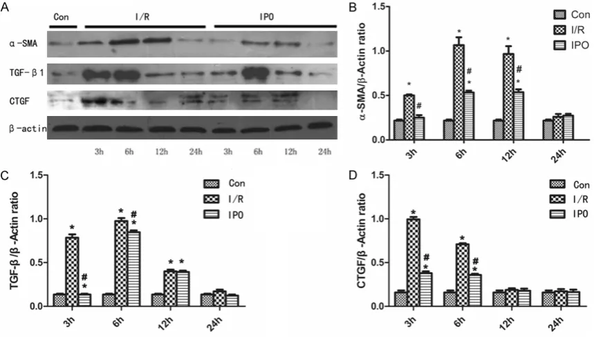
The results of protein extracts of α-SMA, TGF-β, and CTGF in various cell groups of NRK-52E were subjected to Western Blot Analysis are shown in Figure 3. Same amount of the extract-ed protein (20 μg/lane) was loadextract-ed on the same gel. β-actin was used as positive control. After treatment of 3 h, 6 h, 12 h and 24 h at different time points, the expression of α-SMA in the IRI group increased at 3 h, 6 h, 12 h, and then decreased. However, in contrast to the IRI group, the expression levels at 3 h, 6 h, 12 h were significantly suppressed in IPO group. The expression of TGF-β in the IRI group increased at 3 h, 6 h, through the reperfusion stage, and then decreased. Meanwhile, the IPO group ex- pressions similar to the IRI group, although expression levels at 3 h, 6 h were significantly suppressed. Interestingly, the expression mode of CTGF in IRI group is similar to TGF-β, the expression levels at 3 h, 6 h were significantly increased, then decreasing. Compared with the
[image:5.629.101.384.78.304.2]result of combined action of a variety of rea-sons, and is the main pathological basis of end-stage renal disease. When slight renal isch-emia-reperfusion occurs in clinic, the renal function and pathological changes will gradual-ly return to normal with the elapse of time. However, when severe renal IRI occurs, the renal tubular changes are irreversible and sub-sequently manifested as tubular fibrosis, thus resulting in a decline in renal tubular concentra-tion funcconcentra-tion and renal dysfuncconcentra-tion [9-11]. The Previous view was that, during the period of reperfusion, the renal tubular had powerful pro-liferation ability to repair injury and IRI can be completely reversed [12]. However, recently researchers have found that the capacity of proliferation and repair of renal tubular epithe-lial cells is limited, which depends on the sur-vival number of tubular cells and wether the basment membrane is intact. Renal tubular injury is irreversible when the extent of damage exceeds a critical value, and the structure and function of renal tubular injury cannot be fully recovered [13]. Some researchers have found that the moderate fibrosis lesions of renal tubu-lar interstitial disease does not cause an abnor-mal rise in blood urea nitrogen and serum cre-atinine, but may destroy the reabsorption func-tion of urine thus causing increased urinafunc-tion at night and proteinuria [13].
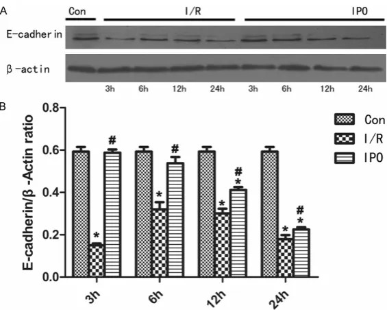
Figure 4. The IPO promotes protective effects on NRK-52E cells and the ex-pression levels of E-cadherin in the three groups (*P<0.05 versus control, #P<0.05 versus I/R. n=4).
control group, the IPO group expression did not change significantly at each time po- int. In this study, we also eval-uated the expression of E-ca- dherin in NRK-52E cells, as shown in Figure 4. The expres-sion levels in the IRI group was decreased at 3 h, while IPO inhibit this change, and the expression levels of IPO had no significant differen- ces compared with the nor-mal control group.
Discussion
The mark of myofibroblasts is α-SMA, and it is the activation forms of fibroblasts, which is also a major source of extracellular matrix collagen ingredients [14, 15]. The α-SMA only expressed in arterial smooth muscle cells in normal renal tissues. However, in the present study, we con-firmed the expression of α-SMA in the IRI group increased at 3 h, 6 h and 12 h. However, in con-trast to the IRI group, the expression levels at 3 h, 6 h and 12 h were significantly suppressed in IPO group. Thus, the present study demonstrat-ed that ischemia-reperfusion injury in vitro can also induce the expression of α-SMA and in- duce renal tubular epithelial cells to acquire mesenchymal polarity as well as the occur-rence of EMT.
During the process of renal interstitial fibrosis, the TGF-β1 was considered to be an important one in many fibrogenic factors. The main func-tion of TGF-β1 was to increase the synthesis of extracellular matrix (ECM), inhibit its degrada-tion and up-reguladegrada-tion of integrin matrix adhe-sion molecules [16, 17]. In multiple down-stream signaling pathways of TGF-β, the Smad signal pathway plays an important role in the process of multiple organ fibrosis, Smad 2 was an pivotal molecule to initialize the signal trans-duction pathway [18]. A large number of investi-gate of TGF-β1/Smad pathway was found that, by antagonism the activation of TGF-β1 can alleviate renal interstitial fibrosis [17]. Yasuda K et al found that through upregulate the expres-sion of TGF-β1 can increase renal tubular extra-cellular matrix, indicating the increased contin-uously of TGF-β1 has the potential to promote fibrosis [19]. In this experiment, we confirmed that the expression levels of TGF-β1 in the IRI group were significantly increased at 3 h, 6 h reperfusion, while its expression in the IPO group was significantly suppressed, shows that IPO can inhibit the expression of TGF-β1 in renal tubular epithelial cells of an ischemia-reperfusion model.
Connective tissue growth factor (CTGF) is matrix protein, plays a crucial role in the pro-cess of pathological fibrosis. In vivo and in vitro experiments have proved that, CTGF expressed in renal tubular epithelial cells, suggesting its involvement in the procedure of renal fibrosis and the course of EMT. The CTGF is a potential biomarker of EMT in chronic allograft nephropa-thy [20, 21]. In the present study, we confirmed
that the expression levels of CTGF in IRI group were obviously enhanced at point of 3 h, 6 h reperfusion, while its expression in IPO group were evidently attenuated, indicating that IPO can restrain the expression of CTGF in renal tubular epithelial cells of an ischemia-reperfu-sion model.
E-cadherin, which expressed in well-differenti-ated and polarized epithelial cells, such as the renal tubular epithelium, is a kind of adhesion protein [22]. Under pathological conditions, when renal tubular epithelial cells lose their E-cadherin, is when epithelial cells are under-going the process of EMT [23]. In this experi-ments, the expression of E-cadherin in IRI group were decreased at 3 h, while the expres-sion of E-cadherin in IPO groups had no signifi-cant changes compared with the normal con-trol group.
In summary, based on the above study, we demonstrated that IPO can relieve renal injury caused by ischemia-reperfusion, and protect the pression of E-cadherin, effectively inhibiting the expression of α-SMA, CTGF and TGF-β1 in renal tubular epithelial cell line NRK-52E cells, indicating the possibility to inhibit the occur-rence of EMT, its protective mechanism may be involvement of the TGF-β1/Smad signal path-way, thus decreasing the tubulointerstitial fibro-sis. However, these findings remain to be con-firmed by future studies. The current study shows that IPO improved the ability of renal tubular epithelial cells to tolerate ischemic-reperfusion injury, particularly to decrease α-SMA, TGF-β and CTGF expression, and pro-tecting the expression of E-cadherin, thus experimental studies in vitro model revealed that IPO may effectively inhibit the occurrence of EMT.
Acknowledgements
This study is supported by the grants from the National Natural Science Foundation of China (No. 30901494 and 2013RMFH012), the Pro- vince Natural Science Foundation of Hubei (No. 2012FFA096), and supported by the Funda- mental Research Funds for the Central Uni- versities (No. 2042014kf0115).
Disclosure of conflict of interest
Address correspondence to: Xiuheng Liu and Guanjun Jiang, Department of Urology, Renmin Hospital of Wuhan University, 238 Jiefang Road, Wuhan 430060, China. Tel: +86 27 88041911-2235; Fax: +86 27 88042292; E-mail: [email protected] (XHL); [email protected] (GJJ)
References
[1] Greenburg G, Hay ED. Epithelia suspended in collagen gels can lose polarity and express characteristics of migrating mesenchymal ce- lls. J Cell Biol 1982; 95: 333-9.
[2] Thiery JP, Acloque H, Huang RY, Nieto MA. Epithelial-mesenchymal transitions in develop-ment and disease. Cell 2009; 139: 871-90. [3] Yang J, Weinberg RA. Epithelial-mesenchymal
transition: at the crossroads of development and tumor metastasis. Dev Cell 2008; 14: 818-29.
[4] Kalluri R, Weinberg RA. The basics of epitheli-al-mesenchymal transition. J Clin Invest 2009; 119: 1420-8.
[5] Liu Y. Epithelial to mesenchymal transition in renal fibrogenesis: pathologic significance, mo-lecular mechanism, and therapeutic interven-tion. J Am Soc Nephrol 2004; 15: 1-12. [6] Iwano M, Plieth D, Danoff TM, Xue C, Okada H,
Neilson EG. Evidence that fibroblasts derive from epithelium during tissue fibrosis. J Clin Invest 2002; 110: 341-50.
[7] Weng X, Shen H, Kuang Y, Liu X, Chen Z, Zhu H, Jiang B, Zhu G, Chen H. Ischemic postcondi-tioning inhibits the renal fibrosis induced by ischemia-reperfusion injury in rats. Urology 2012; 80: 484, e1-7.
[8] Sauvant C, Schneider R, Holzinger H, Renker S, Wanner C, Gekle M. Implementation of an in vitro model system for investigation of reperfu-sion damage after renal ischemia. Cell Physiol Biochem 2009; 24: 567-76.
[9] Menke J, Iwata Y, Rabacal WA, Basu R, Yeung YG, Humphreys BD, Wada T, Schwarting A, Stanley ER, Kelley VR. CSF-1 signals directly to renal tubular epithelial cells to mediate repair in mice. J Clin Invest 2009; 119: 2330-42. [10] Timsit MO, Gadet R, Ben Abdennebi H, Codas
R, Petruzzo P, Badet L. Renal ischemic precon-ditioning improves recovery of kidney function and decreases alpha-smooth muscle actin ex-pression in a rat model. J Urol 2008; 180: 388-91.
[11] Prakash J, Sandovici M, Saluja V, Lacombe M, Schaapveld RQ, de Borst MH, van Goor H, Henning RH, Proost JH, Moolenaar F, Këri G, Meijer DK, Poelstra K, Kok RJ. Intracellular de-livery of the p38 mitogen-activated protein ki-nase inhibitor SB202190 [4-(4-fluorophenyl)-2-(4-hydroxyphenyl)-5-(4-pyridyl)1H-imidazole] in renal tubular cells: a novel strategy to treat
renal fibrosis. J Pharmacol Exp Ther 2006; 319: 8-19.
[12] Suzuki C, Isaka Y, Shimizu S, Tsujimoto Y, Takabatake Y, Ito T, Takahara S, Imai E. Bcl-2 protects tubular epithelial cells from ischemia reperfusion injury by inhibiting apoptosis. Cell Transplant 2008; 17: 223-9.
[13] Gobe GC, Johnson DW. Distal tubular epithelial cells of the kidney: Potential support for proxi-mal tubular cell survival after renal injury. Int J Biochem Cell Biol 2007; 39: 1551-61.
[14] Nouchi T, Tanaka Y, Tsukada T, Sato C, Marumo F. Appearance of alpha-smooth-muscle-actin-positive cells in hepatic fibrosis. Liver 1991; 11: 100-5.
[15] Asanuma H, Vanderbrink BA, Campbell MT, Hile KL, Zhang H, Meldrum DR, Meldrum KK. Arterially delivered mesenchymal stem cells prevent obstruction-induced renal fibrosis. J Surg Res 2011; 168: e51-9.
[16] Okada H, Danoff TM, Kalluri R, Neilson EG. Early role of Fsp1 in epithelial-mesenchymal transformation. Am J Physiol 1997; 273: F563-74.
[17] Fern RJ, Yesko CM, Thornhill BA, Kim HS, Smithies O, Chevalier RL. Reduced angioten-sinogen expression attenuates renal intersti-tial fibrosis in obstructive nephropathy in mice. J Clin Invest 1999; 103: 39-46.
[18] Heldin CH, Miyazono K, ten Dijke P. TGF-beta signalling from cell membrane to nucleus through SMAD proteins. Nature 1997; 390: 465-71.
[19] Azuma C, Tohyama H, Nakamura H, Kanaya F, Yasuda K. Antibody neutralization of TGF-beta enhances the deterioration of collagen fasci-cles in a tissue-cultured tendon matrix with ex vivo fibroblast infiltration. J Biomech 2007; 40: 2184-90.
[20] Qi W, Twigg S, Chen X, Polhill TS, Poronnik P, Gilbert RE, Pollock CA. Integrated actions of transforming growth factor-beta1 and connec-tive tissue growth factor in renal fibrosis. Am J Physiol Renal Physiol 2005; 288: F800-9. [21] Cheng O, Thuillier R, Sampson E, Schultz G,
Ruiz P, Zhang X, Yuen PS, Mannon RB. Con-nective tissue growth factor is a biomarker and mediator of kidney allograft fibrosis. Am J Transplant 2006; 6: 2292-306.
[22] Thannickal VJ, Lee DY, White ES, Cui Z, Larios JM, Chacon R, Horowitz JC, Day RM, Thomas PE. Myofibroblast differentiation by transform-ing growth factor-beta1 is dependent on cell adhesion and integrin signaling via focal adhe-sion kinase. J Biol Chem 2003; 278: 12384-9. [23] Vongwiwatana A, Tasanarong A, Rayner DC,