A refined method to study gene dosage
changes in-vitro using CRISPR/Cas9
Running title: Changing gene dosage with CRISPR
Authors: Teresa P. Raposo1, 2,*, Henry O. Ebili1, 2, 3, Mohammad Ilyas1, 2
1. Division of Cancer and Stem Cells, Faculty of Medicine and Health Sciences, University of Nottingham, United Kingdom
2. Nottingham Molecular Pathology Node, University of Nottingham, United Kingdom 3. Department of Morbid Anatomy and Histopathology, Olabisi Onabanjo University,
Ago-Iwoye, Nigeria *Correspondence:
Abstract
Aims: Gene dosage can have a major impact on cell biology although, hitherto, is has been
difficult to study using in-vitro models. We sought to refine and accelerate the development of “gene dosage” models through using CRISPR/Cas9 (a gene-editing technology) for sequential knockout of gene alleles.
Methods: Our method involved (i) using Cas9 nuclease mRNA rather than expression plasmids, (ii) using a fluorescently labelled FAM-6 tracr complexed with guide RNA and (iii) using High Resolution Melting (HRM) analysis to screen for mutations. HCT116 cells, wild-type for TP53, were transfected with different molarities of FAM-6 tracr labelled and guide RNA targeting different exons of TP53 and selected by Fluorescence Associated Cell Sorting (FACS). Single-cell colonies were then isolated, expanded and tested for mutation in the targeted region by PCR/HRM.
Results: Out of 32 clones tested, 12 have shown aberrant melting by HRM, giving a targetting efficiency of 37.5%. One clone was sequenced and a heterozygous mutation found - in this case comprising a single base deletion in exon 3. mRNA sequencing confirmed the mutation was expressed and western blotting for p53 showed the presence of both wild type and truncated protein bands. Changes in expression of MDM-2 isoforms suggested a functional effect of the induced TP53 mutation.
Introduction
CRISPR (clustered, regularly interspaced, short palindromic repeat) and Cas (CRISPR-associated) proteins are present in bacteria and archea species. In combination, they form a system which recognizes and cleaves foreign nucleic acid derived from phages and plasmids. This system functions as an adaptive immune response to prevent an invading virus from inserting its genome into the host bacterial genome[1]. In one of the most transformative technical developments in recent years, this system has been adapted to allow editing of mammalian genes.
Although there are now many different strategies[2,3], the most commonly used system for gene editing is the CRISPR/Cas9 system. This depends on the helicase and endonuclease activity of wild-type Cas9 protein to create mutations at specific target sites by cleaving double stranded DNA. This is followed by one of two forms of repair (i) non-homologous end joining (NHEJ) repair – a error prone process which is characterized by frequent insertion or deletion of nucleotides resulting in frameshift mutations and early stop codons[4] or (ii) homology directed repair (HDR) in which an exogenous DNA template is used to induce misense mutations[5].
naturally form when mutant and wild type gene sequences are co-amplified. Once a clone has been identified to contain the required mutation, its cells can then be used to target the other allele[8]. One of the disadvantages of CRISPR/Cas9 gene editing is the possibility of inducing additional off-target genetic modifications i.e. mutation of a different gene. This could confound experimental work and would limit use of CRISPR/Cas9 for therapeutic purposes[9]. Off-target mutations could be reduced through limiting the time frame for the activity of Cas9 by transfecting Cas9 mRNA (or its ribonucleoprotein) into the target cells instead of Cas9-expressing plasmid DNA. By electroporating Cas9 ribonucleoprotein directly, several groups have been able to efficiently induce RNA-guided engineered nucleases (RGENs) activity [10,11] and Kim and colleagues have sugessted that in principle, Cas9 protein can be replaced with Cas9 mRNA, which is easily transcribed but not integrated into the host genome[12]. Cas9-transcript could still be detected 96h post-transfection of Cas9-expressing plasmid DNA whereas it was absent for the same time point in the case of Cas9 mRNA transfections [13]. Liang and colleagues have also reported reduced rates of off-target mutations produced by using Cas9 mRNA and ribonucleoprotein versus Cas9-expressing plasmid DNA [14].
Materials and Methods
Cell culture
HCT116, a colorectal cancer cell line previously shown to be wild type for TP53[16]was obtained from the American Tissue Culture Collection (ATCC). It was cultured in high glucose DMEM (Gibco, 41965-062) supplemented with 10% foetal bovine serum, 2mM L-glutamine (Sigma Aldrich) and maintained in a humidified incubator at 37°C in a 5% CO2 atmosphere. Cells were regularly passaged at 70-80% confluency and experiments performed up to passage number 20.
Transfection
Lipofectamine® messengerMAX (Invitrogen, LMRNA001) was employed as a transfection reagent. During the transfection cells were maintained in Opti-MEM I Reduced Serum Medium (Gibco, 31985062).
Wild type Cas9 mRNA, capped and polyA-tailed, at a concentration of 500ng/µL was purchased from Sigma Aldrich (St Louis, MO, USA). Guide RNAs including crRNAs designed to target the first 3 exons of TP53 (Figure 1) as well as the FAM6-labelled and unlabelled tracr RNA oligos were kindly supplied by Dr Gulpreet Balrey (Merck, UK).
Cells were seeded in 24-well plates at a density of 1.2х 105 cells/ well and left to adhere
overnight in a humidified incubator at 37ºC, 5% CO2. Transfection was initiated at 70%
diluted Cas9 mRNA, sgRNA (complexed crRNA and tracrRNA) were added to diluted lipofectamine messenger MAX, gently mixed and incubated for 10 min at room temperature, while protecting it from light. The mixture was added drop-wise to each well and each experiment was performed in triplicate.
The cells were incubated in a humidified incubator at 37ºC, 5% CO2 and after 6 hours
Opti-MEM was replaced with complete DOpti-MEM, 10% FBS, 2mM L-glutamine and incubated for 72 hours. Guide RNA Genomic coordinates ID: NG_017013.2 Length (bp)
GC(%) Target* Sequence 5’-3’
sgRNA1 18311 - 18329 41 48.7 Exon 3 CUGCAUGGGCGGCAUGAACGUUUUAGAGCUAUGCUGUUUUG sgRNA2 17440 - 17458 41 46.3 Exon 1 GCACAUGACGGAGGUUGUGGUUUUAGAGCUAUGCUGUUUUG sgRNA3 17589 - 17608 41 41.4 Exon2 UCGGAUAAGAUGCUGAGGAGUUUUAGAGCUAUGCUGUUUUG
FAM6-tracr
69 42.0 N/A [FAM6]AAACAGCAUAGCAAGUUAAAAUAAGGCUAGUCCGUUAU CAACUUGAAAAAGUGGCACCGAGUCGGUGCU
Table 1 - Guide RNA sequences, respective genomic coordinates, target exonsreferring to transcript variants 5,6 and 7 of
human TP53 and FAM-6 labelled tracr sequence.
Flow cytometry and single cell colony formation
of approximately 10 cells/mL and 100uL was pipetted into each well of a 96-well plate. This equates to seeding 1 cell per well. Two 96-well plates for each sgRNA were seeded with approximately one cell per well to allow the isolation of single cell clones. Each well was supplemented with 100 µL of conditioned media (48h) collected from the respective pool of sorted cells, to enhance the formation of single colonies. The single colonies that formed in a 96-well plate were then expanded successively into larger capacity plates (24-well plates, 6-well plates) and ultimately T25 flasks. At this point, each of the expanded clones was split for DNA extraction and cryopreservation in a solution of 10%DMSO, 90% fetal bovine serum.
PCR for High-resolution melting analysis of single cell clones
Genomic DNA was extracted from cell pellets of each clone using GenElute™ Mammalian Genomic DNA Miniprep Kit (Sigma Aldrich, G1N70) following the manufacturer recommendations. The eluted DNA was quantified using nanodrop 2000 (ThermoScientific) and diluted to 20ng/µL.
Using primers flanking the regions targeted by the three sgRNAs (Figure 1). PCR was performed in triplicates for subsequent high-resolution melting analysis. Briefly, 40ng of genomic DNA template from each clone was added to a PCR mix containing 7.5 µL of 2x PCR master mix hotshot diamond (Clent Life Science, HS002), 1 µL Eva Green dye and 250nM primer. The PCR reaction was performed in a final volume of 15µL on a PeqLab primus thermocycler set at 95°C for 5 min, followed by 40 cycles of 95°C for 20 sec, 60.5° for 20sec and 72°C for 20sec, followed by an extension step at 72°C for 5min, 95°C for 2min for heteroduplex formation and holding at 4°C. The primer sequences used for the PCR are shown in Table 2 and their respective positions are illustrated on Figure 2.
when there are heterozygous mutations as these result in the formation of heteroduplexes which are very unstable [17,18]. PCR products were melted in triplicates by firstly transferring 10µL to a 96-well black hardshell plate (Bio-Rad laboratories, HSP9661) and overlaying with 20µL mineral oil (Sigma Aldrich, M5904). After a short centrifugation for 5 min at 2500 rpm, the melting was performed on a lightscanner (Idaho technologies, Biofire Defense) pre-heated to 62°C. The range of melting temperatures varied with product sizes and sequences analysed, but was set to detect melting peaks from 95-70°C. Exposure was set to ‘Auto’ and data were captured at a ramp rate of 0.10C/sec. The acquired melting data were analysed with the LightScanner Call-IT software version 2.0.0.1.331. Results were viewed as ‘Shifted melt curves’ and ‘Difference curves’ outputs. The criteria for selecting mutant clones were the quality of template DNA, the reproducibility of the aberrant melting patterns on separate PCR runs and the consistency amongst the three replicates.
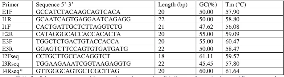
Primer Sequence 5’-3’ Length (bp) GC(%) Tm (°C)
E1F GCCATCTACAAGCAGTCACA 20 50.00 57.90
I1R GCAATCAGTGAGGAATCAGAGG 22 50.00 58.80
I1F CACTGATTGCTCTTAGGTCTG 21 47.62 56.08
E2R CATAGGGCACCACCACACTA 20 55.00 59.09
E3F TGGCTCTGACTGTACCACCA 20 55.00 60.47
E3R GGAGTCTTCCAGTGTGATGATG 22 50.00 58.47
I2Fseq CCTGCTTGCCACAGGTCT 18 61.11 59.57
I3Rseq TGGAAGAAATCGGTAAGAGGTG 22 45.45 57.80
[image:8.595.59.520.433.558.2]I4Rseq* GTTGGGCAGTGCTCGCTTAG 20 60.00 61.64
Table 2 - Primer sequences used for screening and sequencing. F indicates a forward primer and R a reverse primer.
Primers marked seq were used for sequencing. *Spans junction between exons 4 and 5.
RNA extraction, cDNA synthesis and RT-PCR
to12μl nuclease free water. The mixture was heated to 70°C for 5 minutes, immediately cooled to 4°C and then the following were added: 5 μl of 5xM-MLV buffer, 1.75 μl of 10mM dNTPs (ThermoFisher Scientific, R0191), 25 units of RNAse inhibitor (Promega, N2111) and 200 units of M-MLV reverse transcriptase and nuclease free water to make a final volume of 25 μl. The mix was then incubated in a thermocycler for 60 min at 37°C, after which the cDNA was stored at -20°C.
RT-PCR was performed with 1µL of cDNA template, 1 unit of AmpliTaq 360 DNA polymerase (Applied Biosystems, 4398818), 1X AmpliTaq buffer, 400nM of primers (E1F/E4R and E3F/E3R), 200µM of dNTPs and 1.75mM of MgCl2. To prevent any secondary structure on
the template that could have been caused by heteroduplex formation with the induced mutation, betain (Sigma Aldrich, B0300-1VL) at a final concentration of 1.3M and 1.3% DMSO PCR grade (Sigma Aldrich, D9170-1VL) were used in the RT-PCR. The cycling parameters were set at 94°C for 5 min, followed by 10 cycles of touchdown annealing temperature, decreasing 1°C/cycle (94°C – 15 seconds, 65 to 55°C – 30 seconds, 72°C – 40 seconds), 30 cycles of denaturation at 94°C for 15seconds, annealing at 60°C for 30seconds and extension at 70°C for 40 seconds and a final extension at 72°C for 5 minutes with storage at 4°C.
Cloning of PCR products
In order to get the highest quality sequencing data, PCR products containing amplified cDNA from exons 1 to 4 was cloned into a pCR2.1-TOPO vector after purification using GenEluteTM PCR Clean-Up Kit (Sigma Aldrich, NA1020-1KT) according to the manufacturer’s instructions.
For the ligation reaction, 46ng of purified PCR product were added to 10 ng pCR2.1-TOPO vector (Invitrogen, K450001), 1µL salt solution (1.2 M NaCl, 0.06 M MgCl2) and incubated for
ice for 30min and then heat shocked for 30 seconds at 42°C then quickly returned to ice for 5 min. Next, S.O.C. medium (2% Tryptone, 0.5% Yeast Extract, 10 mM NaCl, 2.5 mM KCl, 10 mM MgCl2, 10 mM MgSO4, 20 mM glucose) was added and the mixture incubated for 1 hour at 37°C, shaking at 230rpm before spreading onto pre-warmed selective plates of Luria Broth (LB) agar containing 100ug/mL of ampicillin (Invivogen, fas-am-s), coated with 40uL of 40mg/mL X-gal in dimethylformamide. After overnight incubation at 37°C, 12 white colonies were picked, inoculated in 5mL of LB containing 100µg/mL ampicillin (Invivogen, fas-am-b), incubated for 16h at 37°C and then used for plasmid DNA extraction following the instructions on the Genelute Plasmid Miniprep kit (Sigma Aldrich, PLN350).
Sequencing and analysis
The purified PCR products were diluted to 10ng/µL and cloned plasmids were diluted 100ng/µL in nuclease free water. Sanger sequencing was performed at the DeepSeq facilities in the School of Life Sciences at the University of Nottingham using dye terminator chemistry (BigDye version 3.1) on the 3130xl ABI PRISM Genetic Analyzer . The obtained sequences were analysed with BioEdit software version 7.2.5 and aligned to human genomic or transcript TP53 sequences using Mega software version 7.0.1.8. Translation of gene sequences of wild-type TP53 isoform A (NCBI RefSeq: NG_017013.2, Homo sapiens tumor protein p53 (TP53), RefSeqGene (LRG_321) on chromosome 17) and mutant clone G11A8 with a point deletion, was performed using the online translation tool Expasy (http://web.expasy.org/translate/) and the respective protein alignments were performed on Mega software version 7.0.1.8.
Western Blot
Wild-type and clone G11A8 HCT116 cells seeded at a density of 106 cells/well were
supernatant was collected and quantified by BCA assay (ThermoFisher Scientific, 23225). Thirty µg of protein were mixed with 4x LDS sample buffer (Invitrogen, 11549166), loaded into a 4-12% Bis-Tris Nupage Gel (Invitrogen, NP0321BOX) and run at constant voltage of 150V for 90 min. Protein was transferred onto a nitrocellulose membrane using a semi-dry transfer system (Bio-rad laboratories, Turbo-blot) which was incubated in blocking buffer (5% skimmed milk in TBST (Tris buffer saline, pH 7.6, 0.1% Tween 20)) for 1hour and then hybridised overnight at 4°C with mouse monoclonal TP53 antibody at 1:200 dilution (DAKO, clone do-7, M700101-2) and MDM2 at 1:500 dilution (Abcam, ab3110), diluted in blocking buffer. Mouse anti-human beta-actin (1:2000 dilution in blocking buffer, Sigma Aldrich, A5441) was used as a loading control and the membrane was incubated for 2 hours at room temperature. After 3x washes of 5min each in TBST, the membranes were incubated for 1 hour at room temperature with secondary HRP-conjugated Rabbit anti-mouse IgG (Sigma Aldrich, A6154), diluted 1:1000. After another 3x5min washes with TBST and incubation with ECL prime (GE Healthcare, RPN2236) for 5min, the membranes were scanned with a digital scanner (Li-cor, C-digit) to detect chemiluminescence.
Results
Identification of targeted cell pools
By using flow cytometry to detect FAM-6 labelled tracr transfected in the cells, we were able to detect and identify the molarities showing best FAM-6 labelling in the population of cells succesfully transfected with sgRNAcontaining FAM6-labelled tracr and Cas9 mRNA (Figure 3). The labelled population ranged from 1.67 to 4.55% in the pooled triplicates containing over 3x10⁶
Isolation of single cell clones
In total, 32 colonies were selected for further expansion, genomic DNA extraction and cryopreservation. The 3 pools of selected sgRNA1, sgRNA2 and sgRNA3 initially targeted cells were also cryopreserved to allow a repeated selection of single cell clones in case a mutant clone could not be detected. .
Detection of mutant clones by high-resolution melting (HRM)
HRM was used to screen for the presence of mutant clones isolated from the pools of sg RNA1,2 and 3 targeted cells. The use of normal template genomic DNA from untargeted HCT116 in the PCR reactions served as a baseline control to which shifts in melting temperatures could be compared. Of the 32 clones which were tested, 12 (37.5%) showed aberrant metling (Figure 5).
Confirmation of indel mutation and its expression by sequencing
sequence created as a consequence of the c735delG mutation, when translated, is predicted to produce a truncated protein of 343 amino acids with a molecular weight of 41kDa (Fig 7C).
Confirmation of truncated TP53 protein and its functional effect
Discussion
In cancer biology, changes in gene dosage may have a significant effect on cell biology. Oncogenes such as -catenin depend on “just right” signalling for maximum activity[19], tumour suppressors, such as TP53, would be expected to confer some selective advantage following somatic mutation (prior to loss of heterozygosity) and some tumor suppressors, such as PARK2, appear to mediate effect through haploinsufficiency[20]. We have sought to develop an efficient protocol to establish models for gene dosage studies by taking advantage of recent technical developments in gene editing and combining these with FACS and HRM to enrich for the target population and screen for mutations. Using this methodology, we have induced a heterozygous truncating mutation in TP53 and shown that it causes a change in the pattern of MDM2 expression.
The first step in our method involved introduction of the sgRNA and the Cas9 mRNA into the cells through lipid-based transfection. This would result in a pulse of gene editing activity (lasting until the mRNA was degraded) rather than the constant editing activity which may be expected if inducing CRISPR/Cas9 machinery from a DNA plasmid expression vector. Therefore, the main advantage of this methodology is a reduction of off-targets gene editing as the translated Cas9 protein has a short half-life and reduced editing time[14].
After single cell cloning, the next step was to screen the clones for the presence of somatic
TP53 mutation. Rather than performing genomic cleavage detection (GCD) on targeted cells, we
with double mutations from single mutations. In contrast, GCD could not do this since the mismatches leading to heteroduplex formation will be approximately in the same position whether single or double mutations and this will lead to cleavage fragments of the same size,
Aberrant melting patterns were observed for 37.5% of the clones. Depending on the cell line and transfection method, this is commensurate with the range of efficiencies found by Liang et al targeting HPRT using the Cas9 mRNA format in several established and primary human cell lines (0-70%), even though efficiency of gene editing in HCT116 cell line was not tested[14]. However, in the study by Liang et al, the induced mutations were not tested in individual single cell colonies but were inferred from PCR product cleaved fragment density from PCR performed on pooled colonies, therefore our results are not in comparable terms.
In summary, we have combined several methods to develop a CRISPR/Cas9 -based system for creating models for studying gene dosage effects. The methodology was used to create a heterozygous truncating mutation of TP53 which resulted in changes in MDM2 expression. Our protocol is rapid, it will probably reduce off-target effects, FACS enrichment allows isolation of a single cell clone with a high probability of mutation and HRM allows mutation detection in both the heterozygous and the homozygous state.
Key messages
A gene dosage model was developed by applying a variant of the CRISPR/Cas9
method involving the use of Cas9 mRNA and FAM6 labelled sgRNA.
High-Resolution Melting (HRM) was applied to screen for mutant clones instead of
enzymatic Genomic Cleavage Detection.
A TP53 heterozygous truncating mutation was created, resulting in changes in
MDM2 expression.
The authors would like to thank the technical assistance of Dr Matthew Carlile of the University of Nottingham DNA Sequencing Laboratory, School of Life Sciences, and Nicola Croxall at the Flow Cytometry facility, School of Medicine, Queen’s Medical Centre, Nottingham as well as the support of Dr. Gurpreet Balrey (Merk, UK).
Competing Interest: None declared.
REFERENCES
1 Garneau JE, Dupuis M-È, Villion M, et al. The CRISPR/Cas bacterial immune system cleaves bacteriophage and plasmid DNA. Nature 2010;468:67–71. doi:10.1038/nature09523
2 Long KB, Beatty GL. Harnessing the antitumor potential of macrophages for cancer immunotherapy. Oncoimmunology 2013;2:e26860. doi:10.4161/onci.26860
3 Kelley ML, Strezoska Ž, He K, et al. Versatility of chemically synthesized guide RNAs for CRISPR-Cas9 genome editing. J Biotechnol 2016;233:74–83. doi:10.1016/j.jbiotec.2016.06.011
4 Sampson TR, Weiss DS. Exploiting CRISPR/Cas systems for biotechnology. Bioessays
2014;36:34–8. doi:10.1002/bies.201300135
5 Findlay GM, Boyle EA, Hause RJ, et al. Saturation editing of genomic regions by multiplex homology-directed repair. Nature 2014;513:120–3. doi:10.1038/nature13695
6 Jinek M, Chylinski K, Fonfara I, et al. A programmable dual-RNA-guided DNA endonuclease in adaptive bacterial immunity. Science 2012;337:816–21. doi:10.1126/science.1225829
7 Mali P, Yang L, Esvelt KM, et al. RNA-Guided Human Genome Engineering via Cas9.
8 Ran FA, Hsu PD, Wright J, et al. Genome engineering using the CRISPR-Cas9 system. Nat
Protoc 2013;8:2281–308. doi:10.1038/nprot.2013.143
9 Hsu PD, Scott DA, Weinstein JA, et al. DNA targeting specificity of RNA-guided Cas9 nucleases. Nat Biotechnol 2013;31:827–32. doi:10.1038/nbt.2647
10 Yu X, Liang X, Xie H, et al. Improved delivery of Cas9 protein/gRNA complexes using lipofectamine CRISPRMAX. Biotechnol Lett 2016;38:919–29. doi:10.1007/s10529-016-2064-9
11 Zuris JA, Thompson DB, Shu Y, et al. Cationic lipid-mediated delivery of proteins enables efficient protein-based genome editing in vitro and in vivo. Nat Biotechnol 2014;33:73–80. doi:10.1038/nbt.3081
12 Kim S, Kim D, Cho SW, et al. Highly efficient RNA-guided genome editing in human cells via delivery of purified Cas9 ribonucleoproteins. Genome Res 2014;24:1012–9. doi:10.1101/gr.171322.113
13 Kouranova E, Forbes K, Zhao G, et al. CRISPRs for Optimal Targeting: Delivery of CRISPR Components as DNA, RNA, and Protein into Cultured Cells and Single-Cell Embryos. Hum Gene Ther 2016;27:464–75. doi:10.1089/hum.2016.009
14 Liang X, Potter J, Kumar S, et al. Rapid and highly efficient mammalian cell engineering via Cas9 protein transfection. J Biotechnol 2015;208:44–53. doi:10.1016/j.jbiotec.2015.04.024
15 Naccarati A, Polakova V, Pardini B, et al. Mutations and polymorphisms in TP53 gene--an overview on the role in colorectal cancer. Mutagenesis 2012;27:211–8. doi:10.1093/mutage/ger067
16 Liu Y, Bodmer WF. Analysis of P53 mutations and their expression in 56 colorectal cancer cell lines. Pnas 2006;103:976–81. doi:10.1073/pnas.0510146103
high-resolution melting: a simple and robust method for mutation detection in formalin-fixed paraffin-embedded tissue. J Clin Pathol 2010;63:134–40. doi:10.1136/jcp.2009.070508
18 Ebili HO, Hassall J, Asiri A, et al. QMC-PCRx: a novel method for rapid mutation detection. J Clin Pathol 2017;:jclinpath-2016-204264. doi:10.1136/jclinpath-2016-204264 19 Cho K-H, Baek S, Sung M-H. Wnt pathway mutations selected by optimal β-catenin
signaling for tumorigenesis. 2006. doi:10.1016/j.febslet.2006.05.053
20 Poulogiannis G, Mcintyre RE, Dimitriadi M, et al. PARK2 deletions occur frequently in sporadic colorectal cancer and accelerate adenoma development in Apc mutant mice. Published Online First: 2010. doi:10.1073/pnas.1009941107