Ajax E. George1 Mony J. de Leon2 Cynthia I. Gentes2 Jeff Miller 1.2 Eric London2 Gleb N. Budzilovich3
Steven Ferris2 Norman Chase 1
Received July 24, 1985; accepted after revision January 14, 1986.
Presented in part at the 22nd annual meeting of the American Society of Neuroradiology, Boston, June 1984.
This work was supported by Grant MH 36969 awarded by the National Institute of Mental Health, Dept. of Health and Human Services.
1 Department of Radiology (Neuroradiology), New York University Medical Center, 560 First Ave., New York, NY 10016. Address reprint requests to A. E. George.
2 Department of Psychiatry (Geriatric Study and
Treatment Program), New York University Medical Center, New York, NY 10016.
3 Department of Pathology (Neuropathology), New York University Medical Center, New York, NY 10016.
AJNR 7:561-566, July/August 1986 0195-6108/86/0704-0561
© American Society of Neuroradiology
Leukoencephalopathy in
Normal and PathologiC
Aging:
1. CT
of
Brain Lucencies
561
Central white matter lucencies are commonly seen in CT scans of elderly patients. Reports in the literature have implicated demyelination due to subcortical vascular disease (Binswanger disease) as the cause of these lucencies. Binswanger disease, however, is thought to be rare. Because of this apparent discrepancy we decided to determine the incidence and to attempt to define the clinical significance of the CT white-matter changes in a study population at New York University Medical Center. The studies of 275 normal and demented subjects, ages 23 to 85 years, were reviewed. All subjects received neurologic, psychiatric, and medical evaluation, formal psychometric evaluation of their cognitive status, and a CT scan. CT scans were evaluated for the presence and severity of white-matter changes (leukoencephalopathy). The incidence and severity of white-matter changes increased significantly with age (p < 0.01). Leukoencephalopathy was consistently more common in demented patients than in normal subjects, but the difference was not statistically significant, and the severity of the leukoencephalopathy was not related to the severity of dementia (p > 0.05). Five patients (ages 74 to 95 years) with a clinical diagnosis of Alzheimer disease who had CT evidence of lucencies were examined at autopsy. Neuropathology demonstrated extensive changes of Alzheimer disease in one brain and mild-to-moderate changes in the other four brains; areas of white-matter rarefaction were present in all brains, with microscopic evidence of arteriolar hyalinization. This study demonstrates that leukoen-cephalopathy is strongly related to the aging process and is seen in both "normal" and cognitively impaired individuals who have no other evidence of vascular disease. White-matter lucencies are not necessarily associated with dementia, but their increased incidence in Alzheimer disease patients and the neuropathologic results suggest that their presence may represent a risk factor potentiating the effects of Alzheimer disease.
Improved contrast resolution of currently available CT scanners has resulted in the frequent demonstration of white-matter lucencies in the brains of elderly patients. Evidence to date based on limited autopsy series [1-6] suggests that this leukoencephalopathy reflects the changes of subcortical vascular encephalopathy or Binswanger disease. This entity, originally described in 1894 [7], is thought to be a rare occurrence associated with stroke, hypertension, and dementia [3, 5]. Binswanger himself identified only eight cases in 11 years. The pathologic changes [3, 5] include patches of demyelination and cyst formation associated with hyalini-zation of medullary arterioles. We have noted that CT white-matter lucencies are commonly present in our study patients with clinically diagnosed Alzheimer disease (senile dementia of the Alzheimer type, or SDAT) who have no history of hyperten -sion or stroke, as well as in normal elderly subjects. We therefore undertook clinical, radiologic, and neuropathologic studies of normal and demented subjects in order to determine the incidence of CT white-matter lucencies and to attempt to define the clinical and pathologic significance of these changes.
Subjects and Methods
this screening protocol and using the exclusion criteria described below, all subjects that were diagnosed as normal or as presumed Alzheimer disease (SDAT) were selected for study.
The control subjects were from two cognitively normal groups: an elderly group, consisting of 89 subjects, 44 men and 45 women
(mean age, 69.5 years), and a young group, consisting of 35 men
(mean age, 25.0 years).
The Alzheimer subjects consisted of 151 demented, elderly
pa-tients, 88 women and 63 men (mean age, 72.5 years) who were
diagnosed clinically as having SDAT.
All subjects with history of stroke or with neurologic or CT evidence
of stroke were excluded. Subjects with hypertensive and/or cardiac disease requiring medication other than diuretic drugs were excluded.
Also excluded were subjects with diabetes requiring treatment with insulin or oral medication Those with diabetes under dietary control
were not excluded from the study.
Medical-Neurologic Evaluation
The clinical evaluation included history and physical examination,
routine blood and urine analyses, and a determination of blood pressure. Subjects with blood pressure of 150 systolic over 90 diastolic or less were considered normotensive. Subjects with systolic or diastolic pressure above these levels were considered
hyperten-sive. After complete neurologic evaluation the presence or absence of gait disturbance was assessed and recorded by the examining neurologist. Historic and physical evidence of heart disease, diabetes, or peripheral vascular disease was also tabulated as either present
or absent.
Psychometric Evaluation of Cognitive Status
Each study subject received a psychiatric and neuropsychological test battery, which has been in use in our laboratory for 10 years.
This examination included the Global Deterioration Scale (GDS) [8] and the Guild Memory Test. The Guild includes a measure of vocab-ulary, immediate and delayed recall of paragraphs, paired associates,
digit span, and memory for designs. A score on the GDS that indicated moderate to severe symptomatology (GDS >4) was required for a patient to be included in the dementia group.
CT Examination
CT scans were obtained for all patients and normal controls on the General Electric 8800 CT scanner (General Electric, Milwaukee, WI).
Scans were obtained without intravenous contrast administration with the scanning plane parallel to the canthomeatal line. Scan parameters were mA = 200 to 320, scan time = 9.6 sec, slice thickness = 10 mm. Hard-copy images were obtained at constant window (80 H) and level (40 H) settings.
Subjective assessment for the presence or absence of lucency
was done independently by two observers who were unaware of the patients' clinical status. Subsequently, the observers evaluated the severity of leukoencephalopathy with a seven-point rating scale using hard-copy images. Scans were rated as demonstrating no, minimal, mild, mild-to-moderate, moderate, moderate-to-severe, or severe
leu-koencephalopathy. "Minimal" implied faintly visualized lucencies,
usu-ally in the white matter anterior and lateral to the frontal horns or,
Fig. 1.-CT of mild leukoencephalopathy in
83-year-old woman. Faint lucencies (arrows) involve
white matter adjacent to frontal horns.
occasionally, in the peritrigonal areas. "Mild" (Fig. 1) implied lucencies occupying less than one-third of the visualized white matter of the frontal and peritrigonal areas. "Moderate" implied involvement of approximately one-half of the visualized white matter. "Severe" im-plied involvement of virtually all visualized white-matter regions (Fig.
2).
Neuropathology
As part of a larger ongoing neuropathology study of Alzheimer disease, postmortem examinations were performed on five patients with clinical diagnosis of Alzheimer disease who had CT evidence of leukoencephalopathy prior to death. The brains were fixed in 10% formalin for 3 weeks, in accordance with standard procedure, and then sectioned in the coronal plane. Microscopic examination of multiple brain samples was routinely performed. This included the neocortex with subcortical and deep white matter of the frontal, temporal, parietal, and occipital lobes. Also included for study were the basal ganglia, the nucleus basalis of Meynert, the hippocampi,
the hypothalamus, the thalami, the lower brainstem, and the cerebel-lum. In addition, using the CT for localization, we sampled the specific sites of leukoencephalopathy. All sections were routinely stained with hematoxyline and eosine. Luxol-fast-bluejPAS and silver impregna-tion methods were used in the study of selected blocks.
Results
Incidence of Leukoencephalopathy
[image:2.613.395.560.79.293.2]AJNR:7, July/August 1986 CT OF BRAIN LUCENCIES 563
A
B
c
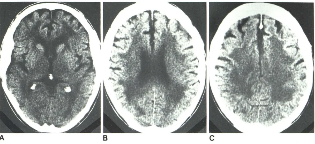
Fig. 2.-CT of severe leukoencephalopathy in 70-year-old man. Virtually ali visualized white matter demonstrates lucencies at basal ganglia (A), ventricular (B), and centrum (C) levels.
incidence of lucencies was found with increasing age. With
the subjects divided into two age groups (55-69 and 70-85 years) for determining the presence or absence of lucencies, we found that in the Alzheimer patients the incidence of
lucency increased from 18% in the 55-69 age group to 35%
in the 70-85 age group (chi square = 4.53 P
<
0.05) (Fig. 3).The incidence of leukoencephalopathy among normal older
patients increased from 7% in the 55-69 age group to 24% in the 70-85 age group (chi square = 5.21 P
< 0
.05). For theAlzheimer patients as compared with the normal controls (Fig.
3), there was a nonsignificant trend toward a higher incidence
of leukoencephalopathy for both the younger (chi square =
2.75 P
>
0.05) and the older (chi square = 1.69 P>
0.05) groups. These results indicate that the incidence of leukoen-cephalopathy increases with increasing age in both normal subjects and in patients with presumed Alzheimer disease.The anatomic distribution of lucencies in the Alzheimer group is shown in Table 2. These data indicate that involve-ment of the frontal white matter is present in 86% of cases with lucencies.
C/inical Correlates of Leukoencephalopathy
For both the normal subjects and patients with presumed
Alzheimer disease, the frequency of various clinical
parame-TABLE 1: Incidence of CT White-Matter Lucencies
No. of No. of
Group
Cases Mean Age (SD) Lucencies
(%)
Young normal 35 25.0 (± 2.0) 0 (0)
Old normal 89 69.4 (± 6.5) 14 (16)
Alzheimer 151 72.6 (± 7.5) 45 (30)
ters is tabulated in Table 3. Note that the incidence of hyper-tension in the normal population with leukoencephalopathy was 57% whereas only 25% of normals without leukoen-cephalopathy were hypertensive. Thus the presence of lucen-cies is twice as frequent in hypertensive normals. This was not the case in the Alzheimer patients, among whom 22%
without lucencies were hypertensive and 24% with lucencies were hypertensive.
The presence of leukoencephalopathy was not associated
with a significant increase in the associated incidence of heart disease, peripheral vascular disease, or diabetes.
We also found that leukoencephalopathy was associated
with a significantly increased incidence of gait impairment in
the Alzheimer disease patients.
Table 4 shows a correlation between leukoencephalopathy severity ratings and the above medical parameters and age
for both normal and Alzheimer groups combined. The leuko-encephalopathy rating (severity) showed no relationship to the degree of dementia. Severity of leukoencephalopathy was
of 50
TOTAL POPULATION 40
30
20
10 45
36
152 (TOTAL N= 151)
55-69 70-85
AGE
Fig. 3.-CT leukoencephalopathy; percent of affected normal individuals
[image:3.612.55.560.79.310.2] [image:3.612.316.559.599.710.2] [image:3.612.53.297.672.734.2]Diffuse 15
significantly related to age and also to the presence or ab-sence of gait impairment. Thus, the older the subject, the greater the likelihood the leukoencephalopathy would be se-vere and the subject's gait would be impaired.
Neuropathology
Neuropathologic findings from five patients clinically diag-nosed as having Alzheimer disease are summarized in Table 5. These patients had CT evidence of lucencies that ranged in severity from mild to severe. Their ages at death ranged from 74 to 95 years and all subjects were severely demented with GDS ratings of either 6 or 7. At autopsy, white-matter rarefaction and focal spongy changes were present in all cases. On histologic examination, all brains demonstrated typical features of subcortical vascular disease; that is, de-myelination, hyalinization of medullary arterioles, and cystic degeneration corresponding to the white-matter lucencies demonstrated by CT. Arteriolar changes ranged from mod-erate to severe.
Alzheimer changes were also present at postmortem in all five subjects. The distribution and severity of senile plaques and neurofibrillary tangles are given in Table 5. One patient (subject 4), a 75-year-old woman with severe CT lucencies at postmortem (Fig. 4) showed extensive evidence of Alzheimer disease as well as extensive concomitant white-matter changes. The patient had had long-standing hypertension. The brain after fixation weighed 1200 grams. The white matter of the centrum semiovale of the anterior two-thirds of both frontal lobes was soft and finely porous. Both hippocampal formations showed approximately 50% reduction in size.
Microscopically, arteriolar hyalinization was moderate. No infarcts or lacunes were present. A single 2-mm hemorrhage,
which was assumed to be of hypertensive origin, was found in the inferior extremity of the right postcentral gyrus. Alz-heimer changes consisted of numerous plaques and tangles in the neocortex. Tangles were also seen in the nucleus basalis and hypothalamic nuclei and in the upper and lower brainstem.
In the other four autopsied brains, Alzheimer changes ranged from mild (subjects 5 and 1) to moderate (subjects 2 and 3). In subjects 5 and 1, a discordance is suggested between the degree of dementia, which was severe, and the severity of Alzheimer changes at autopsy, which was mild (Table 5). For example, in subject 5, a 74-year-old woman, only a few plaques and tangles were present in the cortex with rare tangles in the nucleus basalis. CT leukoencephalop-athy was severe and corresponded to the pathologic findings of moderate-to-severe arteriolar hyalinization and
moderate-Heart disease 14 8 0.02
Peripheral vascular 15 15 0.00
Diabetes 8 0 0.22
Gait impairment 3 8 0.00
Alzheimer patients (n
=
106) (n=
45)Hypertension 22 24 0.05
Heart disease 18 16 0.05
Peripheral vascular 16 24 1.00
Diabetes 6 0 1.07
Gait impairment 20 45 6.81*
• p < 0.05
TABLE 4: Severity Rating of Leukoencephalopathy vs Clinical
Measures, Pearson Product Correlations: Normal Subjects and
Alzheimer Patients Combined (n
=
58)Age
Mental status (GDS) Hypertension Heart disease Peripheral vascular Gait impairment
··p<0.01 'p < 0.05
0.36
*-0.09 (NS) 0.09 (NS) -0.16 (NS) -0.04 (NS)
-0.32
-Note.-GDS = Global Deterioration Scale; NS = nonsignificant.
to-severe leukoencephalopathy. The white matter was finely porous with ill-defined, 4- to 6-mm cysts present bilaterally.
Discussion
Subcortical vascular encephalopathy was recently reported at autopsy [5] in only 3.8% of patients over the age of 60.
We report CT evidence of leukoencephalopathy in 30% of our patient population with clinical diagnosis of Alzheimer disease and in 16% of normal, age-matched controls. We excluded all subjects who had historiC, physical, or CT evi-dence of stroke. The increased incidence of lucencies in the Alzheimer group, though not statistically significant, does raise the possibility that the subjects are predisposed to the dementing process of Alzheimer disease, perhaps causing earlier manifestation of the dementia or more severe dementia than in patients without lucencies. This hypothesis is sup-ported by the neuropathologic findings. All five autopsy pa
[image:4.612.315.562.102.236.2] [image:4.612.53.299.103.149.2] [image:4.612.314.560.309.371.2]AJNR:7, July/August 1986 CT OF BRAIN LUCENCIES 565
TABLE 5: Neuropathology Cases
Case Clinical Severity Alzheimer Plaques Changes in CT
Age Gender Neurofibrillary Tangles
No. of Dementia (NEO/HIP)
(NEO/HIP) Lucencies 1 95 M Severe Mild/Mild Minimal/Mild Mild 2 79 M Severe Moderate/Moderate Minimal/Mild Minimal 3 82 F Severe Moderate/Normal Moderate/Normal Minimal 4' 75 F Severe Moderate/Moderate Minimal/Moderate Severe
5' 74 F Severe Minimal/Minimal Normal/Mild Severe
• Hippocampal cell loss was severe (estimated 40-50%).
Note.-NEO = neocortex: HIP = hippocampus. All subjects prior to death were rated GDS (Global Deterioration Scale) 6 or 7. 6 implies the subject required assistance and had difficulty communicating. 7 implies loss of all verbal and psychomotor abilities. At postmortem, all subjects demonstrated moderate to severe white-matter changes (see text). For Alzheimer changes, minimal = occasional
or rare lesions; mild = scattered lesions; moderate = lesions regularly seen. For CT, minimal = faint lucencies; mild = definite lucencies
involving less than one-third of white matter; severe = diffuse involvement of virtually all white matter.
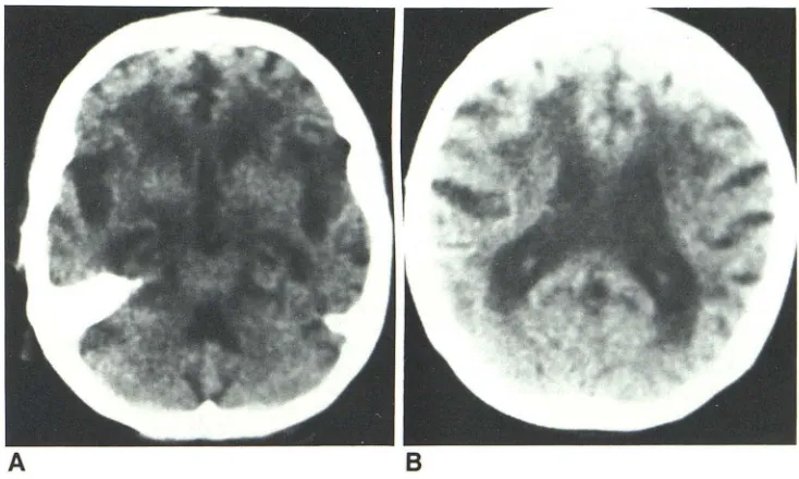
Fig. 4.-CT of neuropathology case 4,
obtained 2 months before death.
Coex-istent Alzheimer disease and
leukoen-cephalopathy; 75-year-old chronically hy-pertensive, demented woman. A, Low
basal ganglionic level. B, Ventricular body
level. At postmortem, both white-matter
and Alzheimer changes were found to be
extensive.
A
discrepancy is suggested in two subjects (1 and 5, Table 5) between the severity of Alzheimer disease changes and the
degree of dementia. At autopsy, subject 5, a 74-year-old
woman, showed only mild evidence of Alzheimer disease and moderate-to-severe leukoencephalopathy. In view of this pa-tient's clinically severe dementia, it is postulated that the white-matter disease contributed to the severity of the de
-mentia. A similar effect is suggested in subject 1, who was a severely demented 95-year-old man. At postmortem, there were mild Alzheimer changes and evidence of moderate white-matter changes. Therefore, in these two cases the presence of leukoencephalopathy may have resulted in the
appearance of dementia earlier or with greater severity than if leukoencephalopathy were not present.
An analysis of the medical profiles of all subjects revealed that the lucencies were associated with double the incidence
of hypertension in normal individuals but not in the Alzheimer group. We postulate that this apparently unexpected result
most likely reflects an inability of the elderly Alzheimer patients to tolerate hypertension superimposed on leukoencephalop-athy and Alzheimer disease. Thus, many patients with this
8
combination may have been either too disabled to participate in the research program or may have sustained a stroke and
therefore were excluded from the study. Seventy-six percent of the Alzheimer patients with lucencies and 43% of the
normal subjects with lucencies were normotensive. The inci -dence is higher than that reported by Goto et al. [3], who noted that only two of 10 autopsied patients with diffuse white-matter disease had been normotensive.
The increased incidence of gait impairment in the patients with Alzheimer disease and leukoencephalopathy is also of interest. Alzheimer patients without lucencies, by virtue of their disease alone, showed more than double the incidence of gait impairment (20%) seen in normal patients. That inci-dence was again doubled if leukoencephalopathy was also
present.
Summary
The studies of 275 normal and cognitively impaired subjects
[image:5.612.189.556.249.469.2]-cardiac disease. Leukoencephalopathy was more common in
patients with presumed Alzheimer disease, showing a
nonsig-nificant trend when compared with normal subjects;
leuko-encephalopathy severity was
not
related to the degree ofdementia. The presence of CT lucencies was associated with
double the incidence of hypertension in normal subjects and more than double the incidence of gait impairment in patients
with Alzheimer disease. Neuropathologic evidence, including
our own study of lucency cases, indicates that CT
leukoen-cephalopathy represents demyelination in association with
subcortical vascular disease. The presence of
leukoenceph-alopathy, though perhaps not itself causing dementia, may
potentiate the dementing effects of Alzheimer disease and
may therefore be a risk factor for dementing disorders.
ACKNOWLEDGMENT
We thank Joan Askew for her assistance in the preparation of this
manuscript.
2. De Reuck J, Crevits J, DeCosta W, Wieben G, VanderEcken H.
Patholgenesis of Binswanger chronic, progressive, subcortical
encephalopathy. Neurology 1980;30:920-928
3. Goto K, Ishiin, Fukasawa H. Diffuse white-matter disease in one
geriatric population. Radiology 1981;141 :687-695
4. Caplan LR, Schoene WC. Clinical features of subcortical arterio
-sclerotic encephalopathy (Binswanger's disease). Neurology
1978;28: 1206-1215
5. Tomonaga BM, Yamamouchi H, Tohgi H, Kameyama M. Clinical
pathologic study of progressive subcortical vascular
encephalop-athy (Binswanger type) of the elderly. J Am Geriatr Soc
1982;30(8): 524-529
6. Zeumer H, Schon sky B, Sturm KW. Predominant white matter
involvement in subcortical arteriosclerotic encephalopathy
(Bin-swanger disease). J Comput Assist Tomogr 1980;4(1): 14-19
7. Binswanger O. Die abregenzung der allgemeinen progressiven
paralyse. Berl Klin Wochenschr 1894;31 :1103-1186
8. Reisberg B, Ferris SH, deLeon MJ. The global deterioration scale
(GDS): an instrument for the assessment of primary degenerative